mercoledì 29 novembre 2006
Gliomatosis cerebri
Findings
Poorly defined, diffusely increased signal is noted on T2-weighted images (Figure 1) involving predominantly the right frontal and parietal lobes. This abnormal signal involves predominantly the white matter but extends to the adjacent cortex, to the insular cortex and inferiorly involves the thalamus and posterior limb of internal capsule to reach the upper mid brain.
These lesions are iso- to hypo-intense on T1WI (Figure 2). For the size of the lesion, there is not much distortion of the anatomy and there is little mass effect on the ipsilateral lateral ventricle with slight shift of midline to the left.
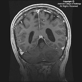
On the post contrast images (Figures 3, 4 and 5), there are only scattered foci of enhancement in the centrum semiovale and parasagittal gray matter (Figure 3). Foci of enhancement likely represent areas of dedifferentiation.
Figure 6: MR Spectroscopy reveals slight increase in the choline peak and slight reduction of NAA peak.
Diagnosis: Gliomatosis cerebri
Gliomatosis cerebri is an uncommon primary brain tumor characterized by diffuse neoplastic proliferation of astrocytes, with relative preservation of underlying brain architecture and sparing of neurons. By definition, it is infiltrative, involving at least two lobes of the brain, frequently bilateral. Usually WHO Grade III, it is differentiated from multifocal gliomas based on continuity of cellular infiltration and lack of clear distinction from adjacent normal brain tissue.
Patients usually present between the 3rd and 5th decades of life. Clinical findings are nonspecific and characteristically mild in comparison with imaging findings. The process shows a relentless progression over time and typically presents with a slow decline in cognitive function.
The areas infiltrated by neoplastic astrocytes include (in decreasing order of frequency) the cerebral hemispheres, basal ganglia, thalami, corpus callosum, brainstem, spinal cord and cerebellum. The tumor typically crosses the midline and involves at least two, usually three lobes of the brain. Hemispheric white matter involvement is typical, but the cortex may also be involved in approximately 20% of cases.
CT may demonstrate normal findings or may show ill-defined asymmetric low density with minimal or no enhancement. MRI is more sensitive, but also often underestimates the extent of disease. The tumor appears as infiltrating hyperintense mass on T2 WI with enlargement of involved structures. Enhancement may indicate malignant progression or a focus of malignant glioma. Vascular proliferation and necrosis are typically absent.
MRS and dynamic contrast-enhanced T2*-weighted imaging help further characterize the tumor.
Stereotactic biopsy may be guided by an enhancing nodule if present, or by the area of maximum Choline/NAA increase.
The prognosis is generally poor with 50% mortality at one year. Steroid therapy may be helpful in the short term. Radiation therapy and chemotherapy are of questionable benefit.
martedì 21 novembre 2006
Pyriform aperture stenosis
Findings
There is narrowing of the anterior nasal passage (Figure 1 and Figure 2) due to overgrowth of the nasal processes of the maxilla (Figure 1 and Figure 2). The nasal septum appears thinned.
Diagnosis: Pyriform aperture stenosis
Pyriform aperture stenosis is an uncommon cause for nasal obstruction in the newborn period. The patient will typically present with dyspnea or apneic spells during feeding or the inability to pass a nasogastric tube. Clinicians will often suspect choanal atresia as this is a more common cause of these symptoms. The diagnosis is easily made with thin section CT images through the maxillofacial region which will demonstrate narrowing or obliteration of the anterior nasal passage with a normal appearing posterior nasal caliber and choana. A pyriform aperture width of less than 11 millimeters is diagnostic of stenosis.
The differential diagnosis for a child with nasal obstruction should include choanal atresia, choanal stenosis, pyriform aperture stenosis, nasopharyngeal encephalocele, sinonasal polyposis or mucous impaction.
A single frontal megaincisor is sometimes associated with pyriform aperture stenosis and is considered within the spectrum of holoprosencephaly. Therefore, when this finding is noted further imaging of the brain should be performed to rule out holoprosencephaly. The premaxillary portion of the maxilla originates from midline mesodermal tissue. The mesoderm is thought to have an inductive effect on the forebrain, causing this association.
lunedì 13 novembre 2006
Progressive Multifocal Leukoencephalopathy (PML)
Findings
MRI shows multiple foci of hypointensity on T1 and hyperintensity on T2 and FLAIR throughout the subcortical white matter of bilateral frontal, temporal and parietal lobes. Similar lesions are seen in left pons and right cerebellar hemisphere. Figure 2 and 3, which are FLAIR images, demonstrate well the involvement of the subcortical U-fibers. There is no significant mass effect associated with these lesions. No appreciable enhancement is seen on postcontrast images (Figure 4, Figure 5, Figure 6, and Figure 7).
Diagnosis: Progressive Multifocal Leukoencephalopathy (PML)
Progressive Multifocal Leukoencephalopathy (PML) is a fulminating opportunistic infection of the brain caused by JC virus. The virus is commonly latent in the central nervous system (up to 80% of adult population is infected) but targets the oligodendrocytes in immune compromised patients. It occurs in AIDS patients with very low (less than 50-100) CD4 counts.
PML typically results in progressive neurologic decline in AIDS patients, but can be the presenting illness.
The need to diagnose and treat PML is urgent in patients with HIV as the infections are synergistic. Brain biopsy was previously required for definitive diagnosis. With positive JCV Polymerase Chain Reaction CSF results and MR findings characteristic of PML, brain biopsy can be avoided in many AIDS patients.
On CT, it presents as focal, bilateral, asymmetric, usually well-demarcated areas of low attenuation, typically in the subcortical and periventricular white matter.
MRI reveals asymmetric, multifocal areas of T1 and T2 prolongation in the periventricular and /or subcortical white matter. Involvement of subcortical U-fibers is common, giving a “scalloped” appearance to the lateral margins of the lesions. Classically, the lesions occur in the parieto-occipital white matter. However, lesions may involve any part of cerebral hemispheres as well as brainstem, cerebellum, and basal ganglia. Mass effect and enhancement are usually absent or mild, if present. Marked mass effect or robust contrast enhancement should suggest alternative diagnoses.
HIV encephalitis is the leading diagnostic possibility in the differential diagnosis of non- enhancing, non-mass-producing white matter lesions in patients with AIDS. White matter changes in HIV encephalitis are usually symmetrical and diffuse. Posterior fossa is also uncommonly involved with HIV encephalitis.
There is no specific therapy for PML, and it was previously considered to be fatal with median survival being around four months. Improved patient survival has been reported with HAART (highly active antiretroviral therapy).
giovedì 9 novembre 2006
Cavernous hemangioma
Findings
Figure 1 and Figure 5: T1 axial and coronal images demonstrate the soft tissue mass iso-intense to the adjacent muscles located predominantly in the extraconal plane with minimal intraconal extension.
Figure 2: T1 coronal post contrast with fat saturation demonstrates enhancement of the cavernous hemangioma.
Figure 4: T1 axial postcontrast image demonstrates peripheral hypointense margin from pseudocapsule (arrow at the site of pseudocapsule).
Figure 3: T2 coronal images demonstrating thin septa within the cavernous hemangioma.
Figure 6: Catheter angiogram in arterial phase does not show any feeding arteries.
Figure 7: Catheter angiogram delayed phase shows pooling of the contrast within the cavernous hemangioma.
Diagnosis: Cavernous Hemangioma
Cavernous hemangioma is the most common congenital vascular malformation of the orbit. Female predominance is appreciated with female to male ratio as high as 7:3. Hormonal factors have been implicated as there is more rapid growth during pregnancy. They grow slowly and are composed of multiple endothelial and smooth muscle lined vascular channels. Cavernous hemangiomas are well circumscribed round or oval lesions with well defined margins. They have a pseudo capsule from the surrounding compressed tissue.
The common clinical presentation is painless proptosis. Diplopia, visual impairment, increased intraocular pressure are seen with larger lesions.
Cavernous hemangiomas are more often intraconal. Larger lesions have extraconal extension. Primary extraconal lesions are also reported. Intraosseous hemangiomas are rare.
CT demonstrates the location of the lesion and micro calcifications. Phleboliths and macroscopic calcifications are rare. Remodeling of the bone is seen in long-standing cases. On MRI homogeneous soft tissue mass, which is isointense to the muscle on T1WI and hyperintense on T2WI with well-defined septations, can be seen especially in large lesions. Hypointense pseudo capsule is very visible along the periphery of the lesion.
Heterogeneous patchy enhancement is seen with contrast with more homogeneous enhancement on delayed imaging. Angiography demonstrates no obvious distinct tumor blush during the arterial phase. Contrast puddling is seen, which extends to the late venous phase, essentially isolated from systemic circulation.
Treatment options include observation only for stable lesions and lesions without significant symptoms. Intralesional laser, cryosurgical and radio surgical techniques are other alternatives in addition to surgery. Surgical resection is indicated for visual disturbance, cosmetic reason, and any symptoms related to mass effect. Recurrence rate is very low after surgery.
venerdì 3 novembre 2006
Lhermitte-Duclos disease (LDD)
Findings
There is a well-defined lesion in the left cerebellar hemisphere, slightly hypointense to the brain parenchyma on T1-weighted images and slightly hyperintense on the T2 sequence (Figure 1 and Figure 2). Cerebellar folia are clearly demonstrated coursing through the lesion. There is no mass effect or surrounding edema. Minimal enhancement is seen on postcontrast administration (Figure 5).
The lesion is bright on diffusion-weighted images as well as on the ADC map (Figure 6 and Figure 7).
Diagnosis: Lhermitte-Duclos disease (LDD)
LDD, also known as dysplastic gangliocytoma of the cerebellum and granular cell hypertrophy, is a rare cerebellar mass lesion which has hamartomatous as well as neoplastic characteristics.
LDD may be sporadic or associated with Cowden disease, a rare autosomal dominant familial cancer syndrome with multiple manifestations, including trichilemmomas, diverse hamartomas, intestinal polyposis, palmoplantar keratoses, oral papillomatosis, and an increased predisposition to breast cancer and thyroid tumors.
Patients usually present as young adults with occipital headaches, ataxia and papilledema.
The lesions usually show unilateral cerebellar hemispheric involvement. CT demonstrates low-density cerebellar mass that may contain calcifications. There may be thinning of the ipsilateral occipital bone.
MRI shows the characteristic “striated” appearance with widened cerebellar folia. Striated pattern of hypointensity on T1-weighted imaging and hyperintensity on T2-weighted imaging alternates with isointense bands of tissue. Postcontrast enhancement is rare. No diffusion disturbance is seen on ADC maps. MR perfusion may show elevated rCBV.
The lesion shows elevated 18-FDG uptake on PET scans.
Mass effect by the lesion may cause hydrocephalus and tonsillar herniation. Surgery is typically required in cases of brainstem compression to reduce mass effect upon the fourth ventricle. Regrowth of the lesion following resection has been reported, so careful follow up is advisable.