giovedì 28 settembre 2006

Fibrous dysplasia (FD) of the skull


Findings

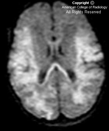
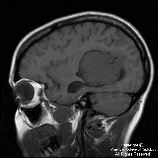
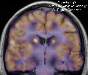
There is expansile lesion involving predominantly the left frontal and sphenoid bones (Figure 1 and Figure 2). The diploic space is widened with displacement of the outer table. This gives a “ground-glass” appearance with small lytic areas (Figure 1) dispersed within. This expansile, predominantly sclerotic lesion is seen to involve the sphenoid and ethmoid bones also (Figure 2 and Figure 3). The nasal cavity is distorted.
The bilateral anterior clinoid processes are involved by this expansile lesion (Figure 2), with mild narrowing of the bilateral optic canals (Figure 2).
No significant soft tissue mass is seen to be associated with the expansile bony lesion.
Figure 4: 3D rendering nicely demonstrates the abnormally deformed calvarium in this patient.


Diagnosis: Fibrous dysplasia (FD) of the skull


FD is an acquired and benign disorder in which the normal medullary space of an affected bone is replaced by fibrous tissue. The disorder usually manifests in childhood and adolescent years, and presents as either an incidental finding in asymptomatic patients or with nonspecific symptoms, such as pain, swelling, or tenderness. Skull involvement can lead to hearing impairment. There are monostotic (involving a single bone) and polyostotic (involving multiple bones) forms. The monostotic form is much more prevalent than the polyostotic form. FD can affect any bone; however. when solitary, the most common bone to be affected is the proximal femur, followed by the ribs and craniofacial bones. The polyostotic form can vary, and usually affects the femur, tibia, pelvis, and bones of the feet. The association of polyostotic fibrous dysplasia with precocious puberty in girls, and cafe-au-lait spots is termed the McCune-Albright syndrome. The combination of intramuscular myxomas and polyostotic fibrous dysplasia is called Mazabraud syndrome.

Because FD is a medullary process, the radiographic appearance consists of a lucency with an epicenter that is intramedullary or eccentric, but not cortical. The lesion can appear cystic and expansile with endosteal scalloping, but is always well defined without breaking through the bony cortex. The lucent lesion is usually described as having a ground-glass quality. Another classic appearance includes the shepherd’s crook deformity, which describes the appearance of the femoral neck that results when the disorder causes varus angulation. FD of the calvarium appears as increased density, usually at the skull base. Paget’s disease can give this appearance as well; Paget’s is generally bilateral, whereas FD is unilateral.

There is rare potential for malignant transformation, especially in the polyostotic form. When radiologic features such as cortical destruction, periosteal reaction, or a soft tissue mass are present at the site of prior FD, malignant degeneration should be suspected. Progression to osteosarcomas, fibrosarcomas, and malignant fibrous histiocytomas has been reported.

Most lesions appear low to intermediate signal on T1-weighted MR images. The signal varies on T2-weighted images, but is generally high in signal, especially with the presence of nonmineralized and cystic components. Enhancement is present, and is usually heterogeneous. Because the MR imaging appearance is variable and can imitate neoplasm, correlation with plain films or CT is helpful when fibrous dysplasia is suspected. MRI is useful for evaluating cranial neuropathy and suspected malignant transformation.

CT shows three overlapping patterns - pagetoid (mixed) pattern in 50%, sclerotic pattern in 25%, and cystic pattern in another 25% of cases. The lesions usually show nonspecific increased uptake of radiotracer on bone scans.

Involvement of the skull may cause cranial nerve dysfunction with visual and hearing impairment. Mucoceles may complicate paranasal sinus ostial bone involvement. The term "leontiasis ossea" describes a rare form of polyostotic disease that involves the frontal and facial bones. Another craniofacial entity, “cherubism,” is a hereditary fibrous lesion of bone, symmetrically involving the mandible and often the maxilla.

giovedì 21 settembre 2006

Pyogenic cerebral abscess


Findings

There is a well-defined, smoothly marginated, thinly rim-enhancing intraaxial mass (Figure 4) with surrounding edema (Figure 3) and mass effect centered in the right deep gray matter. The rim is essentially isointense to white matter on the T1-weighted image (Figure 1), hypointense on the T2-weighted image (Figure 2), and there is homogeneously increased signal intensity of the mass on the diffusion-weighted image (Figure 5).


Diagnosis: Pyogenic cerebral abscess


Most pyogenic cerebral abscesses are located supratentorially at the gray - white junction, secondary to hematogenous spread, and within the frontal and parietal lobes (middle cerebral artery distribution). Size is variable and may range from a few millimeters to many centimeters.

The characteristic imaging findings are illustrated in this case, but the pathogenesis and assoiciated imaging findings have been divided into four stages, which have been extensively published and are as follows:
- early cerebritis
- late cerebritis
- early capsule formation
- late capsule formation

Although hematogenous dissemination from an extracranial infection is the most common source, other potential etiologies to consider include: hematogenous spread from any right-to-left shunt, penetrating trauma, postoperative, and direct extension from a paranasal sinus, meningeal, calvarial, or otic infection.

These relatively uncommon lesions are potentially fatal but treatable, with treatment (often depending of the size of the lesion) typically ranging between systemic antibiotics alone and stereotactic aspiration or drainage. Reports of mortality vary, with published numbers between 0% and 30%. It is important to be aware of the complications of incompletely or inadequately treated abscesses and to look for them on MR.

These include:
- Intraventricular rupture with ventriculitis
- Meningitis with satellite abscess development
- Hydrocephalus
- Local mass effect or herniation.

lunedì 18 settembre 2006

Hypoxic ischemic encephalopathy


Findings

Figure 1 and Figure 2: Axial CT images show linear high attenuation subdural collections consistent with hematomas. No fractures were seen on bone windows.
Figure 3: Axial CT image shows an extracalvarial hematoma.
Figure 4, Figure 5, Figure 6, Figure 7, and Figure 8: DWI shows high signal demonstrating restricted diffusion in the thalami bilaterally (Figure 4). High signal is seen in the lateral temporal parietal regions (Figure 5), and the splenium of the corpos callosum (Figure 6), as well as the body of the corpus callosum (Figure 7), and the high parietal regions (Figure 8).


Diagnosis: Hypoxic ischemic encephalopathy


Hypoxic ischemic injury can present in a variety of patterns, including:
- Periventricular leukomalacia (PVL)
- Cortical watershed ischemic changes
- Deep gray matter ischemia
- Mixed pattern

Hypoxic ischemic injury results from a global hypoxic or ischemic insult to the brain in utero or during birth. Factors that can contribute to neonatal brain injury include placental abruption, meconium aspiration, cord prolapse, dystocia, eclampsia, maternal hypotension, and severe maternal bleeding.

Various patterns of brain injury have been described based upon pathological specimens.

They include:
(1) PVL that does not involve the cortex,
(2) injury to the parasagittal cerebral cortex
(3) involvement of the deep gray matter
(4) mixed injury pattern, which may or may not include hemorrhage.

Because the brain injury is global, the patterns of brain injury are, in general, bilaterally symmetric.

The imaging evaluation of neonates in whom a hypoxic injury is suspected, can include a variety of modalities including ultrasound, CT, and MRI. Ultrasound is a useful modality to detect PVL, which usually affects premature infants. PVL results in response to ischemia in the watershed areas of the brain, and presents as areas of increased white matter echogenicity, predominantly in a posterior distribution. The affected white matter can eventually develop small cystic cavities and, in severe cases, result in atrophy and ventriculomegaly. These findings may be evident on CT and MRI as well.

Whether there is involvement of the cortex or the deep gray matter, usually depends on the degree of the hypoxic insult. Severe injury tends to affect the deep gray structures, including the basal ganglia and thalami, which can show bright T1 weighted signal. Less severe injury will lead to ischemic changes in the cortical watershed areas. These affected areas will also show restricted diffusion. The corresponding areas are hypodense on CT, secondary to the presence of edema.

giovedì 14 settembre 2006

Cerebellopontine angle (CPA) vestibular schwannoma


Findings

Figure 1: Noncontrast-enhanced T1-weighted coronal MR image of the brain demonstrates a heterogeneous, well-defined, iso- to hypointense mass in the right CPA, which has some mass effect on the brain stem. The lesion fails to demonstrate high T1 signal to suggest fat or hemorrhage.
Figure 2: Contrast-enhanced T1-weighted coronal MR image of the brain demonstrates the mass in question to avidly, albeit heterogeneously, enhance.
Figure 3: Noncontrast-enhanced T2 image demonstrates the mass to have heterogeneously increased T2 signal. This is characteristic for a schwannoma. In comparison, a meningioma should not demonstrate increased T2 signal.


Differential diagnosis for CPA masses:

Most likely:
- Vestibular schwannoma (90%)
- Meningioma
- Epidermoid
- Arachnoid cyst

Less likely:
- Aneurysm
- Metastasis
- Ependymoma
- Superior extension of glomus jugulare
- Vertebrobasilar dolichoectasia


Diagnosis: Cerebellopontine angle (CPA) vestibular schwannoma


Schwannomas are typically benign, slow growing tumors that arise from the schwann cells, which envelop and myelinate cranial, spinal, and peripheral nerves. These cells are the peripheral analog of oligodendrocytes, which serve the same function within the brain substance. In the skull, the most common location for schwannomas is within the CPA, where they arise from the vestibular portion of the eigth cranial nerve. Patients often present with tinnitus and hearing loss. Curiously, most patients do not demonstrate clinical symptoms of vertigo, despite vestibular nerve involvement. This is thought to be secondary to the slow-growing nature of the tumor, which allows for chronic compensation.

While small (less than 2 cm) schwannomas demonstrate a nonspecific, homogeneous appearance, they can become heterogeneous (as in our case), cystic, and even hemorrhagic as they enlarge. MR is the imaging modality of choice and typically demonstrates a round mass, which forms an acute angle with the petrous bone. On noncontrast-enhanced T1 images, the mass is iso- to slightly hypointense to brain matter and enhances intensely with gadolinium administration. The mass is hyperintense on unenhanced T2 images. As the mass actually arises from the internal auditory canal (IAC) and extends into the CPA, some secondary radiographic signs may suggest the diagnosis. These include erosion and flaring of the ipsilateral IAC, an IAC measuring wider than 8 mm, as well as a greater than 2 mm difference between the bilateral IACs. Many schwannomas may also be incidentally found on CT scans, where they are iso- to hypoattenuating with immutably intense intravenous contrast enhancement. However, MRI is so specific that a diagnostic biopsy is rarely performed.

A common imaging dilemma arises from determining whether a CPA mass reflects a schwannoma or a meningioma, both of which can grow along the eigth cranial nerve, and are the two most common diagnoses. A few differentiating points may help. First, as mentioned, a schwannoma is T2 hyperintense in contradistinction to a meningioma, which will be T2 iso- or hypointense. Secondly, in 81% of cases, a vestibular schwannoma will form an acute angle with the petrous bone, while a meningioma will form an obtuse angle. And finally, a classic dural tail, which may be found with a meningioma, will invariably be absent with a schwannoma.

While most of these neoplasms arise spontaneously, anywhere from 5% to 20% of patients will have an underlying disorder, such as neurofibromatosis type 2, predisposing them to the neoplasm. This should especially be considered if multiple masses are present or if there is a young age of presentation. Treatment options include surgical removal, fractionated stereotactic radiosurgery, or monitoring. There is a less than 5% recurrence rate.

venerdì 8 settembre 2006

Intraventricular meningioma


Findings

Figure 1: Axial CT image shows a heterogeneous, lobulated mass arising in the atrium of the left lateral ventricle with dense calcification.
Figure 2: On an axial T1-weighted MR image, the mass is isointense relative to gray matter.
Figure 3: On an axial T2-weighted MR image, the mass shows even more heterogeneity, with peripheral areas of marked hypointensity, few cystic areas and prominent vascular flow voids.
Figure 4: Contrast-enhanced axial T1-weighted MR image shows intense enhancement of the mass.
Figure 5: On sagittal T1, there is dilatation of left temporal horn, likely due to trapping of CSF.


Diagnosis: Intraventricular meningioma


The most useful indications for the specific diagnosis of intraventricular tumors are location of the tumor within the ventricles, and age of the patient

Intraventricular meningioma is a rare but well-described tumor, most often located in the trigone of the lateral ventricle. It constitutes approximately 0.5% to 2% of all intracranial meningiomas. Still, this tumor is one of the more common intraventricular neoplasms in the adult population, and in some reports, a meningioma is the most common atrial mass to manifest in an adult patient. Majority of patients present in the 4th to 6th decades of life, with a female to male ratio of approximately 2:1. It is found only occasionally in patients under the age of 40. Neurofibromatosis should be suspected when such a tumor is found in children. In the pediatric population, almost one-fifth of all meningiomas occur within the ventricular system.

Intraventricular meningiomas are believed to arise from the arachnoidal cap cells trapped within the choroid plexus, the tela choroidea, or the velum interpositum. Correspondingly, the trigone of the lateral ventricle is the most common site.

Nearly all of these tumors have benign histologic characteristics.

The appearance on imaging studies is similar to that of other meningiomas, being sharply defined and globular. On CT scans, these tumors are usually hyperdense and may contain foci of calcification (50 %). Meningiomas are iso- or hypointense on T1-weighted MR images, and iso- or hyperintense on T2-weighted images. Contrast enhancement is strong and homogeneous in noncalcified portions of tumor on both CT and MR studies, and areas of necrosis and cystic change may be present.

As with other intracranial meningiomas, preoperative embolization may be efficacious in limiting blood loss, and may even produce a reduction in size of the mass before surgery.

The main differential diagnosis of a tumor located in the trigone of the lateral ventricle should include choroid plexus papilloma in patients under 10 years of age; low-grade gliomas, such as ependymoma, oligodendroglioma, and low-grade astrocytoma in patients between 10 and 40 years of age; and metastases and meningioma after the 4th decade of life.

venerdì 1 settembre 2006

Mesial temporal sclerosis (MTS)


Findings

Figure 1: Oblique coronal T1-weighted MRI shows asymmetry of the hippocampus, consistent with left-sided volume loss. There is also generalized atrophy of the left temporal lobe.
Figure 2: Oblique coronal FLAIR MRI shows T2 hyperintensity in the left hippocampus.
Figure 3: Axial FDG-PET of the brain shows decreased metabolic activity of the entire left temporal lobe, most marked medially.
Figure 4: FDG-PET of the brain shows decreased metabolic activity of the left temporal lobe.
Figure 5: Fused oblique coronal PET-MR again shows decreased metabolic activity of the left temporal lobe, including the hippocampus.


Diagnosis: Mesial temporal sclerosis (MTS)


MTS is a highly epileptogenic lesion that encompasses changes in the hippocampus, amygdala, and adjacent entorhinal cortex. It is the most common abnormality found in the temporal lobes of patients with temporal lobe epilepsy and a common cause of medically intractable seizures.

Clinically, patients present with intractable partial complex epilepsy with onset in childhood. The electroencephalogram (EEG) shows evidence of unilateral temporal lobe seizure onset.

Histopathologically, there is neuronal loss, gliosis, and sclerosis of the involved areas. In the hippocampus, the cornu ammonis (CA) 1 through 4 areas are involved, and various patterns of involvement are categorized into different subgroups. Predominant involvement of the CA 1 and CA 4 areas is classic Ammon's horn sclerosis and is the most common subgroup.

Imaging evaluation commonly consists of MRI and FDG-PET. T1-weighted oblique coronal images perpendicular to the long axis of the hippocampus reveals hippocampal volume loss. Increased T2 signal of the hippocampus is presumably due to gliosis. Additionally, there may be loss of internal architecture of the hippocampus and unilateral atrophy of the mammillary body, columns of the fornix, amygdala, and white matter bundle in the parahippocampal gyrus. Computer-aided volumetric analysis and MR spectroscopy can be helpful. PET shows hypometabolism of a wide area that includes lateral temporal neocortex, and may extend outside the temporal lobe.

Studies have shown that the presence of lateralizing hypometabolism, corresponding volume loss, and lateralizing EEG findings can reliably prognosticate an excellent seizure surgery outcome. Prior to surgery, the Wada intracarotid amobarbital test is performed to lateralize speech and memory dominance. Surgical options include resection of a seizure focus, partial lobectomy, or interruption of seizure pathways. Most commonly, an anterior mesial partial temporal lobectomy is performed.