venerdì 28 settembre 2007

Pineal germinoma


Findings

Figure 1: Axial noncontrast CT scan demonstrates a lobulated mass of increased attenuation arising from the pineal gland. This is causing dilatation of the lateral and third ventricles.
Figure 2: Sagittal T1 demonstrates a lobulated mass arising from the pineal gland of slightly decreased signal intensity when compared to grey matter.
Figure 3: Axial FLAIR image shows mild increased signal in the region surrounding the pineal mass, representing edema.
Figure 4: Sagittal postcontrast T1 demonstrates heterogenous enhancement, which is mostly peripheral, of a pineal mass. Subtle leptomeningeal enhancement along the anterior aspect of the brainstem and the cerebral vermis is also seen.
Figure 5: Axial postcontrast T1 demonstrates heterogenous enhancement, which is mostly peripheral, of a pineal mass. Obstructive hydrocephalus is present with mild ventricular dilatation.


Diagnosis: Pineal germinoma


Pineal region tumors account for only 0.3-2.7% of intracranial tumors with germinomas making up the largest portion of the region’s tumors, approximately 40%. Additionally, germinomas are the most common of the germ cell tumors, with a large portion, 90%, presenting in patients less than 20 years of age. They tend to show a substantial male predominance. Patients with pineal germinomas may present with Parinaud syndrome (upward gaze paralysis with altered convergence). Additionally, diabetes insipidus can be a presenting sign with suprasellar germinomas prior to abnormal imaging findings.

Germinomas are more frequently located in the suprasellar region (50-60%), and less commonly located in the pineal region (30-40%). This is in contradistinction to the commonly held opinion that they occur more frequently in the pineal region. Synonyms for germinoma include: dysgerminoma, extra-gonadal seminoma, and atypical teratoma.

NECT scan of a germinoma usually reveals a mildly hyperattenuating mass that may have calcifications in or around the tumor. Both contrast enhanced CT and MRI will demonstrate intense enhancement (which is often speckled in appearance on MRI). Iso to hyperintense signal is seen on both T1 and T2 weighted MRI images. The tumor marker PLAP (Placental Alkaline Phosphatase) tends to be elevated in both the serum and the CSF of patient’s with a germinoma.

Several important diagnoses should be considered when faced with pineal region pathology. Pineal parenchymal tumors, pineoblastomas and pineocytomas, may demonstrate calcifications in or around the tumor, however this occurs three to four times more frequently with germinonas. Other lesions to consider in the differential include other germ cell tumors and tectal gliomas. Serum and CSF tumor markers, including ß-HCG and alpha-fetoprotein, may be beneficial in narrowing the differential.

Dissemination of germinomas arising from both locations into the CSF is common. Consequently, MRI of the entire neuroaxis is recommended prior to surgery. Germinomas have a relatively good prognosis because of their sensitivity to radiation and chemotherapy; 5-year survival approaches 90%.

CNS effect of hepatic cirrhosis


Additional clinical information: Patient has hepatic cirrhosis.


Findings

There is T1 shortening of the basal ganglia, internal capsules and corticospinal tracts bilaterally. No area of abnormal contrast enhancement. There is mild cerebellar atrophy. The ventricles are normal in size and configuration. There are normal vascular flow-voids. There is no mass or abnormal fluid collection.

Differential diagnosis of T1 hyperintensity of basal ganglia:
- Cirrhosis
- TPN (manganese deposition)
- Calcification
- Products of hemoglobin breakdown (methemogbobin)
- Melanoma
- Lipid materials

Differential diagnosis (in general, abnormal intensity of basal ganglia):
- Metabolic etiologies
- Mitochondrial disorders (Leigh Syndrome)
- Methylmalonic acidemia
- Wilson's disease
- Hallervorden-Spatz
- Toxins (CO, methanol, cyanide)
- Hypoxia


Diagnosis: CNS effect of hepatic cirrhosis (Toxic-metabolic effect on basal ganglia and white matter, thought to be secondary to metal deposition)


Key points

T1 hyperintensity on MRI indicates increased fat or metallic deposition. Normally, the liver removes excess metals for storage or excretion; failure leads to hemochromatosis or Wilson's disease, in 2 examples. Another example is portosystemic shunting, as a result of liver disease (or in the case of a TIPS shunt to relieve portal hypertension). In these cases, the basal ganglia undergo metallic deposition. In the case of cirrhosis, deposition of manganese and copper lead to the bright T1 signal in the basal ganglia. In fact, degree of signal intensity quantitatively relates to the tissue concentration of manganese in the basal ganglia.

lunedì 24 settembre 2007

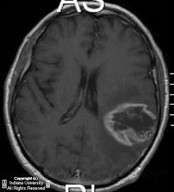
Glioblastoma multiforme


Findings

CT shows a large region of primarily white matter mass-like with hypodense center, with mild mass-effect in the high left frontoparietal region. MRI confirms a mass within the left frontoparietal region. This mass demonstrates thick, irregular peripheral post contrast enhancement and surrounding edema. There is no restricted diffusion within the central region of necrosis. There is no decreased T2 rim signal to suggest a fibrotic capsule. There is mild mass-effect on the left lateral ventricle and minimal left-to-right midline shift.


Diagnosis: Glioblastoma multiforme (biopsy proven)


Each year approximately 17,000 primary brain tumors are diagnosed in the United States. Sixty percent of these tumors are gliomas. Glioblastoma multiforme (GBM) is the most common and most malignant of the gliomas. The GBM is a heterogeneous tumor composed of poorly differentiated astrocytes. Most commonly, GBMs affects adults and involve the cerebral hemispheres. Although much less likely, GBMs can affect the brain stem and spinal cord in children. GBMs can transform from lower-grade astrocytomas or anaplastic astrocytomas. However, in most instances, GBM arises new, without a precursor lesion. The GBM can spread via several routes including: white matter tracts, across the midline via commissures such as the corpus callosum, subependymal seeding of the ventricles, and CSF seeding of the subarachnoid space.

In this case, the differential diagnosis includes both neoplasm (most likely glioblastoma multiforme) or abscess. Both diagnoses were considered almost equally after the initial head CT. The MRI provided some new information, but still was not be conclusive for either neoplasm or abscess. Specifically, the lesion demonstrated a central region of necrosis but lacked restricted diffusion, making abscess less likely. Similarly, the lesion lacked a decreased T2 rim signal to suggest a fibrotic capsule, which also made abscess a less likely diagnosis. Because imaging was not conclusive for neoplasm or abscess, stereotactic biopsy was performed and revealed glioblastoma multiforme.


Radiologic overview of the diagnosis

Usually heterogeneous low-density mass (seen on CT)
Intense contrast enhancement
Hemorrhage and central necrosis are common
Calcification is uncommon
Abundant vasogenic edema
Mass effect
Bihemispheric spread via the corpus callosum or commissures (the classic "Butterfly lesion")
If peripheral location – often demonstrate broad dural base and dural tail (appearance similar to an extra-axial lesion)
CSF seeding (leptomeningeal drop metastases)

martedì 18 settembre 2007

Nasopharyngeal carcinoma


Findings

Figure 1: Axial CT image displayed at bone window reveals the destructive lesion at the skull base involving the basi-sphenoid, basi-occiput, pterygoid plates and extending across the midline.
Figure 2: Axial T2-WI reveals soft tissue mass in the right lateral pharyngeal recess of the nasopharynx, involving the right veli palatine muscle and effacement of the Fossa of Rosenmüller.
Figure 3: Axial T2 WI reveals the mass to extend superiorly involving the right pterygopalatine fossa and the right pterygoid recess. The right masticator space appears spared.
Figure 4: Axial T2 WI reveals the mass to extend to involve the right sphenoid sinus and petrous apex, and into the right cavernous sinus, encasing and narrowing the right cavernous carotid artery.
Figure 5: Axial contrast-enhanced T1-weighted image obtained at level of nasopharynx shows fullness and enhancement in region of right Rosenmüller's fossa.
Figure 6 and Figure 7: Axial contrast-enhanced T1-weighted images reveal extension into the sphenoid sinus, petrous apex, pterygopalatine fossa and right cavernous sinus.
Figure 8: Coronal contrast-enhanced T1-weighted MR image reveals invasion of nasopharyngeal cancer into right cavernous sinus with encasement of internal carotid artery.
Figure 9: Coronal contrast enhanced T1-weighted image reveals an enhancing mass in sphenoid sinus region and increased enhancement (compared with normal left side) extending through the right foramen ovale.


Diagnosis: Nasopharyngeal carcinoma


Most nasopharyngeal malignancies in adults are carcinomas, usually of the squamous cell variety (80%). Squamous cell carcinomas (SCC) of the nasopharynx are relatively rare in the United States and much more common in Asia, being endemic in Southern China. A strong correlation between Epstein-Barr virus and nasopharyngeal cancer is well documented. Genetic predisposition and association with consumption of salted fish and preserved foods is also suspected.

Nasopharyngeal squamous cell cancer usually presents before age 50 and is more common in males (M:F=2.5:1). It usually presents late because of its infiltrative pattern. The most common presentation is an asymptomatic neck mass due to metastatic adenopathy. Serous otitis media may result from eustachian tube dysfunction resulting in otalgia and unilateral conductive hearing loss. Nasal obstruction and epistaxis are other common symptoms. Perineural spread and intracranial extension can cause headaches and cranial nerve deficits.

Nasopharyngeal cancers usually present as a poorly marginated nasopharyngeal mucosal space mass with deep extension and invasion. They tend to grow along the path of least resistance: along submucosal and soft tissue planes, along neurovascular bundles and intracranially through neural foramina at the skull base. Nodal metastases are present in 90% of cases at presentation. Distant metastases to the bones, lung or liver are seen in less than 10% of cases.

These tumors show squamous differentiation with intracellular bridges or keratinization. There are three main histologic subtypes- keratinizing, non-keratinizing and undifferentiated carcinoma.

Biopsy is the way to establish the diagnosis since imaging cannot distinguish among nasopharyngeal malignancies. Imaging plays a crucial role for mapping out the spread of tumor and planning radiation therapy/ surgical resection. Enhanced MR imaging is the best tool for evaluating intracranial extension via direct, perineural or perivascular routes. Thin section CT helps to evaluate bone invasion. CECT is preferred for evaluation of cervical metastatic nodes, except retropharyngeal adenopathy which is better seen on MRI. These tumors are strongly FDG avid.

XRT is the mainstay of treatment with combined XRT and chemotherapy used for advanced disease.

Cryptococcal meningitis


Findings

There is nodular thickening and T2-weighted hyperintensity of the callosal splenium with associated diffusion restriction. There is subtle leptomeningeal enhancement.

Differential Diagnosis:
- Embolic infarcts
- Abscess / multifocal infection
- Marchiafava-Bignami syndrome
- Diffuse axonal injury
- Lymphoma


Diagnosis: Cryptococcal meningitis


Key points

Cryptococcus neoformans is a ubiquitous yeast that grows abundantly in soil containing bird (especially pigeon) droppings. Disseminated infection occurs mainly in the immunocompromised, with the lungs and central nervous system most commonly affected. Cryptococcosis risk factors include; AIDS, steroid therapy, immunosuppression induced after organ transplantation, lymphoproliferative disorders, diabetes, tuberculosis, and sarcoidosis. Approximately 30-50% of patients with cryptococcal meningitis, however, show no evidence of immunosuppression (as in this case).

Nonspecific symptoms include:
- Fever
- Headache
- Changes in level of consciousness (somnolence, confusion, stupor or coma)
- Dizziness
- Visual disturbances
- Seizures

Diagnosis relies on CSF Indian ink preparation, culture, and antigen titers.


Radiologic overview of the diagnosis

Manifests as meningoencephalitis with gyral enhancement and intraparechymal lesions. Intraparechymal lesions include non-enhancing gelatinous pseudocysts (often in the basal ganglia) and enhancing cryptococcomas. Cerebral infarction can result from chronic infection. Brain lesions may worsen with Amphoterician-B treatment, causing concern for treatment failure. Hydrocephalus and diffuse atrophy can also be seen, yet could be related to an underlying disease process such a HIV.

giovedì 13 settembre 2007

Infected second branchial cleft cyst


Findings

There is a well circumscribed low attenuating, likely cystic, mass located posterior to the submandibular gland, lateral to the carotid space, and anteromedial to the sternocleidomastoid muscle. The mass demonstrates slight rim enhancement, with mild inflammation of the adjacent fat.

Differential diagnosis:
- Infected second branchial cleft cyst
- Necrotic malignant adenopathy
- Lymphangioma
- Thymic cyst


Diagnosis: Infected second branchial cleft cyst


Key points

Most common type of branchial cleft anomaly – accounts for 75% of branchial cleft anomaly in children and >90% in adults
Results from failure of obliteration of the cervical sinus
Usually not associated with other anomalies
Four subtypes depending on location: Bailey classification – type II is the "classic" location as described below
May have associated fistulas

Clinical Presentation:
- Presents as painless mass at the lateral aspect of the neck
- Mass may be chronic, and may become larger with respiratory infection
- May be painful if infected
- Age of presentation between 10 and 40 year old
- No sex or race predilection
- Treatment is complete surgical excision, with good prognosis
- Controversial if malignancy can arises from branchial remnant


Imaging

Usually a round or oval shaped cystic mass
Located at the anterior triangle or angle or mandible
Lateral to carotid space, posterior to the submandibular gland, and anteromedial to the sternocleidomastoid muscle
Variable in size, may be up to 10 cm

Contrast enhanced CT:
- Cystic density mass with non enhancing wall
- If infected, wall will be thicker and may enhance, with inflammation of adjacent soft tissues

MRI:
- Cystic mass isointense to fluid on T1 and T2
- May become hyperintense on T1 if infected
- Will demonstrate wall enhancement if infected

mercoledì 12 settembre 2007

Mucopolysaccharidoses (MPS) / Hurler disease


Findings

Figure 1: T1-weighted image showing dilated perivascular spaces.
Figure 2: Abnormal T2-signal in surrounding white matter. T2-weighted image showing dilated perivascular spaces.


Diagnosis: Mucopolysaccharidoses (MPS) / Hurler disease


Mucopolysaccharidoses (MPS) are inherited disorders of metabolism which are characterized by the inability to break down glycosaminoglycan (GAG) because of an enzyme deficiency. This results in an accumulation of toxic intracellular substrate. There are multiple MPS, classified from MPS 1-9, depending upon the specific enzyme deficiency. The prototype is MPS 1H, which is Hurler disease. This is caused by alpha-L-Iduronidase deficiency.

On noncontrast CT there is macrocrania. The dilated perivascular spaces are rarely visible on CT. There is progressive hydrocephalus and atrophy. On contrast enhanced CT there is often enhancing pannus associated with the ligaments and dura at the craniocervical junction.

A classic diagnostic imaging finding on MRI is dilated perivascular spaces (PVS) or Virchow-Robin spaces. This will appear as dark on T1WI and bright on T2WI. The dilated PVS are usually under 5 mm, but can be larger, and range from one to many. The most common locations are the corpus callosum and peritrigonal white matter, but can occur anywhere. On T2WI, there is often increased signal of the white matter surrounding the dilated PVS, which is a result of gliosis, edema, demyelination or dysmyelination. On FLAIR, there may be increased signal surrounding the PVS.

domenica 9 settembre 2007

Hemangioblastoma


Findings

There is a right parasagittal lesion along the medial aspect of the cerebellum. The cystic mass has pronounced mass-effect on the dorsal brainstem and tectum. Extending from the cyst wall along one of the septations is an enhancing nodule.

Differential diagnosis:
- Hemangioblastoma (given patient age, the most likely diagnosis)
- Medulloblastoma
- Pilocytic astrocytoma


Diagnosis: Hemangioblastoma


Key points

The hemangioblastoma (HB) is a vascular neoplasm of unknown etiologycular. Cerebellar hemangioblastoma is the most common primary brain neoplasm involving the infratentorial space in adults. This benign tumor is readily curable by surgery. More than 85% of hemangioblastomas occur in the cerebellum, with the remainder occurring in the spinal cord, medulla, and cerebrum in a 4:2:1 ratio. Approximately 10% of posterior fossae masses are hemangioblastomas. Men are more commonly affected than females, and patients are usually young adults. The common symptoms are headache, nausea, vomiting, ataxia, and vertigo. In 40% of patients, polycythemia is present secondary to increased erythropoietin produced by the tumor (more common in solid tumors). A spinal hemangioblastoma may present with subarachnoid hemorrhage.

The classic findings of an HB are that of a cystic mass with a solid mural nodule (60% of cases), which is highly vascular and has serpentine signal voids of feeding vessels. However, 40% of HBs are solid. Less commonly, these tumors may be purely cystic. Overall, HB is a rare tumor representing 1-2.5 % of primary CNS neoplasms; 10-20% of all cases are associated with with von-Hippel-Lindau syndrome; converesly, 45% of patients with VHL develop HBs.

Treatment: Treatment frequently requires removal of the mural nodule only, since the cyst is not truly neoplastic. Preoperative embolization aids in resection of the highly vascular tumors. Prognosis is very good with 5-year a survival rate of more than 85%.


Radiology

Angiography: Vascular nodule with intense, prolonged stain; +/- avascular cyst.
CT: Low-density cyst with strongly enhancing mural nodule that abuts a pial surface (75%)
MRI: Cyst slightly hyperintense to CSF on T1WI; hyperintense to brain on T2WI; mural nodule variable but typically enhances strongly

giovedì 6 settembre 2007

Acute subdural hematoma


Findings

There is acute subdural hematoma along the tentorium. This extends along the posterior interhemispheric fissure. The ventricles are prominent out of proportion to the sulci raising the possibility of obstructive hydrocephalus. The fourth ventricle is normal in size. No definite tectal mass is appreciated.

Differential diagnosis: "Bright tentorium"
- Subdural hematoma
- Epidural hematoma
- Aneurysmal bleed
- Metastatic disease to the cerebellum with local bleeding
- Isodense primary CNS tumor (meningioma) with focal bleeding

Other subdural fluid collection
- Hygroma (clear CSF, no encapsulating membranes)
- Effusion (xanthochromic fluid from extravasation of plasma from outer membrane; 20% evolve into chronic SDH)
- Empyema (peripheral enhancement, restricted diffusion centrally)

Epidural Hematoma
- Biconvex extra-axial collection
- Often associated with fracture
- May cross dural attachments, limited by sutures
- Pachymeningopathies (thickened dura)

Chronic meningitis (may be indistinguishable)
- Post-surgical (shunt, etc)
- Intracranial hypotension ("slumping" midbrain, tonsillar herniation)
- Sarcoid (nodular, "lumpy-bumpy")

Tumor
- Meningioma, lymphoma, leukemia, metastases
- Dural based, enhancing mass
- ± Skull involved


Diagnosis: Acute subdural hematoma


Key points

Acute (± 6 hrs-3 days) hemorrhagic collection in subdural space
Diagnostic clue: Crescent-shaped, homogenously hyperdense on CT, extra-axial collection that spreads diffusely over affected hemisphere
May cross sutures, not dural attachments
May extend along falx & tentorium
Compresses & displaces underlying brain
Recurrent, mixed-age hemorrhage in a child raises suspicion of non accidental trauma!
CT density & MR signal intensity vary with age & organization of hemorrhage
Protocol advice: Use wide window settings (150-200 HU) to identify small SDH

Etiology
- Trauma most common
Stretching & tearing of bridging cortical veins as they cross subdural space to drain into dural sinus
Both nonimpact as well as direct injury
Trauma may be minor, particularly in elderly

- Less common etiologies include
Dissection of intraparenchymal hematoma into subarachnoid, then subdural space
Aneurysm rupture
Vascular malformations: Dural AVF, AVM, cavernoma
Coagulopathy

Predisposing factors
- Atrophy
- Shunting (leads to increased traction on superior cortical veins)
- Arachnoid cyst (middle fossa most common site)

Epidemiology: SDH found in 10-20% imaged & 30% autopsy cases following craniocerebral trauma
Associated abnormalities: > 70% of aSDH have other significant associated traumatic lesions