
Findings
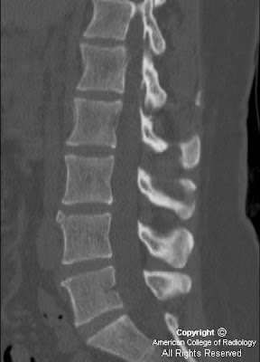
Figure 1 and Figure 2: Axial image from a CT of the lumbar spine and a sagittal reconstruction demonstrate a well-corticated, triangular bone fragment anterior to the superior end plate of L4. This is a classic finding of a limbus vertebra in a classic location. Note the well corticated nature of the bone fragment on the axial view.
Diagnosis: Limbus vertebra
Limbus vertebra is closely related to Schmorl nodes and is a relatively common finding in adults. It is a result of remote injury to a skeletally immature spine, which causes anterior intraosseous herniation of part of the nucleus pulposus through an unfused ring apophysis prior to fusion of the apophysis to the vertebral body. The apophysis fragment then remains separated from the vertebral body, and eventually develops into a well corticated, triangular fragment of bone, usually adjacent to the anterosuperior corner of a mid lumbar vertebral body. Less commonly, it can also be found near the anteroinferior aspect of a mid cervical vertebral body.
In children, the diagnosis can be trickier, since the well corticated fragment might not yet be present, and only an irregular, destructive appearing process is seen, typically involving the superior end plate of a mid lumbar vertebral body
This finding is commonly misinterpreted as a fracture, infection, or tumor when the patient presents for imaging due to back pain or following trauma, causing undue grief to the patient and the treating physician. Limbus vertebra is typically not the cause of the patient’s pain and is an incidental finding. It is important for the radiologist to be well aware of this entitiy in order to preclude further unnecessary intervention or invasive diagnostic procedures.
Nessun commento:
Posta un commento