giovedì 30 aprile 2009
Marchiafava-Bignami syndrome
Findings
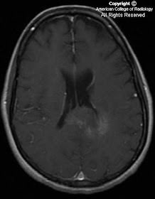
CT Head reveals hypo attenuation of the splenium and posterior aspects of the corpus callosum. Brain MRI reveals T2 prolongation of the entire corpus callosum, subcortical white matter, medial cerebellar hemispheres, and ventral medulla. There is a small right occipital subdural hematoma. There was no post-contrast enhancement.
Differential diagnosis:
- Marchiafava-Bignami syndrome
- Lymphoma
- Demyelinating disease
- Viral encephalitis
- Wernicke's encephalopathy
- Glioblastoma multiforme
Diagnosis: Marchiafava-Bignami syndrome
Key points
MB is primary degeneration of the corpus callosum. Other white matter tracts may be involved (as in this case).
First described in 1903 by two Italian pathologists, who found necrosis of the corpus callosum on autopsy in 3 alcoholic men that had recently died from seizures.
Initially thought to be caused by excess red wine consumption, now known to be related to vitamin B deficiency, generally seen in alcoholics.
Patients present with confusion, neurocognitive defects, and seizures.
Most patients with acute symptoms go into coma and die.
Chronic symptoms are characterized / accompanied by chronic dementia.
MB typically affects the body of the corpus callosum first, followed by the genu, and finally the splenium. Other white matter tracts may be involved.
On cytopathology, the middle layer of the corpus callosum is most affected.
On MR, disease revealed by areas of low T1 signal and high T2 and FLAIR signal. Signal intensity in the body of the corpus callosum at times extending into the genu and adjacent white matter.
Lesions do not enhance, unlike in glioblastoma, lymphoma, or encephalitis.
On CT, lesions are hypoattenuating.
martedì 28 aprile 2009
Rathke cleft cyst
Findings
Figure 1 and Figure 2: Sagittal and coronal T1 weighted images demonstrate a well-defined, high-signal round structure in the suprasellar region with a low signal mural nodule. The high signal material is likely due to hemorrhagic or proteinaceous material within the cyst.
Figure 3 and Figure 4: Axial and coronal T2 weighted images demonstrate an isotense suprasellar structure with a low signal mural nodule.
Figure 5: Post-contrast T1 weighted image demonstrates no enhancement of the Rathke cleft cyst with some lateral displacement of the enhancing stalk.
Diagnosis: Rathke Cleft Cyst
Rathke cleft cysts (RCC) are nonneoplastic cysts which arise from remnants of the Rathke pouch. The cyst is lined with a single layer epithelium. On pathlogy, inflammatory changes may be present. The cyst may contain cerebrospinal fluid density material as well as thick mucoid debris, hemorrhage, hemosiderin as well as colloid. RCCs stain positively for cytokeratins.
Most RCCs are found incidentally as they are asymptomatic. RCCs can produce symptoms by causing pituitary dysfunction, visual disturbance or headache. RCCs may rarely produce sphenoid sinusitis or compress the optic chiasm and/or hypothalamus. RCCs have a slight female predominance. Some RCCs will decrease in size or spontaneously resolve while others display stable size and MR signal over time. If RCCs are deemed to be symptomatic, cyst aspiration or excision may be necessary. They recur in up to 1/3 of patients.
CT imaging characteristics of RCCs are well defined intra- or suprasellar masses which are fluid attenuation or mixed density. Cyst wall calcification is present in 10-15% of patients. The T1 appearance of RCCs varies bases upon cyst content. Five to 10% may have a fluid-fluid level. Intracystic mural nodules are present in 75%. T2 signal characteristics also vary with cyst content, with 75% demonstrating a low signal intracystic nodule. RCCs do not demonstrate internal enhancement, however the adjacent compressed pituitary enhances.
Differential diagnostic considerations for Rathke cleft cysts include:
- Craniopharyngioma (differentiated by the degree of calcification and enhancement)
- Cystic pituitary adenoma (rarely have calcification, demonstrate heterogeneous signal intensity and may demonstrate rim and/or nodular enhancement)
- Colloid cysts as well as pars intermedia cysts are also diagnostic considerations
- Other non-neoplastic cysts derived from arachnoid, dermoid, or epidermoid elements
Right petrous apicitis (Gradenigo's syndrome clinically)
Findings
CT shows an expansile, destructive lesion in the right petrous apex, which is bright on T2 weighted MR images, and has an enhancing soft tissue component on post contrast T1 weighted-images.
Differential diagnosis:
- Petrous apicitis
- Cholesterol granuloma
- Trapped fluid
- Chondrosarcoma
- Cholesteotoma
- Lymphoma
Diagnosis: Right petrous apicitis (Gradenigo's syndrome clinically)
Key points
Synonyms for petrous apicitis include apical petrositis and confluent apical petrositis.
Petrous apicitis typically occurs via spread of infection from mastoids through air cells into petrous apex.
Clinical presentation is variable; most have otorrhea and pain. Patients can have cranial neurophaties.
In kids, petrous apicitis is often a sequelae of acute otomastoiditis. In adults, it is more often due to chronic infection.
Gradenigo's syndrome is acute otomastoiditis, trigeminal (CN 5) nerve neuritis, and Abducens (CN 6) palsy (CN 6 controls the lateral rectus muscle).
Temporal bone CT and MRI are the best imaging modalities for making the diagnosis.
On CT, findings of petrous apicitis include a destructive lesion with both cortical and trabecular loss in the petrous apex.
The best clue to diagnosis is trabecular destruction in an opacified petrous apex, which is best appreciated on CT.
On MR, findings of petrous apicitis are a thick walled, enhancing area of soft tissue with fluid in the petrous apex.
Nuclear medicine Gallium-67 scan with SPECT can improve the sensitivity for disease detection and can be used to monitor response to treatment.
Petrous apicitis was a common, severe and life-threatening infection in the pre-antibiotic era. It is now relatively uncommon.
Inflammation/infection from the petrous apex can spread to meninges and cause meningitis or can lead to cavernous thrombosis.
Treatment requires antibiotics and often surgical drainage.
giovedì 23 aprile 2009
Adrenal leukodystrophy
Findings
Figure 1: Axial non contrast CT demonstrates symmetric low attenuation in the temporal-parietal white matter. Note involvement of the splenium of the corpus callosum.
Figure 2 and Figure 3: Axial T1 and T2 images demonstrate symmetric T1 hypointensity and T2 hyperintensity in the temporal-parietal white matter corresponding to the low attenuation on CT. Note relative sparing of the subcortical U-fibers.
Figure 4: Axial post gadolinium T1 weighted image demonstrates abnormal hypointensity involving the temporal-parietal white matter, with enhancement of the leading edge which represents active demyelination.
Figure 5: MR spectroscopy demonstrates increased choline and decreased NAA, findings that are non specific but consistent with adrenal leukodystrophy. Note the presence of lactate an indicator of necrosis.
Diagnosis: Adrenal leukodystrophy
Adrenal leukodystrophy is a hereditary disorder caused by impaired beta-oxidation of very long chain fatty acids (VLCFA). Various forms exist, each named according to the age of presentation, including neonatal, childhood, adolescent and adulthood. Another variant, in which there is severe involvement of the spinal cord rather than the cerebrum, is termed adrenomyeloneuropathy (AMN).
The disease is caused by a mutation of the ALD gene which codes for a peroxisomal membrane protein. Transport of very long chain fatty acids into the peroxisome is consequently restricted. This results in reduced supply of shorter chain fatty acids within the peroxisome available for synthesis of complex lipids and proteins, which are myelin components.
The defective myelin is more easily broken down by the body’s normal physiologic functioning and the resulting pathophysiologic processes which include a severe inflammatory demyelination that predominates in the cerebral white matter and axonal degeneration that predominates in the posterior fossa and spinal cord.
These processes manifest radiologically as three zones in the cerebral white matter:
- Zone A, a central burned out zone containing only astrogliosis
- Zone B, an inflammatory zone peripheral to the central zone containing perivascular inflammatory cells and demyelination where axons are preserved
- Zone C, site of demyelination without inflammation.
Clinically, patients suffering from the childhood variant of ALD develop normally initially, but then present with behavioral changes and progressive visual, auditory and motor dysfunction between the ages of 4-10. Diagnosis is usually made by laboratory evaluation of very long chain fatty acid levels once there is clinical suspicion. Treatment options are limited and aimed at lowering levels of VLCFA’s through dietary restriction, consumption of Lorenzo’s oil and lovastatin therapy. Bone marrow transplantation has also shown to be of benefit in some cases. Imaging is helpful as an adjunctive tool to evaluate the extent of disease at presentation and to assess for evolution of the disease during treatment.
The classic picture of ALD is that of confluent, bilateral periatrial (parietal-occipital) deep white matter signal abnormality with sparing of the subcortical u-fibers. The pathological basis for the signal abnormality is a combination of demyelination and gliosis. The signal abnormality demonstrates a “leading edge” of enhancement which reflects blood brain barrier breakdown at sites of active demyelination at the margins of the signal abnormality. Calcifications along the trigone have also been noted as late sequelae.
Loes et al. have described five patterns of involvement, which when considered in the context of patient age at presentation, may give a clue to prognosis:
- Pattern 1 describes involvement of the parietal-occipital white matter and splenium and is associated with rapid progression with younger age of presentation and with enhancement.
- Pattern 2 is that of frontal white matter and genu involvement, and has a similar prognosis to that of Pattern 1.
- Pattern 3 is that of corticospinal tract involvement, usually seen in adults and demonstrates slower progression.
- Pattern 4 is that of corticospinal tract involvement associated with abnormality of the cerebellar white matter, which is seen in adolescents and is also associated with slower progression.
- Pattern 5 is that of concomitant frontal and parietal-occipital white matter abnormality, seen mostly in children and having a rapidly progressive course.
Proton MR spectroscopy demonstrates decreased NAA and myo-inositol and increased choline, glutamine, glutamate and lactate. Decreased NAA/Cr and NAA/Ch ratios and increased Ch/Cr ratios are characteristic. The classic imaging findings are virtually pathognomonic with limited differential diagnosis. Another peroxisomal disorder, acyl CoA oxidase deficiency, has similar imaging findings, but the clinical presentation is much different.
lunedì 20 aprile 2009
Vein of Galen malformation
Findings
Ultrasound shows that the sulcal pattern is normal. There is no hydrocephalus. There is no hemorrhage. There is a round structure in the midline, anterior to the occipital horns of the lateral ventricles which has color-flow. This measures up to 2.8 cm. There is no extra-axial fluid.
CT shows a large arteriovenous malformation containing numerous large direct fistulous from the bilateral anterior cerebral arteries as well as numerous smaller connections involving choroidal, pericallosal, and thalamoperforator arteries. There is marked dilatation of the vein of Galen measuring approximately 2.2 x 2.0 cm in the axial plane and 2.4 x 2.5 cm in the sagittal plane.
Diagnosis: Vein of Galen malformation
Key points
The vein of Galen is located under the cerebral hemispheres and drains the anterior and central regions of the brain into the sinuses of the posterior cerebral fossa. Aneurysmal malformations of the vein of Galen (VGAM) typically result in high-output congestive heart failure or may present with developmental delay, hydrocephalus, and seizures.
VGAM results from an aneurysmal malformation with an arteriovenous shunting of blood. The congenital malformation develops during weeks 6-11 of fetal development as a persistent embryonic prosencephalic vein of Markowski; thus, VGAM is actually a misnomer. The vein of Markowski actually drains into the vein of Galen.
Associated findings include cerebral ischemic changes such as strokes or steal phenomena that result in progressive hemiparesis. Hemorrhage from the malformation can occur, although this is not a common finding. Finally, the malformation may result in mass effects, causing progressive neurological impairment. Alternatively, the malformation may cause obstruction of the cerebrospinal fluid (CSF) outflow and result in hydrocephalus.
venerdì 17 aprile 2009
Mandibular ossifying fibroma
Findings
Bubbly, expansile lesion centered at the inferior aspect of the midline and right parasymphyseal mandible. Contains internal cystic regions and areas of coarse calcified matrix or septation with expansile margin predominantly involving the buccal cortex. There is mild remodeling of the lingual cortex. There is no bony dehiscence or significant soft tissue component.
Differential diagnosis:
- Ossifying fibroma
- Odontoma
- Fibrous dysplasia
- Odontogenic myxoma
- Ameloblastoma
- Hemangioma
- Cementoma
Diagnosis: Mandibular ossifying fibroma
Key points
Ossifying fibromas are one of the fibro-osseous lesions, which also include cementifying ossifying fibroma, cementifying fibroma and fibrous dysplasia.
Distinction is difficult because of overlapping clinical, radiological and pathological criteria.
Usually arise from the periodontal ligament, and are most commonly found in the mandible.
Presents in middle age, often asymptomatic. Can cause paresthesia from nerve compression.
Round or oval periapical lesion with variable lucency and matrix calcification depending on spectrum. Usually no cortical disruption.
Conservative curettage is the treatment.
mercoledì 15 aprile 2009
Pelizaeus Merzbacher disease (PMD)
Findings
Figure 1: The coronal T2 image from age 5 demonstrates diffuse T2 hyperintensity in the cerebral white matter.
Figure 2, Figure 3, and Figure 4: Coronal T2 images from ages 8, 10, and 15 demonstrate gross stability of white matter signal abnormalities.
Figure 5 and Figure 6: Axial pre- and post- contrast images from age 15 demonstrate no abnormal enhancement in the white matter.
Diagnosis: Pelizaeus Merzbacher disease (PMD)
Pelizaeus-Merzbacher disease (PMD) is a rare leukodystrophy that is more properly categorized as a dysmyelinating disorder than a demyelinating disease. Demyelinating disorders such as adrenal leukodystrophy usually result from inborn errors in metabolism. Instead, PMD arises from a genetic defect affecting PLP-1, the primary protein in myelin.
The patient in this case was diagnosed with classic PMD which is the most common of the four forms of the disease. Classic PMD is an X-linked recessive disorder that results most frequently from the duplication of the PLP-1 gene. This genetic error causes a diffuse hypomyelination of the cerebral white matter.
Patients with classic PMD present in infancy with nystagmus, spasticity, and other motor problems. Because of this early presentation, cerebral palsy is the most common misdiagnosis for PMD patients as in this patient. As the patient becomes older, cognitive deficits are more conspicuous and further motor deficiencies may arise. The life span of patients with classic PMD is variable with many succumbing in adolescence and early adulthood while some other patients may live into the sixth decade.
The very slowly progressive MRI findings of classic PMD are quite specific for the disease especially when sequential exams are available for comparison. The cerebral white matter usually appears diffusely low in intensity on T1 images. Findings are more striking on T2 images where the hypomyelinated white matter remains diffusely hyperintense. The corpus callosum is notably atrophic. Myelination is usually present in the brainstem, thalamus, and posterior limbs of the internal capsules. Abnormal enhancement is not seen in PMD as opposed to some demyelinating disorders such as adrenal leukodystrophy in which enhancement is seen at the periphery of the active demyelination. In classic PMD, the MRI findings tend to be stable or mildly progressive over years since the primary problem is hypomyelination rather than demyelination.
Right sialolithiasis with sialodochitis, sialoectasia and sialadenitis
Findings
Near the right Warthin duct orifice, there is a signal void consistent with a duct stone. Proximal to that, there are segmental dilatations and stenoses consistent with sialodochitis, or chronic inflammatory change of the duct. Near the mylohyoid turn of the duct, there is another signal void consistent with a stone and proximally, there is a stenosis. In the superficial submandibular gland, there are patchy areas of T2 prolongation consistent with dilated ducts, or sialoectasia. In the deep portion of the submandibular gland, there are no secondary or tertiary ducts because they have closed off due to chronic inflammatory change. There is T2 prolongation of the right submandibular gland, in relation to the left, consistent with sialadenitis.
Diagnosis: Right sialolithiasis with sialodochitis, sialoectasia and sialadenitis
Key points
The submandibular gland is the second largest in the mouth. It has relatively viscous secretions, which can predispose to the formation of duct stones.
The submandibular gland has a superficial portion, which is superficial and inferior. The deep portion is deep to the mylohyoid muscles and superior to the superficial lobe. Wharton's duct arises from the deep portion of the gland.
Conventional sialography, with contrast injection of Wharton's duct under fluoroscopy, is a limited evaluation of the duct and the gland, if, as in this case, there is complete obstruction of the distal duct by a stone or stricture. Conventional sialography requires cannulation of the duct, which could be technically difficult. While CT is the best imaging modality to identify sialolithiasis, MR sialography gives the best evaluation of the gland. Sumi et al. established that MR sialography has good correlation between CT imaging findings and clinical findings.
Acute sialadenitis can be bacterial or viral. Purulent material can be milked from the duct. Complications can include formation of a fistula to the skin, an abscess or trismus, if the parapharyngeal space is involved. Also, rarely, Ludwig angina, with involvement of the submental and sublingual space, can occur.
Chronic sialadenitis presents with persistent colicky pain with eating. It is not as painful as acute sialadenitis. This is often associated with sialolithiasis.
Sialolithiasis is usually associated with the submandibular gland (70%). The stones are calcium phosphate or calcium carbonate. Formation may be related to calcium salt precipitation, epithelial duct injury or dehydration. Surgical excision is usually needed.
Autoimmune sialadenitis is usually associated with Sjögren's syndrome. MR findings include T2 prolongation of the parotid glands consistent with diffuse parenchymal destruction.
lunedì 13 aprile 2009
Interhemispheric cyst
Findings
There is agenesis of the corpus callosum. There is a multiseptated, left parasagittal midline cyst. Possible communication with the third ventricle. No associated enhancement is present. The left lateral ventricle appears dilated. No midline shift is present.
Differential diagnosis:
- Interhemispheric cyst
- Holoprosencephaly
Diagnosis: Interhemispheric cyst
At surgery: No definite communication with the ventricular system was found at surgery, thus categorizing the lesion as an interhemispheric cyst – type 2.
Key points
The association between an interhemispheric cyst and agenesis of the corpus callosum has been reported, although the etiology of the cyst is uncertain (arachnoid cyst, neuroepithelial cyst, or extensions of the ventricular lining).
Classification was most recently described by Barkovich et al based on appearance and communication with the ventricular system in 25 patients. Type 1 cysts communicate with the ventricular system while Type 2 cysts do not. Lack of communication with the ventricles was determined by visualizing a wall of the loculated cyst on MRI.
Classification of agenesis of the corpus callosum with interhemispheric cyst:
- Type 1a:
Cyst: Isointense to CSF (MR), unilocular.
Communication with lateral ventricles only.
Macrocephaly, hydrocephalus, Dandy-Walker malformation.
Males.
- Type 1b:
Cyst: Isointense to CSF (MR), unilocular.
Communication with and obstruction of third ventricle.
Macrocephaly, thalamic fusion without subcortical heterotopia.
Males > Females.
- Type 1c:
Cyst: Isointense to CSF (MR), unilocular.
Communication with lateral and third ventricles.
Microcephaly, cerebral dysplasia or hypoplasia.
Males.
- Type 2a:
Cyst: Isointense to CSF (MR), multilocular.
No communication with lateral or third ventricles.
Macrocephaly, hydrocephalus.
Male.
- Type 2b:
Cyst: Hyper attenuation (CT), hyper intense (T1W MR), multilocular.
No communication with lateral or third ventricles.
Aicardi syndrome, subependymal heterotopia, polymicrogyria, seizures, hypoplastic falx cerebri, uni- or bilateral ventriculomegaly, developmental delay.
Female.
- Type 2c:
Cyst: Isointense to CSF (MR), multilocular.
No communication with lateral or third ventricles.
Subcortical heterotopia, developmental delay.
Male.
venerdì 10 aprile 2009
Hallervorden Spatz disease
Findings
Figure 1 : Axial T2 weighted image at the level of the deep gray nuclei demonstrates the classic “eye of the tiger” sign, with T2 prolongation in the medial globi pallidi and hypointensity in the peripheral aspect of the globi pallidi.
Figure 2: Axial SPGR image at the same level demonstrates hypointensity in the globi pallidi as a result of the susceptibility effect from iron deposition.
Diagnosis: Hallervorden Spatz disease
Hallervorden Spatz disease is a rare neurodegenerative condition whose exact pathogenesis is unknown. Abnormal accumulation of iron in the brain, specifically in the globus pallidus and substantia nigra, is felt to play a predominant role in the neurodegeneration caused by the disease. A mutation in the pantothenate kinase gene leads to enzyme deficiency and subsequent accumulation of cysteine and iron chelates. The excess iron is deposited in the basal ganglia and damages neuronal structures.
Symptoms begin early in life, typically in early adolescence, and include extrapyramidal and gait abnormalities. Slow movements, rigid extremities, dystonia, tremors and speech abnormalities are typical findings. No cure exists, and therapy is aimed toward neurological symptoms. The typical clinical course is that of rapid progression, with death in early adulthood in most cases.
Imaging findings are usually classic, and typically described as the “eye-of-the-tiger sign”. This results from symmetric T2 prolongation in the globi pallidi with peripheral hypointensity and hypointensity in the substantia nigra. There is no associated enhancement of the areas of abnormal signal. The neural damage from abnormal iron accumulation results in diminished NAA on spectroscopy. The areas of iron deposition would be more conspicuous on susceptibility weighted images. In cases where the classic history and imaging findings do not lead to the diagnosis, other pathologies that preferentially involve the globi pallidi should be considered. These include metabolic derangements such as methylmalonic acidemia, Kearns-Sayre syndrome, Canavan disease and toxic and ischemic encephalopathies caused by anoxia, carbon monoxide and cyanide poisoning.
mercoledì 8 aprile 2009
Branchial cleft cyst
Findings
On CT imaging, there is a well circumscribed predominantly cystic lesion at the level of the hyoid within the right neck soft tissue. The lesion has a sharp margin with the sternocleidomastoid muscle and is located superficial to the carotid artery. No adjacent stranding or inflammatory changes.
On ultrasound imaging, an anechoic structure with small amount of debris is noted within the right neck with some internal septations. No flow is noted within the lesion.
Differential diagnosis:
- Branchial cleft cyst
- Lymphangioma
- Thymic cyst
- Suppurative jugulodigastric node
- Cystic vagal schwannoma
- Cystic malignant adenopathy
Differential diagnosis considerations
Lymphangioma are multilocular, trans-spatial collections that fill the available spaces. If they are unilocular and located in typical position of second branchial cleft cyst, it may be very difficult to differentiate between the two. One characteristic that helps differentiate the two, is that lymphangiomas are infiltrative in nature while branchial cleft cysts are well defined and round.
Thymic cysts are usually located inferior within the cervical neck and are centered in the lateral visceral space.
Suppurative jugulodigastric node presents as a thick walled ovoid mass at the angle of the mandible.
Cystic vagal schwannoma has a thick, enhancing wall and is usually centered behind the carotid space causing displacement of the internal jugular vein and internal carotid artery.
Cystic malignant adenopathy presents as a necrotic mass with a thick, enhancing wall. Cystic metastasis (e.g. papillary thyroid carcinoma) and necrotic metastases (e.g. squamous cell carcinoma) as well as necrotic tuberculous lymphadenitis can have a cystic appearance similar to second branchial cleft cyst, therefore it is very important to differentiate the two and ultrasound fine needle aspiration with fluid analysis can provide useful information.
Diagnosis: Branchial cleft cyst
Discussion
The branchial apparatus is a precursor to many head and heck structures. The second branchial cleft arch overgrows the second, third, and fourth clefts and forms a cavity called the cervical sinus. During the seventh week of gestation, the cervical sinus is normally obliterated. However if there is incomplete involution, a congenital squamous epithelial lined cyst (branchial cleft cyst), sinus tract or fistulae can form. There are four subtypes of branchial cleft cyst, described below, of which greater than 90% are second branchial cleft cyst.
Classification
- Type I: anterior to the sternocleidomastoid, beneath the platysma muscle.
- Type II: adjacent to the internal carotid artery, often adherent to the internal jugular vein.
- Type III: extends between the internal and external carotid artery to the lateral pharyngeal wall.
- Type IV: lies against the lateral pharyngeal wall and may extend into the skull base.
Majority present as a painless, compressible lateral neck mass; however, they can become tender, enlarged, inflamed or develop abscesses, especially during periods of upper respiratory tract infection.
The size of cysts are variable and range from a few centimeters to > 5 cm. Most are ovoid or rounded in shape; some present with a focal rim of cyst extending to the carotid bifurcation, which presents as the "Notch sign" which is pathognomonic for second branchial cleft cyst.
These cyst can occur anywhere along the line from the tonsillar fossa to the supraclavicular region and more commonly are seen in the following areas:
- Posterolateral to submandibular gland, most are at or immediately caudal to the mandible angle.
- Lateral to carotid space.
- Anteromedial to sternocleidomastoid muscle.
- Some unusual locations for cyst include:
- Superiorly within parapharyngeal space or carotid space.
- Inferior along anterior surface of infrahyoid carotid space.
- Fistulous tract between the external and internal carotid arteries to faucial tonsil.
Treatment is usually surgical resection.
Controversy exists as to the possibility of carcinoma arising within the branchial remnant squamous epithelial lining or not. However, metastatic squamous cell carcinoma to regional lymph nodes that masquerades as branchial cleft cyst is more common.
Imaging findings
CT or MRI an easily suggest diagnosis. Contrast is used to help differentiate cystic from solid mass.
Contrast enhanced CT:
- Low density cyst with nonenhancing wall and surround soft tissue if noninfected.
- If infected, wall appears thicker and enhances with surround soft tissues appearing as dirty cellulites.
MRI:
- On T1W imaging: cyst is isointense to CSF. If cyst is recurrently infected, it may contain hyper intense contents secondary to increase protein concentration.
- T2W imaging: Hyperintense cyst, no discernible wall.
- FLAIR imaging: Cyst is iso or hyper intense to CSF.
- T1W imaging + Contrast: Peripheral wall enhancement if infected.
Ultrasound:
- Anechoic thin-walled cyst with through transmission.
- May be hyper echoic with internal debris or even pseudo-solid in appearance.
- If infected, see thickened cyst wall.
giovedì 2 aprile 2009
Tumefactive demyelination
Findings
Figure 1, Figure 2, Figure 3, and Figure 4 demonstrate a hypointense T1, hyperintense T2 weighted expansile mass involving the splenium of the corpus callosum. The mass has well defined borders and partially effaces the atrium (trigone) of the left lateral ventricle. There is mild patchy enhancement (Figure 4). DWI images demonstrate increased signal throughout, but only the even more hyperintense rim (Figure 5) demonstrates true restricted diffusion on ADC images (Figure 6). The remainder of the mass is increased signal on ADC images, indicating increased diffusivitiy.
Diagnosis: Tumefactive demyelination
A noncomprehensive list of lesions involving the corpus callosum includes the following: masses such as lipomas, lymphoma, glioblastoma and astrocytoma, and metastases; demyelinating diseases such as multiple sclerosis, progressive multifocal leukoencephalopathy, and Marchiafava-Bignami disease; vascular lesions such as infarctions, vascular malformations; and trauma. More focal lesions with restricted diffusion involving the splenium can also be seen in patients with epilepsy receiving antiepileptic drugs and in the setting of hypoglycemia. Narrowing the differential possibilities to a few relevant entities requires clinical history and detailed analysis of the imaging characteristics. Many of the disease processes mentioned have some unique characteristics that may help favor a particular diagnosis. However, in spite of a full clinical history and an arsenal of imaging tools, a biopsy may ultimately be necessary, as was the case with this patient.
The diagnosis in this case was tumefactive demyelination, which may be thought of as a solitary demyelinating lesion measuring greater than 2 cm and with characteristics that mimic neoplasm. Because of this, tumefactive demyelinating lesions are commonly diagnosed as such only after biopsy. Several imaging characteristics suggestive of such a lesion have been described. These include a large lesion with circumscribed borders and little or no mass effect or surrounding edema. Demyelinating lesions in general also tend to demonstrate an incomplete ring of enhancement, with the nonenhancing component facing the gray matter. The enhancing rim is felt to represent the leading edge of demyelination, and therefore usually faces the white matter. Some foci also have been shown to have decreased perfusion in some studies, which would help distinguish them from glioblastoma and lymphomas.
The abnormality in our case does illustrate a few of these characteristics, such as a circumscribed border and lack of surrounding edema. The lesion did demonstrate increased diffusivity on DWI/ADC maps, which would argue against a highly cellular tumor like lymphoma and a high grade glioma. Active foci of demyelination may demonstrate restricted diffusion. In our case, the thin peripheral rim of restricted diffusion may represent an active edge of demyelination. The mass effect on the atrium of the left lateral ventricle is a confounding factor that can be associated with tumefactive demyelination or neoplasms. Metastases usually elicit a large amount of edema which was noticeably absent in this case. Infarction is also unlikely given the patient’s age and based on the imaging characteristics of the abnormality. Infarctions involving the corpus callosum are uncommon because of the ample blood supply from the anterior and posterior circulation. Unfortunately, none of these features is specific enough to warrant watchful waiting, and therefore a biopsy was performed.