mercoledì 28 ottobre 2009

Carotid cavernous fistula


Diagnosis: Carotid cavernous fistula


Discussion

Carotid Cavernous fistula (CCF) is a direct communication between the intracavernous portion of the carotid artery and the venous cavernous sinus. Usually is ipsilateral.
Most often results from significant trauma, penetrating or nonpenetrating.
Spontaneous rupture happens in the elderly.

Spontaneous rupture of CCF also associated with:
- Osteogenesis imperfecta
- Ehlers-Danlos syndrome
- Psuedoxanthoma elastica

Clinical presentation: acute onset pulsating exophthalmos, orbital bruit, dilated conjunctival vessels, and glaucoma. No pain whereas psuedotumor is almost always associated with pain.


Radiology

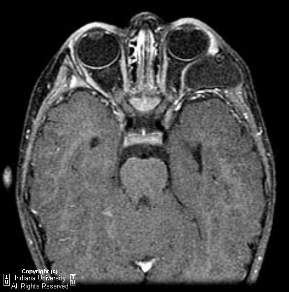
Unilateral diffuse enlargement of extra ocular muscles.
Proptosis.
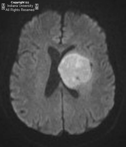
Dilation of Superior ophthalmic vein (SOV) and venous structures within the carotid cavernous sinus due to backpressure. Our case was unusual in that the venous engorgement was on the opposite side of the CCF.
Irregularity of SOV may represent thrombus (not seen in this case).
Bowed convexity to the Cavernous sinus usually unilateral (our case demonstrated right cavernous sinus bowing due to the venous engorgement from the opposite carotid-very atypical)
US: reversal of flow in SOV.
Carotid Angio: filling of ophthalmic veins due to decompression of arterial flow with retrograde filling of orbital veins.
Repair: important to determine the extent of contralateral flow in the cavernous sinus without the CCF if carotid must be sacrificed. Glue embolization of the SOV and coiling are the primary treatments.
Diagnosis first by CT orbit findings then proceed to conventional angiogram for diagnosis and treatment.
Treatment: glue embolization and coiling. Approach varies but may go through the SOV from the internal jugular or direct approach by interventional neuroradiologist or neurosurgeon through the SOV as in our case.


Pearls

With carotid cavernous fistula look for enlarged superior opthalmic vein.(SOV) in addition to unilateral rectus enlargement and proptosis.
Need cerebral angiogram to definitively diagnose-may not see enlarged carotid cavernous sinus on CT.
SOV lives between the superior rectus and the optic nerve.
Treatment of CCF is with glue embolization and coiling

martedì 27 ottobre 2009

Chloroma


Findings


Sagittal T1 image of the lumbosacral spine demonstrates decreased marrow signal throughout the lumbosacral vertebral bodies consistent with marrow infiltration secondary to the patient's leukemia.
Axial T2 image through the level of the sacrum demonstrates an isointense lesion originating in the right sacral ala which is invading the S1 and S2 exiting nerve roots. Incidental note is made of a left sacral foraminal Tarlov cyst.
Axial T1 pre and post contrast images demonstrate enhancement of this sacral mass.


Diagnosis: Chloroma


Granulocytic sarcomas are extramedullary masses composed of granulocytic cell precursors, occurring in patients with leukemia or other myeloproliferative disorders. First described by Burns in 1811, they were given the name "chloroma" by King in 1853, due to the green color produced by the high levels of myeloperoxidase in these cells. Recently there has been a shift away from using the term "chloroma", as these cells can have varying amounts of myeloperoxidase and can produce other colors as well. Granulocytic sarcomas most commonly occur in patients with AML, however are still rather rare, occurring in only 2.5-9.1 % of patients with AML. Children are more commonly affected than adults with 60% occuring in patients younger than the age of 15. While granulocytic sarcomas most commonly develop after the diagnosis of AML has been made, they can precede the hematologic leukemia, making their diagnosis more difficult. In these instances, biopsy is necessary to make the diagnosis.

Granulocytic sarcomas often occur in multiple locations at the same time. These tumors have a predilection for the orbits and subcutaneous tissues, but have been reported in the paranasal sinuses, bowel, breast, bone, lungs, peritoneum and pleural space. In the central nervous system, chloromas appear as extra-axial masses which are contiguous to meninges or ependyma. It is believed that intracranial granulocytic sarcomas arise from dural and subarachnoid veins and surrounding adventitia whereas spinal granulocytic sarcomas spread via the perivenous arachnoid space. On unenhanced CT, granulocytic sarcomas appear isodense or hyperdense to brain. On MR, granulocytic sarcomas are hypointense or isointense on T1, isointense or hyperintense on T2 and typically demonstrate homogeneous enhancement. Intracranial chloromas are often associated with mass effect and edema. When granulocytic sarcomas involve the spine, they are typically extradural or intradural extramedullary lesions. Very rarely, these tumors can cause compression of the spinal cord, caude equina or nerve roots. The differential diagnosis for granulocytic sarcomas when they occur in or around the spinal canal includes lymphoma, intraspinal abscess, plasmacytoma, metastatic disease and other primary bone tumors.

There is dispute as to whether patients with granulocytic sarcomas have worse prognoses. Treatment of granulocytic sarcoma is usually with systemic chemotherapy. If the mass is resistant to chemotherapy, localized radiation or surgery may be considered. While treatment is usually successful, recurrence does occur in up to 23% of patients.

giovedì 22 ottobre 2009

CNS Lymphoma


Findings

CT: There is a hyper dense mass in the left frontal lobe with compression of the left lateral ventricle and mild midline shift.
MR: There is a uniformly enhancing hypercellular mass in the medial left frontal lobe, extending to the left basal ganglia. Mass surrounds the left lateral ventricle and has associated cerebral edema best demonstrated along its lateral margin.


Differential diagnosis:
- Lymphoma
- Glioblastoma
- Abscess


Diagnosis: CNS Lymphoma


Key points

Malignant primary CNS neoplasm made up of B lymphocytes
1 – 7% of all primary brain tumors
90% of lesions are supratentorial. Commonly frontal and parietal lobes, periventricular with subependymal involvement.
Presents with neurological symptoms, such as headache or seizure.
Typically occurs in the 5th-6th decade, males greater than females.
Immunocompromised individuals are predisposed to all forms of lymphoma, including the CNS form.
Deep gray nuclei commonly affected.
Like GBM, often involves and crosses the corpus callosum.
On CT, hyper dense and relatively uniform in appearance.

MR:
Lesions tend to be homogeneous iso/hypo intense to cortex on unenhanced T1 and T2 weighted images.
May see "blooming artifact" from blood products or calcium on T2*GRE.
Strong homogeneous enhancement.

Differential considerations:
- GBM enhances heterogeneously, unlike lymphoma.
- Abscess usually has a rim that is hypo intense on T2 weighted images, and reveals restricted diffusion.
- Toxoplasmosis also involves the basal ganglia, but subependymal spread is not an associated finding.
- PMNL is a white matter disease, and is typically non-enhancing.

mercoledì 21 ottobre 2009

Hirayama disease


Findings

Figure 1 and Figure 2: Neutral position cervical spine MR images demonstrate mild focal atrophy at C5-C6. Low-lying cerebellar tonsils are also present, although this finding is unrelated to Hirayama disease.
Figure 3: Flexion cervical spine MR image demonstrates increase in the posterior epidural space and a crescent shaped epidural mass at C3-C6.
Figure 4: Extension cervical spine MR demonstrates narrowing of posterior epidural space.


Diagnosis: Hirayama disease


Hirayama disease is a rare condition that primarily affects males aged 15-25 of Southeast Asian decent, although it also can be seen in patients of other heritage. It presents with an insidious onset of asymmetric hand and wrist weakness, muscular atrophy, cold paresis, and exertional fasciculations. The disease progresses for approximately 5 years and then stabilizes. This is a lower motor neuron disease with both acute and chronic denervation identified on EMG in the C7-T1 myotomes. There are no upper motor neuron and generally no objective sensory deficits, although paresthesias can occur.

The specific cause of Hirayma disease is unknown. The prevailing theories are based on the presumption of differential growth of the dura and the osseous spinal canal during puberty. The dura is most strongly anchored near the foramen magnum and the coccyx. Usually, there is enough dural slack to accommodate changes in the length of the spinal canal in cervical flexion. In patients with Hirayama disease, however, it is believed that a relatively short dural sac becomes taut in flexion, pulling the dural sac and spinal cord anterior. This results in cord atrophy, possibly from repeated mechanical trauma or from circulatory insufficiency. Correlative pathological data is limited, but ischemic changes in the anterior horn cells at C7-T1 have been described in autopsy data. Selective involvement of the anterior horn likely results from increased sensitivity of this area compared to white matter to ischemia.

Early in the disease, a cervical collar can be used to mitigate progression. Patients that do not respond to cervical collar treatment and demonstrate compression of the cervical cord against the subjacent vertebral body can be treated with duraplasty and posterior spinal fusion.


Imaging

There are several characteristic radiological features of Hirayma disease. In a neutral position, a cervical spine MR often demonstrates atrophy of the lower cervical spinal cord in the anteroposterior direction, which can be subtle. Some authors have reported that cord atrophy is often asymmetric, predominantly affecting the side corresponding the weakest limb. There can be loss of attachment of the posterior dura to the vertebral lamina or occasionally T2 signal change in the atrophic cord.

On flexion views, the dural sac moves anteriorly, and a crescent shape mass forms posterior to the cord. Interestingly, while the forward movement can be pronounced in the progressive stage of the disease, this finding can be subtle or absent in older, affected patients. Myelography can also demonstrate the forward shift of the cervical spinal cord with flexion, although this examination is technically difficult. The crescent-shaped mass is believed to be due to internal vertebral venous congestion, since it disappears in the neutral position, can have flow voids, and reportedly has demonstrated flow on cinematographic MR. This crescentic mass typically demonstrates high signal on both T1 and T2 weighted images.

giovedì 15 ottobre 2009

Vestibular schwannoma


Findings

There is a unilateral 4mm x 3mm x 2mm enhancing ovoid lesion in the left auditory canal near the left CPA.

Differential diagnosis:
- Vestibular schwannoma
- Epidermoid cyst
- Meningioma
- Arachnoid cyst
- Aneurysm
- Facial nerve schwannoma
- Metastatic lymphoma


Diagnosis: Vestibular schwannoma


Key points

Benign tumor of Schwann cells that encircle the vestibulocochlear nerve in the CPA and IAC
Most common mass in CPA
Often presents with progressive hearing loss
100% enhance; appears as "filling defect" in CSF on T2
Rarely associated with dural tail or arachnoid cyst
Unilateral enhancing CPA mass is vestibular schwannoma until proven otherwise
Usually slow growing with peak age 40-60yrs
If complete hearing loss has occurred, resection will not help restore hearing

CHARGE syndrome


Findings

Posterior orbital globe defects of bilateral colobomas (Figure 1).
Bilateral choanal atresia, predominantly osseous component (Figure 2). Notice the bilateral air-fluid levels within the nasal cavity, which is classic for choanal atresia as fluid cannot drain posteriorly into the nasopharynx. There is deformity of both pinnae (Figure 3 and Figure 4).
Deformities of the semicircular canals (Figure 5).


Diagnosis: CHARGE syndrome

CHARGE is an acronym for the major clinical features of the syndrome (ocular Coloboma (75-90%), Heart defects (50-85%), Atresia of nasal choanae (35-65%), Retardation of growth/development, Genital anomalies (50-70% with male predominance), Ear anomalies (>90%). Additional associated features include facial dysmorphism, anosmia, auditory and vestibular anomalies, hypothalamo-hypophyseal dysfunction, and urinary tract anomalies. CHARGE syndrome occurs sporadically with an estimated prevalence of 1:10,000. Many patients with CHARGE syndrome have been genetically linked to mutations in the CHD7 gene, which encodes a protein within the chromodomain helicase DNA-binding (CHD) family of proteins responsible for gene expression regulation via chromatin remodeling. The ubiquitous expression of CHD7 protein helps explain the pleiotropic effects of patients with CHARGE syndrome.

Diagnosis of CHARGE syndrome initially required 4 of the cardinal 6 signs described by its acronym, with at least one sign being choanal atresia or coloboma. The diagnostic criteria were updated by Blake et al in 1998 to include brainstem anomalies, facial dysmorphism, and characteristic ear anomalies. More recently, and relying on the clinical triad of Coloboma-Choanal Atresia-semicircular Canal Anomalies, Verloes has proposed dividing the syndrome into typical, partial/incomplete, and atypical CHARGE syndromes based on 3 major signs (ocular coloboma, choanal atresia, hypoplastic semi-circular canals) and 5 minor signs (rhombencephalic dysfunction, hypothalamo-hypophyseal dysfunction, abnormal middle/external ear, mediastinal organ malformation, mental retardation).

Bilateral choanal atresia is usually osseous but may be membraneous and is usually detected in the newborn since neonates are obligate nose-breathers during feeding. Treatment is surgical with a transnasal or transpalatal approach to open the choanae. Ocular colobomas are often small and may only be detected via a funduscope. Vision is usually not impaired. Semicircular canal aplasia/dysplasia results in loss of nystagmus response to auditory caloric stimuli.

mercoledì 14 ottobre 2009

Intraorbital epidermoid


Findings

There is a well-circumscribed cystic lesion in an extraconal position within the left lateral orbit which traverses into the left temporal fossa. The lesion demonstrates fluid intensity with restricted diffusion and minimal capsular enhancement. There is mass effect on the lateral rectus and posterior left globe.

Differential diagnosis
- Intraorbital epidermoid
- Sebaceous cyst
- Venolymphatic lesion
- Lacrimal gland malignancy
- Rhabdomyosarcoma
- Dermoid


Diagnosis: Intraorbital epidermoid


Key points

Cyst-like mass lesion of the orbit resulting from congenital epithelial inclusion
Well demarcated, typically extraconal, mass with fluid-like or mixed contents
Most show thin, definable wall. Wall usually enhances post contrast
May demonstrate restricted diffusion
May demonstrate adjacent bony remodeling
65-75% occur in frontozygomatic suture in superolateral orbit
Clinical: usually painless mass; 10-15% present with rupture and secondary inflammation.
Acute inflammation mimics cellulitis or inflammatory rhabdomyosarcoma.
Mass effect may result in diplopia.
Surgical resection is curative.
Entire cyst must be removed to prevent recurrence.
Dermoids typically contain fat and appear heterogeneous, epidermoids have features similar to fluid and appear more homogeneous.

venerdì 9 ottobre 2009

Leigh disease


Findings

Noncontrast axial FLAIR images through the brain demonstrate increased T2 signal hyperintensity within the bilateral putamen and caudate heads as well as in the gray matter structures of the midbrain. Corresponding noncontrast T1-weighted images show decreased signal within the same structures. Follow-up axial FLAIR image one year later demonstrates increased hyperintensity within with putamen and caudate heads indicating progression of disease.


Diagnosis: Leigh disease (subacute necrotizing encephalomyelopathy)


Leigh disease (subacute necrotizing encephalomyelopathy) is a progressive neurodegenerative disorder that results from an inherited (autosomal recessive or X-linked) mutation within mitochondrial DNA. This results in chronic energy deprivation within the CNS leading to necrosis, gliosis, demyelination, spongiosis, and/or capillary proliferation.

Age of onset is usually less than 2 years old, but juvenile and even adult forms exist. Patients may present with a wide variety of neurologic symptoms ranging from muscle weakness, dystonia, vision loss, ataxia, tachypnea, and seizures. Death generally occurs within a few years after symptom onset usually from respiratory failure. Laboratory findings may include elevated CSF lactate.

Imaging characteristically demonstrates symmetric involvement of the putamen with increased T2 signal and decreased T1 signal on MRI. Other gray matter structures may also be involved including the corpus striatum (caudate nucleus and globus pallidus), thalami, periaqueductal gray matter, and other gray matter structures within the brainstem. White matter changes on imaging are atypical but usually manifest as areas of periventricular increased T2 hyperintensity if present. In addition, MR Spectroscopy adds diagnostic value as elevated lactate levels may be seen within the affected structures such as the basal ganglia.

giovedì 1 ottobre 2009

Medulloblastoma


Findings

The head CT shows an approximately 2 cm mildly hyperdense mass in posterior fossa, slightly eccentric to left. There is mild hydrocephalus of lateral and third ventricles, with small 4th ventricle suggesting a 4th ventricle obstructing mass. The head MRI shows a mass arising from the 4th ventricle with mild increased T2 signal and enhancement. Extensive leptomeningeal thickening and enhancement.
The MRI of the cervical and thoracic spine shows complete encasement of spine cord (intradural, extramedullary) with abnormal enhancing soft tissue (“sugarcoating”). Abnormal increased T2 signal of entire cord compatible with myelomalacia.

Differential diagnosis
- Medulloblastoma
- Ependymoma
- Pilocytic astrocytoma
- Atypical teratoid/rhabdoid tumor


Diagnosis: Medulloblastoma


Key Points

WHO Grade IV PNET tumor
Comprises 15-20% of all pediatric tumors, 30-40% of all posterior fossa tumors (most common site)
1/3 will have subarachnoid metastatic disease at presentation
Presentation usually nonspecific: headache, ataxia, vomiting
Usually 6-11 years old, M>F by 2-4:1
Treated with combination of surgical excision and adjuvant chemotherapy


Radiologic overview of the diagnosis


Classically: Round dense mass arising from 4th ventricle (roof)
CT: Almost always hyper dense, 50% with cysts/necrosis, calcifications in 20%
Dilated lateral and 3rd ventricles secondary to obstruction

MRI:
- T1WI = hypo intense to gray matter
- T2WI = isointense to gray matter
- DWI = restrictive diffusion
- T1 + Contrast = almost always enhancing, usually heterogenous
- Leptomeningeal and spinal metastasis are seen in up to 1/3 of disease, so MUST image entire spinal column