


Findings
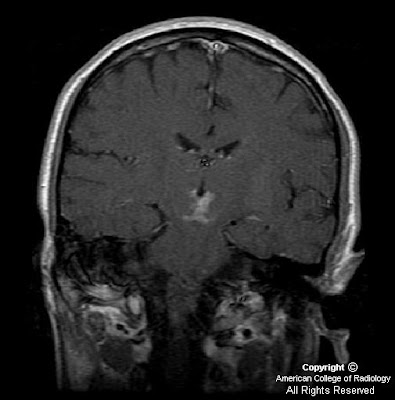
Figure 1: Coronal DWI image demonstrates restricted diffusion within bilateral medial inferior thalami and superior midbrain.
Figure 2: Coronal DWI image, obtained 12 days later demonstrates relatively decreased diffusion hyperintensity.
Figure 3: Coronal post-contrast coronal T1 weighted image demonstrates new enhancement involving bilateral inferior medial thalami and superior midbrain, secondary to breakdown of blood-brain barrier.
Figure 4: Diagram illustrating normal paramedian thalamic mesencephalic arterial supply, with many perforating vessels arising from bilateral P1 segments of the PCA. (Reprinted with permission from AJNR)
Figure 5: Diagram illustrating the variant, “artery of Percheron” a single perforating blood vessel arising from one P1 segment.
Diagnosis: Acute infarction secondary to occlusion of artery of Percheron
The thalami and the midbrain receive their blood supply from both the anterior and posterior circulations, and several variations in this supply are known to exist. The anterior circulation usually supplies the anteroinferior aspects of the thalami and midbrain, with thalamoperforator arteries arising from the posterior communicating arteries. The posterior circulation usually supplies the medial aspects of the thalami and midbrain via branches arising from P1 segments and the lateral and superior aspects with branches arising from P2 segments of the posterior cerebral arteries. Most of the perforating branches from the P1 segments have an ipisilateral distribution (78%); bilateral or even contralateral distributions may be observed in 22% of individuals.
Percheron studied the variations of this arterial supply and its distributions and described three different variations involving the paramedian thalamic-mesencephalic arterial supply: (1) small branches arising from both P1 segments, (2) an asymmetrical common trunk arising from a P1 segment (this variation is called the artery of Percheron), (3) or an arterial arcade emanating from an artery bridging the two P1 segments. In the second type, a common trunk arising from one of the P1 segments provides bilateral distribution. Occlusion of this trunk results in bilateral infarctions in the middle aspects of thalami and brain stem.
The thalami contain strategic nuclei and integrate several important cortical functions. Thus, infarcts at the mesencephalic-diencephalic junctions may result in complex clinical syndromes, with patients exhibiting a wide range of symptoms varying from motor deficits to behavioral and sensory alterations. The third nerve palsy demonstrated in this particular patient may have been due to involvement of the midbrain at the level of the Edinger-Westphal nucleus.
Performing conventional angiography may not be indicated, because lack of visualization of the artery does not exclude its presence (because it is occluded).
Nessun commento:
Posta un commento