giovedì 26 agosto 2010
Germinal matrix hemorrhage
Findings
Hyperechogenic mass like hemorrhage into the right caudothalamic groove with ventricular extension, extension into the septum pellucidum, and resultant hydrocephalus; Grade 3 intraventricular germinal matrix hemorrhage.
Diagnosis: Germinal matrix hemorrhage
Key points
Risks for germinal matrix hemorrhage:
- Less than 32 weeks gestational age
- Under 1500g
- 20-25% risk in premature, low birth weight infant
Germinal matrix
- Proliferating cells; lines the periventricular spaces of the lateral ventricles and third ventricle
- Largest at 24-26 weeks, then involutes
- Begins involuting at the level of the third ventricle
- Involuted completely at 36 weeks
Mechanisms behind germinal matrix hemorrhage-
- Venous bleed
- From cardiovascular instability from lack of autoregulatory mechanism in the preterm infant
- Watershed bleed
- Nearly exclusively occurs in the first 1 wk of life, 50% on day 1
- Identifying and grading germinal matrix hemorrhage
- Blood is always as dense as choroid plexus
- Classic hemorrhage in the caudothalamic groove looks like a mass. May extend into the ventricles
Classification:
- Grade 1: Classic case caudothalamic groove looks like a mass
- Grade 2: Intraventricular extension, usually no long term sequelae
- Grade 3: Dilated ventricles
- Grade 4: Parenchyma, actually from venous hemorrhagic infarct
martedì 24 agosto 2010
Non-hemorrhagic pituitary apoplexy
Findings
A mixed signal (mildly hyper intense on T1-weighted and mixed hyper/hypo intense on T2 weighted sequences) lobular mass with thin rim-enhancement arises from the sella, extending superiorly above the diaphragm sellae (“snowman sign”), deviating the optic chiasm upwards. There is no diffusion restriction, and no susceptibility artifact (on B0 DWI sequences) to suggest hemorrhage. The clivus, and bony sella are intact. A thick rim of dural enhancement dorsal to the clivus is noted.
Diagnosis: Non-hemorrhagic pituitary apoplexy (with underlying macroadenoma)
Key points
Pituitary apoplexy (PA) (e.g. "necrosis") refers to hemorrhage or infarction of the pituitary.
Commonly there is a pre-existing macroadenoma (65-90%).
Acute clinical syndrome includes HA, visual defects/ophthalmoplegia, AMS, and variable endocrine deficiencies.
Acute imaging features show enlarged gland (T1/T2 isointense) with peripheral enhancement, with or without hemorrhage.
Subacutely – gland T1/T2 hyper intense, and chronically – appears as "empty sella" (filled with CSF) with T1 hypo / T2 hyper signal
T2*GRE sequence is very sensitive in looking for hemorrhagic products.
Sometimes there is subarachnoid hemorrhage.
Thickening/enhancement of adjacent dura (50%) and sphenoid mucosa (80%)
Differential diagnosis includes: Pituitary macroadenoma (non-hemorrhagic), craniopharyngioma, Rathke's cleft cyst, pituitary abscess, primary intrapituitary hemorrhage, or giant thrombosed intrasellar aneurysm.
Patients often suffer from long-term pituitary hormonal insufficiency.
Treatment of PA includes: Steroids, fluid/electrolyte replacement, and sometimes surgical decompression.
Discussion of disease
Pituitary adenomas are benign, slow-growing tumors which arise from the adenohypophysis, classified as either micro (< 10mm, 40 %) or macro (> 10 mm, 60%). However, occasionally leptomeningeal metastases can be seen. Patients present with indolent onset of headache, bitemporal hemianopsia (from optic chiasm compression), endocrinologic symptoms.
Pituitary apoplexy (PA), or pituitary necrosis, is usually caused by hemorrhage or infarction of the gland. There can, however, be bland necrosis (without either). The clinical syndrome is of somewhat acute onset with headache, visual defects/ophthalmoplegia, altered mental status, and variable endocrine deficiencies. Pre-existing macroadenoma is very common (65-90%). There is often associated subarachnoid hemorrhage. Thickening / enhancement of the adjacent dura and sphenoid sinus mucosa is very common. Sheehan’s syndrome is a rare peri-partum or post-partum (can be up to 15-20 yrs after index pregnancy) infarction of the pituitary with loss of anterior pituitary hormonal function. Long-term pituitary insufficiency is very common after PA. Treatment may include steroids, fluid/electrolyte replacement, or even surgical decompression.
Radiologic overview
CT (macroadenoma) – isodense to grey matter; fill and expand the sella turcica. CT is excellent for evaluating for sphenoid sinus invasion / bony destruction. Hemorrhage 10%, rarely calcify; pituitary apoplexy appears as hyper dense (acute), or as an "empty sella" if chronic, and very characteristic rim-enhancement.
MR – sellar mass without separate identifiable gland (mass is the gland) with "figure of 8" or "snowman" configuration (coronal), from constriction is caused by diaphragma sellae. Pituitary apoplexy has similar findings, with signal changes based presence/age of hemorrhage. Chronic PA will appear as an "empty sella" (T1 hypo, T2 hyper = filled with CSF). There is thickening / enhancement of adjacent dura in 50% of cases (seen in the presented case), and thickening / enhancement of sphenoid sinus mucosa 80% of the time.
Florid Cemento Osseous Dysplasia
Findings
Figure 1, Figure 2, Figure 3, and Figure 4: Mandibular and maxillary cystic lesions with central calcification, bony expansion and selective involvement of the periapical regions are noted.
Diagnosis: Florid Cemento Osseous Dysplasia
Florid cemento-osseous dysplasia is a slow growing benign lesion which is extremely aggressive and infiltrative. It involves the periapical regions of both the maxilla and mandible with a diffuse distribution of mixed lucent-opaque osseous changes. The lesions are benign fibro-osseous lesions, which histologically, represent normal bone replaced by highly cellular fibrous connective tissue and cementum. Cemento-osseous dysplasia predominantly affects females greater than males (10-14:1) with a predilection for African American patients. Treatment of these lesions should be further radiographic follow-up without surgery unless the lesions become symptomatic, necrotic, or super-infected. Odontoma would also be in the differential as it is a hamartomatous malformation composed of odontogenic tissues. These lesions are usually discovered in the 2nd decade of life and can cause impaction, malpositioning or resorption of adjacent teeth. Ameloblastoma is the most common odontogenic tumor arising from follicular epithelium, dental lamina or enamel. It is a benign lesion but extremely aggressive and infiltrative. It most commonly occurs as an expansile lesion in the posterior mandible in the region of the third molar within the 3rd to 5th decades of life. The unicystic variant occurs in adolescents and has a soap bubble like appearance and involves the ramus and posterior body of the mandible. Large tumors may erode the bony cortex and infiltrate adjacent soft tissues. More malignant processes such as osteosarcoma exhibit a more pronounced periosteal reaction, bone destruction and the presence of a soft tissue mass. Multifocal osteosarcoma is rare. Also, chronic diffuse osteomyelitis is less likely, as it is usually unilateral, associated with pain and fever, exhibits poorly defined borders and would not be exclusively confined to tooth bearing areas.
giovedì 19 agosto 2010
Joubert syndrome
Findings
There is agenesis of the vermis. The interpeduncular fossa is deep. There is a cerebellar cleft. There is prominence and thickening of the superior cerebellar peduncle. There is a bat wing configuration of the fourth ventricle.
Diagnosis: Joubert syndrome
Key points
Joubert syndrome is an autosomal recessive genetic disorder that affects the cerebellum and midbrain. There is agenesis/hypogenesis of the cerebellar vermis and malformation of the midbrain and brainstem.
With poor development of the cerebellar vermis and brainstem, patients can present with ataxia, hypotonia, and hyperpnea. One of the first clinical signs of Joubert is episodic hyperpnea or apnea in an infant. Breathing can reach upwards of 160 breaths/minute. Mild mental retardation is often seen, though some may have normal intelligence. Patient health and growth are usually not affected. Patients with Joubert benefit from physical, occupational, and speech therapy. Infants with respiratory abnormalities require respiratory monitoring.
Radiologic overview of the diagnosis
The initial radiologic manifestations are often seen on fetal ultrasound with increased nuchal lucency being the most apparent finding. Fetal or Brain MR is the best modality to evaluate the presence of Joubert syndrome.
The classic findings are a hypoplastic/aplastic vermis, thick superior cerebellar peduncles, a deep interpeduncular fossa, and an abnormal midbrain. This configuration resembles a molar tooth and is often referred to as the "molar tooth" sign. The fourth ventricle also takes on a bat wing configuration. The midbrain is usually decreased in AP dimension. In this case, there is complete agenesis of the vermis and the molar tooth sign can be seen with thickening of the superior cerebellar peduncles. The corpus callosum and pars intermedia is thickened and may reflect a compensatory response by the brain adapting to the lack of a vermis.
mercoledì 18 agosto 2010
Spinal Cavernous Angiolipoma
Findings
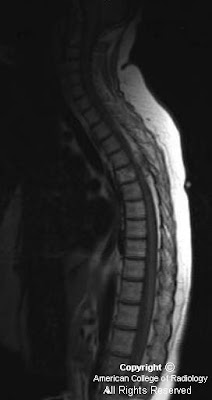
Figure 1: Sagittal T1 image of the thoracic spine demonstrates a mixed signal soft tissue epidural mass posterior to the cord at the T4-T5 level causing posterior impression upon the thecal sac. There is also a T1-bright infiltrating lesion within the T5 vertebral body.
Figure 2 and Figure 3: Sagittal post-contrast T1 imaging shows an enhancing lesion with both bony and soft tissue components. This was localized to the right paraspinal soft tissues and there is compression of the cord. Bony involvement is seen in the vertebral bodies of T11 and L1 and in the posterior elements of multiple other thoracic levels.
Diagnosis: Spinal Cavernous Angiolipoma
Spinal angiolipomas are a rare entity that are histologically benign. These fatty tissue lesions have been quoted in the literature as representing 0.14-1.2% of all spinal axis tumors. Spinal angiolipomas are typically found in the thoracic vertebrae owing to the spine's regional variation in blood supply. The vast majority of epidural noninfiltrating angiolipomas are posterior or posterolateral in location. Patients are more commonly female and usually present in the fourth or fifth decades of life. Symptoms are slow and progressive over several months, with cord compression commonly seen at presentation. Other commonly seen symptoms include: back pain, sphincter dysfunction, progressive paraparesis, lower extremity paresthesias, and hyperreflexia. In rare cases, symptomatology may be acute due to factors such as spontaneous hemorrhage, venous thrombosis, or “steal” phenomenon.
Spinal angiolipomas are divided into two subtypes: the more common noninfiltrating type and the more aggressive (but still histologically benign) infiltrating type. An angiolipoma is termed infiltrating based on bony involvement.
Classically, spinal angiolipomas are T1 hypointense enhancing epidural lesions with or without bony involvement. The intraosseous component is usually heterogenous on T1 imaging. MR is frequently obtained due to patients symptoms of possible cord compression, and it is important to rule this entity out. Differential diagnosis would include hemangiomas, metastatic melanoma and lipid-rich metastases like liposarcoma and the clear cell variant of renal cell carcinoma. On CT, these are indistinguishable from hemangiomas. When viewed in bone windows, the typical “corduroy” appearance with linear streaks of high attenuation can be seen as well as the more stippled foci of increased density seen on axial imaging. To distinguish the two, a pathologic diagnosis is essential
The mainstay of treatment for patients with neurologic symptoms is cord decompression. When possible, surgical excision is preferred with an excellent prognosis for these patients. Heavy bleeding is typical, especially with the infiltrating type owing to the rich vascularity of these lesions. Typically, the neurosurgeon will have multiple blood products available at the time of surgery. For the case discussed above, the estimated blood loss at the time of surgery was 1800cc with 5 units of packed red blood cells administered.
giovedì 12 agosto 2010
Aneurysm of the right cervical internal carotid artery causing vocal cord paralysis
Findings
CT imaging through the neck demonstrates findings suggestive of right vocal cord paralysis.
Figure 1 shows dilation of the right pyriform sinus.
Figure 2 shows thickening and medial positioning of right aryepiglottic fold.
Figure 3 shows dilation of the laryngeal ventricle.
Figure 4: CT through the skull base reveals a rounded well defined brightly enhancing mass in the right carotid space displacing the carotid and internal jugular vein.
Figure 5: MR images demonstrate flow void on T2 weighted images.
Conventional angiography confirms an aneurysm of right internal carotid artery.
Diagnosis: Aneurysm of the right cervical internal carotid artery causing vocal cord paralysis
Unilateral vocal fold paralysis (UVFP) occurs from a dysfunction of the recurrent laryngeal or vagus nerve innervating the larynx. Clinical presentation includes characteristic hoarseness often accompanied by swallowing disabilty, weak cough, and sometimes shortness of breath. Its is important to note that a high vagal lesion results in both a recurrent laryngeal nerve and superior laryngeal nerve palsy with the latter resulting in significant anesthesia of the pharynx and increasing the risk for aspiration
CT scanning or MRI should be performed as part of a workup for a unilateral vocal fold paralysis (UVFP) of unknown etiology. The imaging should include the entire path of the vagus/recurrent laryngeal nerve involved. For left unilateral vocal fold paralysis (UVFP), imaging should extend from the base of skull to the mid chest (arch of the aorta) with right sided vocal fold paralysis including the base of the skull to the clavicle.
Unilateral vocal cord paralysis can be reliably identified on cross sectional imaging. Characteristic findings include:
1. Medial positioning and thickening of the ipsilateral aryepiglottic fold.
2. Ipsilateral pyriform sinus dilatation.
3. Ipsilateral laryngeal ventricle dilatation.
4. Fullness of the ipsilateral true vocal cord.
5. Anteromedial positioning of the ipsilateral arytenoid cartilage.
Causes of vocal cord paralysis are varied with nearly 25% classified as toxic or idopathic. Post-surgical cord paralysis is another important consideration often without specific imaging findings. Identifiable causes include mass compression and or malignant invasion. Although rare, aneurysm or pseudoaneurysm of the cervical internal carotid artery should be considered in the differential diagnosis of a carotid space mass. Because of the intimate proximity of structures within the carotid space, it is sometimes difficult to definitively determine the epicenter of an enhancing lesion. However, as the resolution of cross-sectional imaging improves, preoperative characterization is increasingly possible. A demonstrable arterial connection, arterial enhancement, peripheral calcification, and flow void on MR imaging is suggestive of the diagnosis.
martedì 10 agosto 2010
Pleomorphic Adenoma of the lacrimal gland
Findings
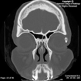
Completely ossified left lacrimal gland with no soft tissue mass, associated superficial inflammatory change or proptosis.
Diagnosis: Pleomorphic Adenoma of the lacrimal gland
Pleomorphic adenomas (benign mixed tumors) are the most common major salivary gland tumor often found in the parotid glands. When seen in minor salivary glands, the hard palate and the upper lip are the most common locations. It is, however, the most common epithelial tumor of the lacrimal gland. Approximately 50% of lacrimal masses are secondary to epithelial tumors; the other 50% are due to lymphoid and inflammatory causes. It will usually present as a palpable lacrimal fossa mass or proptosis. Other lacrimal gland tumors include: germ cell (dermoid and epidermoid), lymphoma/leukemia, metastasis, and sarcoma.
Pleomorphic adenomas are composed of epithelial and connective tissue components. Lesions may have a variable histology with growth patterns in sheets, strands, or islands of spindle and stellate cells with a myxoid configuration occasionally predominating. Cystic degeneration, squamous metaplasia, calcification or ossification may be observed in lacrimal gland pleomorphic adenomas. There may or may not be lytic remodeling or erosion of adjacent bone depending upon the chronicity of the tumor; adjacent bone abnormality does not necessarily confer malignancy. A small percentage of pleomorphic adenomas may undergo malignant transformation.
venerdì 6 agosto 2010
Sinonasal and orbital lymphoma
Findings
Figure 1: Coronal image from CECT shows enhancing mass involving the maxillary and ethmoid sinuses and left orbit.
Figure 2: Coronal image from CECT shows mass in maxillary sinus causing dehiscence of floor of left orbit.
Figure 3: Axial image from CECT shows enhancing mass in ethmoid sinuses and intraconal portion of left orbit causing proptosis.
Diagnosis: Sinonasal and orbital lymphoma (Diffuse Large B-cell Lymphoma)
Malignant neoplasms of the nasal cavity and paranasal sinuses occur primarily in the 5th to 6th decade. Exposure to industrial fumes, leather tanning and even wood dust have been implicated in the carcinogenesis of certain malignant sinonasal tumors. Interestingly cigarette smoking and heavy alcohol consumption have not been linked to sinonasal cancers despite there strong association with other head and neck cancers.
Lymphomas of the sinonasal tract are uncommon neoplasms that can be clinically and radiologically difficult to distinguish from destructive non-malignant processes such as aggressive sinusitis or benign neoplasms. Primary sinonasal lymphoma is rare in western populations, but is more common among Chinese and Japanese populations. Approximately 2000 cases of sinonasal cancer are diagnosed each year in the United States. Lymphoma accounts for less than 5% of all sinonasal cancers and less than 1% of all head and neck cancers.
Low grade lymphomas usually present with a nasal cavity or paranasal sinus mass associated with obstructive symptoms. High grade lymphomas are more likely to present with aggressive signs and symptoms including nonhealing ulcer, cranial nerve manifestations, facial swelling, epistaxis, or pain. Of note, the high grade B-cell lymphomas tend to present with soft tissue or osseous destruction, particularly of the orbit with associated proptosis, whereas T-cell lymphomas are associated with nasal septal perforation and/or destruction.
In Asian populations Natural Killer or T-cell (NK/T-cell) immunophenotypes are more common then B-cell where-as in western populations, there seems to be a slight B-cell predominance. Prognostically NK/T-cell lineage lymphomas have a worse outcome then B-cell. The reason of this discrepancy is not clear but may be related to NK/T-cell frequent expression of an energy-dependent efflux pump capable of transporting chemotherapeutic drugs out of the cell.
Computed tomography (CT) and magnetic resonance imaging (MRI) play complementary roles in determining the full extent of a sinonasal neoplasm. CT is useful to detect bone erosion but can be limited to differentiate the extent of the tumor particularly as the mass approaches the periobita. MRI provides excellent delineation of tumor from surrounding inflammatory tissue and secretions within the sinuses which often accompany obstructive masses. Most sinonasal tumors are highly cellular and give intermediate signal intensity on both T1 and T2 weighted imaging and demonstrate diffuse enhancement.