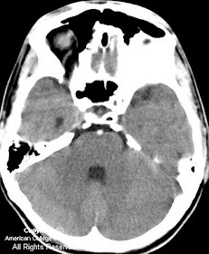

Diagnosis: Basilar thrombosis
Basilar artery occlusion is found in 1 of every 160 autopsies. Thrombosis of an underlying atherosclerotic, high-grade basilar stenosis is encountered in most of these cases. Embolic occlusion, dissecting aneurysm, trauma, and arteritis are less frequent causes. The site of occlusion is most commonly encountered in the lower third of the basilar artery. Clinically, many patients have a history of hypertension, diabetes mellitus, and previous stroke. The neurological signs and symptoms of basilar artery occlusion vary and may include coma, locked-in syndrome, ocular bobbing, palatal myoclonus, and the crossed paralysis syndromes (eg, Millard-Gubler syndrome of facial palsy and contralateral hemiparesis).
The treatment window of opportunity that resolves or improves the neurological effects of basilar artery occlusion appears to be longer than that for occlusions of the anterior circulation. This might be explained at least in part by the retrograde blood supply to the basilar via the posterior communicating arteries. However, recruitment of pial collaterals during the chronic development phase of the basilar artery stenosis prior to thrombosis likely contributes to this ischemia-resistant phenomenon. Examples have been reported of basilar revascularization and subsequent improvement or normalization of neurologic function following endosurgical recannulization even after 48 hours of occlusion.
Recent management strategies involving both IV and IA thrombolytics have shown significantly improved clinical outcomes compared to previous, mostly supportive regimens. IV thrombolysis using tissue plasminogen activator (tPA) effectively reduces the mortality of basilar thrombosis by more than half (40% from 90%) with recannulization occurring in 52% of cases. IA thrombolysis offers advantages over IV intervention in that lower dosages of pharmacologic agents and mechanical thrombolysis can be used together to macerate, dissolve, and if necessary, physically remove the clot by use of microsnares, balloons, stents, and other devices. Endosurgical treatment has a greater recannulization rate of 80%, a noteworthy statistic when considering that these patients often have higher risk than IV thrombolysis candidates owing to their referral beyond the time window for IV therapy.
Nessun commento:
Posta un commento