

Findings
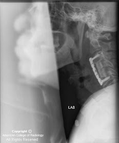
Lateral radiograph of post-operative day 3 demonstrates significant prevertebral soft tissue swelling and gas that is intervally worse from prior radiograph (Figure 1).
Lateral radiograph following emergent reexploration demonstrates interval improvement in prevertebral soft tissue swelling and gas collection (Figure 2).
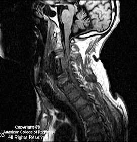
Sagittal T2-weighted images (SE Figure 3, FLAIR Figure 4, and STIR Figure 5) demonstrate a focal collection, hypointense on SE and STIR and isointense on FLAIR anterior to the cord. The collection extends from C3 to C5 (Figure 3, Figure 4, and Figure 5). The cord is compressed and displaced posteriorly.
Axial T2-weighted image of the cervical spine at the level of the inferior endplate of C2 demonstrates a preserved canal with no compression upon the cord (Figure 6).
Axial T2 and axial gradient images demonstrate a collection ventral to the cord with significant cord compromise (Figure 7 and Figure 8).
Diagnosis: Spinal epidural hematoma
Spinal epidural hematoma is being increasingly recognized on magnetic resonance imaging (MRI) in the setting of trauma. Most studies note a male dominance and a reported mean age of 41-52 years. Many causative factors have been implicated including trauma, coagulopathies, rupture of arteriovenous malformations, vertebral body hemangiomas, hypertension, and pregnancy.
Presenting symptoms often include acute radicular pain and rapid onset of paraplegia. The appearance on CT is usually a high-attenuation lesion, which can present both posterior and anterior to the cord. MR is the imaging modality of choice because of excellent soft-tissue contrast resolution, the ability to survey large regions of the spine, and the ability to determine the degree of thecal sac and spinal cord compression. The MR appearance is variable but most commonly is iso- to slightly hyperintense on T1-weighted sequences in comparison to spinal cord, and hyperintense with areas of hypointensity on T2-weighted sequences. Gradient imaging is invaluable in the setting of suspected epidural hematoma because if its excellent ability to delineate blood and blood products.
This case represents a classic appearance of a post-surgical spinal epidural hematoma, which is rarely encountered, and was proven surgically with emergent evacuation secondary to progressive symptoms of weakness.
Nessun commento:
Posta un commento