mercoledì 26 luglio 2006
Ganglioglioma
Findings
Within the medial subcortical portion of the anterior right temporal lobe, there is a 1.5 x 1.6 x 1.3 cm intraaxial lesion that demonstrates increased signal intensity on T2 and FLAIR images (Figure 2, 3, 5 and 6) and relatively low-signal intensity on T1-weighted images (Figure 1). The lesion has well-circumscribed and slightly-lobulated margins. No significant perilesional edema.
Postcontrast images do not reveal any appreciable enhancement (Figure 4 and 7).
This lesion appears to be located inferior and mostly anterior to the right amygdala, and expands the right parahippocampal gyrus medially and the right fusiform gyrus inferiorly.
Diagnosis: Ganglioglioma
Ganglioglioma is a well-differentiated, slowly growing neuroepithelial tumor composed of neoplastic neuronal and glial elements. Majority of gangliogliomas (85%) are low grade (WHO I). It occurs most commonly in children and young adults. Patients usually have a long-standing history of seizures and headaches.
Gangliogliomas are most commonly located in the superficial cerebral hemispheres, especially the temporal lobes. These tumors are usually firm, well-circumscribed masses and may expand the involved cortex. On CT, these have an appearance of low-density or cystic masses. Focal enhancement is seen in 50% of the cases and calcification in approximately 35%. MRI demonstrates these hemispheric lesions in a cortical location, being hyperintense on T2- and hypointense on T1-weighted images. Gadolinium enhancement is variable, often focal or nodular. PET typically shows decreased activity, indicating tumor hypometabolism. In children under 10 years, gangliogliomas can be larger and more cystic.
These tumors have an excellent prognosis with complete surgical excision. The majority (80%) of the patients are seizure-free after surgery. Malignant transformation of ganglioglioma is unusual.
mercoledì 19 luglio 2006
Atypical meningioma
Findings
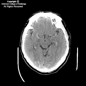
CT without contrast demonstrates an extra-axial heterogeneous mass with solid and cystic components in the left frontal lobe adjacent to the falx. There is calcification along its medial attachment with the falx.
Figure 2: CT with contrast demonstrates homogeneous enhancement of the solid component of the left frontal lobe mass.
Figure 3: Axial T1-weighted MRI with gadolinium contrast demonstrates homogeneous enhancement of the solid peripheral component.
Figure 4: Sagittal T1-weighted MRI demonstrates a hypointense mass in the left frontal lobe. There is curvilinear increased signal intensity along the periphery of the mass, which represents calcifications as demonstrated on CT.
Diagnosis: Atypical meningioma
World Health Organization (WHO) Classification of Meningiomas
- Meningioma (typical benign)
- Atypical meningioma
- Anaplastic (malignant) meningioma
The peak incidence of atypical meningioma occurs in the fourth decade, whereas benign meningiomas show a peak incidence in the fifth decade. Men are affected equally as women with atypical meningioma. In contradistinction, women are affected more frequently with benign meningioma. The most common anatomical location of atypical meningioma is in the parasagittal region (43.7%), followed by the cerebral convexities (15.6%). The recurrence rate of atypical meningioma is 28% within two years, compared to 9.3% for benign meningioma. Atypical meningiomas represent an intermediate category of tumor that has a higher relative risk for recurrence.
Neuroradiologic features of meningiomas
The typical meningioma is a homogeneous, hemispheric, markedly enhancing extraaxial mass located over the cerebral convexity, in the parasagittal region, or arising from the sphenoid wing. However, typical meningiomas and several histologic variants of meningioma can have unusual or misleading radiologic features that may not be suggestive of meningioma. Unusual imaging features, such as large meningeal cysts, ring enhancement, and various metaplastic changes (including fatty transformation), can be particularly misleading. Because meningiomas are so common, the radiologist must be aware of their less frequent and uncharacteristic imaging features in order to suggest the correct diagnosis in atypical cases.
lunedì 10 luglio 2006
Dural arteriovenous fistula with engorgement of pial vessels and venous hypertension
Findings
Figure 1, 2, and 3: The images show engorged pial vessels.
Figure 4, 5, 6 (angio-CT) and 7 (angio-CT reconstruction): Multiple engorged pial vessels are seen throughout.
Diagnosis: Dural arteriovenous fistula with engorgement of pial vessels and venous hypertension
Adult dural arteriovenous fistulas usually present in middle age or older. Dural AVFs represent 10% to 15% of all intracranial vascular malformations. They are arteriovenous shunts located in the dura that usually involve large dural venous sinuses. Arterial supply is usually through meningeal branches of the external carotid, internal carotid, and vertebral arteries. Drainage is via dural venous sinuses or other dural or leptomeningeal channels. They can grow anywhere, but are often found near the skull base.
Adult dural AVFs are usually acquired. These can be secondary to trauma, infection, hypercoaguable states, obstructing neoplasm, and vascular disease. Once the draining dural vein is obstructed, neoangiogenesis ensues, leading to the development of microfistulas in the dural venous sinus wall. Transcalvarial channels may develop from an external carotid, internal carotid, or vertebral artery.
Tentorial and dural AVFs associated with retrograde leptomeninegeal venous drainage are considered to be aggressive lesions. These are more prone to hemorrhage, encephalopathy, and neurologic deterioration.
Digital subtraction angiography is the test of choice to pinpoint the dural and transosseous feeding arteries. Treatment options include endovascular obliteration of the dural AVF, surgical resection, and stereotactic radiosurgery.
lunedì 3 luglio 2006
Subependymal (periventricular) heterotopia
Findings
Figure 1 and Figure 2: Increased density in the periventricular region. This is consistent with subependymal gray matter.
T1 axial (Figure 3), T1 sagittal FLAIR (Figure 6), and T2 coronal FSE (Figure 4) images demonstrate isointensity to normal gray matter on all sequences. T1 post-gad image (Figure 5) shows no abnormal enhancement.
Differential diagnois:
- Subependymal (periventricular) heterotopia
- Subepedymoma
- Hamartoma (tuberous sclerosis)
- Subependymal hemorrhage
- Vascular malformation
Diagnosis: Subependymal (periventricular) heterotopia
Heterotopia, simply defined, means a collection of neurons in an abnormal location, due to an arrest in migration.
There are three clinically useful classifications of gray matter heterotopia:
- subependymal heterotopia
- band heterotopia
- subcortical heterotopia
Subependymal, which is the most common of the three, tends to present with epilepsy in the second decade of life. Subcortical and band heterotopia tend to present with epilepsy earlier in life (first decade). Additional brain anomalies may also be associated.
Initial imaging modality is typically CT in a postepileptic patient. Findings include increased density in a periventricular distribution without enhancement or calcification. Proper window settings are essential for proper diagnosis. Low level and window allow for greatest contrast. All three are most easily diagnosed with MRI, which demonstrates isointensity to normal gray matter on all sequences without postgadolinium enhancement.
Subependymal heterotopia (SEH) can occur in isolation or with other neurologic and metabolic disorders.
Anatomically, it is subdivided into three subtypes
- unilateral focal
- bilateral focal
- bilateral diffuse.
All types typically present with epilepsy in the second decade of life with disorders of cognition being proportionate to the area of heterotopia. MRI shows small areas of gray matter, which is isointense to mature gray matter, protruding into the ventricular lumen. X-linked causes have been found in many of the bilateral diffuse cases, resulting in more dramatic neurodevelopmental disorders in males. The same causative gene mutation has been associated with Ehlers-Danlos syndrome.
Band heterotopia is diagnosed by visualization on MRI of a smooth layer of gray matter separated from the cortex and ventricles by white matter. This may also be called “double cortex.” Neurodevelopmental disability is proportionate to the thickness of the heterotopic “band.” The manifestations of this x-linked disease are more drastic than SEH; males suffer from lissencephaly, while females suffer from band heterotopia. Clinically, females typically present with epilepsy in the first decade of life.
Subcortical heterotopia is anatomically classified into two forms
- nodular form
- curvilinear form.
Much less is known about this classification of heterotopia than the others mentioned. Patients present with varying degrees of mental and motor delay and epilepsy in the first decade of life. Imaging shows various sizes of clusters of improperly migrated neurons throughout the white matter. Many theories regarding the cause of this subtype are currently being studied.