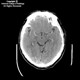


Findings
Figure 1, 2, and 3: The images show engorged pial vessels.
Figure 4, 5, 6 (angio-CT) and 7 (angio-CT reconstruction): Multiple engorged pial vessels are seen throughout.
Diagnosis: Dural arteriovenous fistula with engorgement of pial vessels and venous hypertension
Adult dural arteriovenous fistulas usually present in middle age or older. Dural AVFs represent 10% to 15% of all intracranial vascular malformations. They are arteriovenous shunts located in the dura that usually involve large dural venous sinuses. Arterial supply is usually through meningeal branches of the external carotid, internal carotid, and vertebral arteries. Drainage is via dural venous sinuses or other dural or leptomeningeal channels. They can grow anywhere, but are often found near the skull base.
Adult dural AVFs are usually acquired. These can be secondary to trauma, infection, hypercoaguable states, obstructing neoplasm, and vascular disease. Once the draining dural vein is obstructed, neoangiogenesis ensues, leading to the development of microfistulas in the dural venous sinus wall. Transcalvarial channels may develop from an external carotid, internal carotid, or vertebral artery.
Tentorial and dural AVFs associated with retrograde leptomeninegeal venous drainage are considered to be aggressive lesions. These are more prone to hemorrhage, encephalopathy, and neurologic deterioration.
Digital subtraction angiography is the test of choice to pinpoint the dural and transosseous feeding arteries. Treatment options include endovascular obliteration of the dural AVF, surgical resection, and stereotactic radiosurgery.
Nessun commento:
Posta un commento