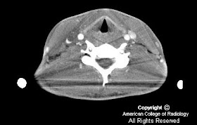
Findings
Figure 1: The CT scan of the neck demonstrates low density material within the right internal jugular vein. This is consistent with internal jugular vein thrombosis.
The CT scan of the chest demonstrates numerous peripheral nodules, many with central cavitation, as well as bilateral pleural effusions and a left lower lung consolidation. The presence of multiple peripheral nodules, some of which are cavitary, as well as consolidation and pleural effusions, is very consistent with septic embolic disease. In this patient, the septic emboli are arising from a thrombophlebitis of the internal jugular vein.
Diagnosis: Lemierre syndrome
Lemierre syndrome, also known as postanginal sepsis, is thrombophlebitis of the internal jugular vein with metastatic septic emboli, generally following an acute oropharyngeal infection.
The disease typically progresses in a stepwise fashion, starting with a primary infection, which is usually a pharyngitis of the palatine tonsils and peritonsillar tissue (in almost 90% of cases). The infection then invades the parapharyngeal space, usually via lymphatic vessels. The posterior parapharyngeal space contains the internal jugular vein, common carotid artery, cervical sympathetic trunk, and cranial nerves IX to XII. Although the infection can involve any of these structures, the most common consequence is internal jugular vein thrombophlebitis. Clinically, patients may have pain or swelling of the neck, sometimes with associated trismus or spasm of the sternocleidomastoid muscle. Once the internal jugular vein is involved, the infection can spread hematogenously to any part of the body, but most frequently affects the lungs, with metastatic infection occurring in 80% of cases, and to the joints, particularly the hips, shoulders, and knees, in 16% of cases. Although findings of pulmonary septic emboli, including multiple peripheral cavitating nodules or pulmonary infarction, are the classic presentation of Lemierre syndrome, less specific pulmonary manifestations, such as noncavitating pulmonary infiltrates and pleural effusions, are frequently seen as well.
Treatment involves a prolonged course of high-dose intravenous antibiotics. Prior to the use of antibiotics, this disease carried a mortality rate of 90%. Although the incidence of this disease has decreased substantially, delay in treatment is associated with significant morbidity and mortality. Therefore, familiarity with this syndrome is important so the diagnosis can be made quickly and appropriate antibiotic coverage started. The constellation of internal jugular vein thrombosis with pulmonary findings, even if they are not clearly septic emboli, should immediately lead to the consideration of Lemierre syndrome as the diagnosis until proven otherwise. Although a clinical history of recent pharyngitis, or evidence of an inflamed pharynx is helpful, as Hall warned in 1939, “Be not deceived by a comparatively innocent-appearing pharynx, as the veins of the pharynx may be carrying the death sentence for your patient.”
Nessun commento:
Posta un commento