

Findings
Axial CT scan at the level of third ventricle demonstrates a well-defined hyperdense mass in relation to the posterior third ventricle showing peripheral calcification (Figure 1).
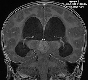
Figure 2 demonstrates hydrocephalus with transependymal flow seen as confluent low attenuation in the periventricular regions.
The fourth ventricle is normal in size (Figure 3).
The mass demonstrates minimal, heterogeneous, increased signal on the T2-weighted image (Figure 4) as well as heterogeneous enhancement on postcontrast images (Figure 5). Sagittal postcontrast T1-weighted image (Figure 6) demonstrates mass effect on the midbrain tectum, which is displaced inferiorly. The internal cerebral veins are seen as tubular enhancing structures superior to the lesion (Figure 7).
Diagnosis: Pineoblastoma
Pineal region tumors are uncommon, but are more often seen in children compared with adults. Germinomas and astrocytomas account for the majority of pineal region masses. Pineal parenchymal tumors constitute less than 15% of pineal neoplasms. Pineoblastomas are highly malignant, primitive neuroectodermal tumors of the pineal gland that are typically found in children 2 to 3 years of age. Pineocytomas, on the contrary, are slow growing pineal parenchymal tumors of adults.
Patients usually present with signs of elevated intracranial pressure, ataxia and/or Parinaud’s syndrome (palsy of the upward gaze, dissociation of light and accommodation, and failure of convergence). Unlike germ cell tumors, there is no elevation of serum tumor markers in pineal parenchymal tumors.
Pineoblastomas are WHO Grade IV tumors that are poorly marginated, demonstrate peripheral calcification, as well as hyperdensity and heterogenous enhancement of the solid components. Peritumoral edema is characteristically mild. In contradistinction, germinomas demonstrate central “engulfed” calcification and uniform enhancement. Both pineoblastomas and germ cell tumors can demonstrate CSF dissemination.
The treatment for pineoblastoma includes surgical ressection, cranio-spinal radiation, as well as chemotherapy. The prognosis is dismal in most cases.
In “trilateral retinoblastoma”, pineoblastoma may develop in patients with familial and/or bilateral retinoblastoma.
Nessun commento:
Posta un commento