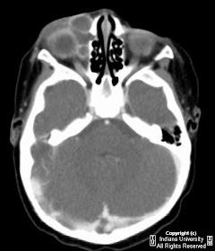

Findings
Right sided preseptal soft tissue inflammation with rim enhancing preseptal and extraconal fluid collections. No definitive evidence of intraconal spread of infection, extension to the orbital apex or adjacent bony erosion.
Differential Diagnosis:
- Cellulitis with abscess
- Subperiosteal abscess
- Ethmoid mucocele
- Dacryocystocele
- Congenital/Developmental lesions: lymphangioma, dermoid cyst
- Subperiosteal hematoma
- Orbital pseudotumor
- Orbital rhabdomyosarcoma
Diagnosis: Orbital cellulitis and abscess secondary to dacryocystitis
Key points
Dacryocystitis occurs secondary to stagnation of fluid within the lacrimal sac resulting in bacterial overgrowth.
Acute dacryocystitis commonly presents as tender preseptal cellulitis and is treated with antibiotics while chronic cases usually present as painless purulent reflux from the lacrimal sac and is treated with definitive dacryocystorhinostomy.
Acute dacryocystitis most often results in preseptal cellulitis; however, orbital extension with abscess is also a known complication which can result in severe visual compromise.
Key radiologic features
MRI and CT are imaging modalities of choice, with MRI preferred when evaluating for possible intra-cranial extension.
Edema of the orbital soft tissues.
Low density fluid collection with rim enhancement.
Associated myositis represented by swelling and/or abnormal enhancement of the extra-ocular musculature.
Nessun commento:
Posta un commento