giovedì 30 ottobre 2008

Microtia


Findings

Figure 1: On the right, the mastoid air cells are under pneumatized. There is no identifiable external auditory canal.
Figure 2: A thick bony plate is visualized in the expected area of the external auditory meatus.
Figure 3: The facial nerve is identified.
Figure 4: There is a small amount of bone in the attic of the middle ear cavity but no formed malleus or incus is identified. This suggests rudimentary and/or hypoplastic ossicles. However, a normal morphology and location of the stapes is seen. The internal auditory canal is normal in caliber.
Figure 5: The apical and basal turns of the cochlea are within normal limits.


Diagnosis: Microtia


Microtia, which is a congenital deformity of the pinna and occurs more commonly in boys, is seen 1 in 8,000-10,000 births. It can be unilateral or bilateral. The etiology is unknown, but related to a variety of genetic factors. It is associated with other congenital syndromes, such as Townes-Brocks syndrome, Nager syndrome, and Miller syndrome. Some clinicians consider microtia to be a manifestation of the oculo-auriculo-vertebral spectrum (OAVS), where there are also facial, vertebral, and renal abnormalities. Renal abnormalities usually warrant a renal ultrasound, looking for abnormalities such as renal agenesis, hypoplasia, and crossed ectopia.

Microtia can be graded as follows:
- Grade I: A slightly small ear with identifiable structures and a small but present external ear canal
- Grade II: A partial or hemi-ear with a closed off or stenotic external ear canal producing a conductive hearing loss
- Grade III: Absence of the external ear with a small peanut vestige structure and an absence of the external ear canal and ear drum
- Grade IV: Absence of the total ear or anotia

Imaging of microtia is largely performed to form a template for further management options. Initially, a CT scan of the head (and/or temporal bones) is used to evaluate the exact middle and inner ear anatomy. Further staging by CT is recommended in order to avoid lesions to the facial nerve which most often can be displaced from its original location in patients with microtia, and also to assess for prognosis. In those patients being evaluated for surgical intervention, the following structures should be evaluated for planning surgery: the external auditory canal, bones (temporal, zygomatic, and the mandibular condyle), vessels (carotid canal, sigmoid sinus, and the jugular bulb), tensor tympani muscle and grade of mastoid pneumatization, ossicles, cochlear turns, vestibule, semicircular canals, facial nerve canal, and finally, the internal auditory canal. These pre-operative images substantially reduce the risk of facial nerve palsy, bleeding and worsening of hearing.

On axial CT images of the head, one can evaluate for either stenosis or complete atresia of the external auditory canal. At the same time, bone erosion by a secondary cholesteatoma or epidermoid cyst can be identified. One study showed the tympanic and/or mastoid portions of the temporal bone as commonly hypoplastic on CT images of patients with microtia. Dysplasia of the mandibular condyle and the zygomatic arch were also shown. Changes of the ossicles are frequently present and include dysplastic shape, diminution, thickening, axis rotation, or complete absence. Occasionally, the lateral semicircular canal is hypoplastic in patients with severe middle ear involvement. The internal auditory canal is rarely dysplastic in patients with microtia.

Microtia repair is complex. Testing is done initially to determine whether hearing is normal. If hearing is normal and a canal is not visible externally, a CT will be done to determine whether a rudimentary canal exists. The earliest age surgery can be attempted is three years, but will vary according to the graft material used. In some cases, patients may have to wait as long as 6 years of age. Exploration involves cautiously avoiding the facial nerve while drilling a canal through solid bone. A cartilage framework, usually derived from costal cartilage, is created and anchored beneath the skin of the mastoid area. Once it is well attached to the surface skin, a post-auricular crease is created in a second operation.

martedì 28 ottobre 2008

Vanderknaap disease - Megaloencephalic leukoencephalopathy with subcortical cysts (MLC)


Findings

Figure 1: Subcortical CSF intensity cyst in superior frontal and parietal lobe. Normal cerebellum.
Figure 2: CSF intensity subcortical cyst in superior parietal and anterior temporal lobe.
Figure 3: Diffusely swollen white matter (blue arrow). Preserved gray matter. CSF intensity subcortical cyst in anterior temporal lobe.
Figure 4: Diffusely swollen white matter. Preserved gray matter.
Figure 5: Cavum septum pellucidum. CSF intensity subcortical cyst in superior frontal and anterior temporal lobe. Normal basal ganglia.
Figure 6: Cavum septum pellucidum. CSF intensity subcortical cyst in superior frontal and anterior temporal lobe. Normal basal ganglia.


Diagnosis: Vanderknaap disease - Megaloencephalic leukoencephalopathy with subcortical cysts (MLC)


Vanderknaap disease is newly described rare leukoencephalopathies includes:
- 1. MLC - Megaloencephalic leukoencephalopathy with subcortical cysts
- 2. VWM - Leukoencephalopathy with vanishing white matter
- 3. WML - White matter disease with lactate
- 4. H-ABC- Hypomyelination with atrophy of the basal ganglia(BG) and cerebellum

Megaloencephalic leukoencephalopathy with subcortical cysts (MLC) is a rare leukoencephalopathy with an autosomal recessive mode of inheritance. The disease is relatively prevalent among Turkish people and in a certain Asian-Indian community, the Agarwal ethnic group.


MRI Features

In MLC the cerebral hemispheric white matter is diffusely abnormal and swollen. There are almost invariably subcortical cysts in the anterior temporal region, often also in the frontal and parietal subcortical regions The cysts are bilateral. The cysts tend to become larger with age and may increase in number. In some patients they become very large, The signal intensity of the contents of the cysts is always similar to that of CSF. Cortical gray matter structures,corpus callosum and basal nuclei are always normal. A patent and enlarged cavum septi pellucidum and cavum vergae are often present.

Similar white matter changes with swelling have been reported in Canavan disease, Alexander disease, L-2-hydroxyglutaric aciduria, and merosin-deficient congenital muscular dystrophy. However, in Canavan disease, as a rule, MRI demonstrates additional involvement of the thalamus and globus pallidus, not found in MLC patients. Special MRI findings in Alexander disease are a more prominent sparing of parieto-occipital white matter and often also sparing of the U fibers throughout. Basal ganglia and brain stem structures are typically involved. Cavitation starts in the deep frontal white matter. None of these features is present in MLC. In L-2-hydroxyglutaric aciduria MRI shows additional involvement of caudate nuclei, putamen, dentate nuclei, and severe atrophy of the cerebellar vermis, not observed in MLC. The MRI abnormalities observed in merosin-deficient congenital muscular dystrophy are very similar to those observed in MLC.

giovedì 23 ottobre 2008

Subependymal giant cell astrocytoma (SEGA)


Findings

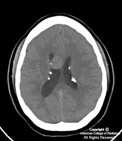
Figure 1: Axial noncontrast CT image shows an intraventricular mass near the foramen of Monro with foci of calcification, as well as several calcified subependymal nodules along the lateral ventricular surface. Hydrocephalus is also present with enlargement of the right lateral ventricle.
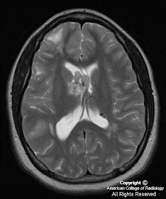
Axial T2-weighted and FLAIR MR images demonstrate a mass near the foramen of Monro with heterogenous, somewhat hyperintense signal compared to white matter. Intense homogeneous enhancement is seen on contrast-enhanced axial and coronal T1 weighted images. Subependymal nodules are seen along the lateral ventricles. Multiple foci of increased signal are seen on FLAIR images in the subcortical regions representing parenchymal tubers.


Diagnosis: Subependymal giant cell astrocytoma (SEGA)


Subependymal giant cell astrocytomas are intraventriclar neoplasms that occur in 15% of patients with tuberous sclerosis. Tuberous sclerosis (Bourneville’s disease) is a neurocutaneous phakomatosis characterized by an autosomal dominant pattern of inheritance presenting with the classical clinical triad of mental retardation, seizures, and adenoma sebaceum (although only 30% present with all three features).

The disease is characterized by hamartomatous tumors and malformations affecting multiple organ systems, the CNS being the most commonly involved, with seizure being the most frequent presenting clinical sign of the disorder. Other than the CNS manifestations, patients may present with renal angiomyolipomas, cardiac rhabdomyomas, and cystic lung disease indistinguishable from lymphangiomyomatosis.

Hamartomatous brain lesions include cortical tubers, white matter heterotopias, subependymal nodules, and the subependymal giant cell astrocytoma. Histologically, cortical tubers, white matter lesions, and subependymal nodules are identical lesions composed of disordered neurons, glia, and giant cells mostly of the astrocyte type, only differing in size and location. Subependymal nodules are usually easily identified with CT due to frequent calcification (>90%) and usually do not enhance thus helping to distinguish, but not entirely exclude a SEGA from a subependymal nodule. Cortical tubers are less likely to calcify and appear as low attenuation lesions at CT, demonstrate increased signal intensity on T2-W images, and rarely enhance. White matter lesions are seen as curvilinear or straight bands of increased T2 signal extending from the ventricles. SEGAs are characterized by slow growth and a benign biological behavior (WHO grade I), likely arising from the degeneration of subependymal nodules. On CT, SEGAs are iso-to slightly hypoattenuating intraventriuclar masses located near the foramen of Monro, with calcification and secondary hydrocephalus being common findings. On MR imaging, SEGAs exhibit hypointense signal compared to white matter on T1-weighted images, heterogenous hyperintensity on T2-weighted images, with intense homogenous enhancement (except for calcified areas). Because MR enhancement cannot always reliably distinguish between subependymal nodules and a SEGA, larger size (>1cm) and interval growth of a mass on annual follow-up CT or MR are considered better indicators of a SEGA rather than a benign subependymal nodule. Therefore, annual surveillance MR imaging is recommended in patients with tuberous sclerosis.

lunedì 20 ottobre 2008

Multiple post traumatic pseudomeningoceles secondary to brachial plexus avulsion with atrophy and myelomalacia of the spinal cord


Findings

Right-sided cyst-like structures are demonstrated at C7-T3. At T1-T2 and T2-T3 these coalesce to form a larger cystic structure overlying the right lung apex.
Additional history: Young adult with history of traumatic brachial plexus injury 2 years ago, now with new onset weakness.

These findings are compatible with the patient's history of previous traumatic right brachial plexus nerve root injury. Since the prior examination, these "pseudomeningoceles" have significantly increased in size. There is a focal 1.5 cm atrophy of the spinal cord with a 6 mm in length area of increased T2 signal is demonstrated within the spinal cord at T1-T2 level consistent with myelomalacia likely the result of the patient's previous nerve root injury.


Diagnosis: Multiple post traumatic pseudomeningoceles secondary to brachial plexus avulsion with atrophy and myelomalacia of the spinal cord


Key points

Cervical nerve root avulsions are the result of traction injuries of the upper extremities that tear the roots from the spinal cord. The roots are absent on the ipsilateral side and the spinal cord is pulled towards the contralateral side.
Pseudomeningoceles are associated with this type of injury. They result from tears in the arachnoid or dura and can easily be identified by MR or CT myelography.
They can be asymptomatic, or present in an acute or delayed fashion with symptoms of myelopathy.
A very rare complication is development of spinal cord herniation.

venerdì 17 ottobre 2008

Ruptured intracranial dermoid


Findings

Fat density lesion within the pericavernous region in the region of Meckel’s cave extending inferiorly into the posterior fossa. Scattered lipid droplets throughout the subarachnoid space.

Differential diagnosis:
- Dermoid cyst
- Epidermoid cyst
- Craniopharyngioma
- Teratoma
- Lipoma


Diagnosis: Ruptured intracranial dermoid


Key points

Congenital inclusion cyst.
Epidemiology: Rare; <0.5% of primary intracranial tumors.
30-50 y/o.
Presentation (uncomplicated): HA (32%), Seizure (30%), rarely with DI, hypopituitarism, visual defects.
Benign, slow growing.
Rare malignant transformation into SCCa.
Increased risk of rupture with increased size.
Significant M&M with rupture: chemical meningitis, seizures, coma, vasospasm, etc.
Fat density distinguishes from epidermoid (usually CSF density).
Scattered lipid droplets in subarachnoid/ventricles with rupture.
20 % have capsular calcium.
Most supratentorial, midline or near midline.
Other sites: spine, orbit.

martedì 14 ottobre 2008

Spinal foreign body granuloma


Findings

The T2 sagittal and axial images (Figure 2 and Figure 3) show a posterior intradural nodule with low signal intensity which is compressing the spinal cord anteriorly causing cord edema. The sagittal T1 (Figure 1) sequence shows that the nodule is isointense to hypointense which is non specific. It also confirms its extramedullary location.
The axial T1 postcontrast (Figure 4) sequence shows peripheral contrast enhancement with a central hypointense non enhancing area. This could be associated with the presence of central calcification, metal within an inflammatory nodule or the non enhancing central “dot” described in some nerve sheath tumors.


Diagnosis: Spinal Foreign body granuloma


Intradural extramedullary lesions include meningiomas, nerve sheath tumors (schwannomas and neurofibromas), drop metastasis and foreign body granulomas. Meningiomas are globular lesions. They are more common in elderly females. They are usually located in the thoracic region. These tumors are isointense to slightly hypointense on T1. They enhance and can have a dural tail. On T2 they are isointense to hyperintense and can demonstrate low signal on T2 related to intalesional calcification. Meningiomas originate from the denticulate ligament.

Nerve sheath tumors are usually isointense on T1 and hyperintense on T2. They also enhance and can show a central non enhancing “dot”. Hemorrhage and cystic degeneration has been associated with nerve sheath tumors more than with meningiomas. They are slow growing and can erode the bone. Nerve sheath tumors are associated with neurofibromatosis 1 and 2.

Intradural extramedullary metastasis could be the result of drop lesions from primary brain neoplasms such as medulloblastomas or ependymomas or the result of hematogenous spread from solid organ tumors.

Foreign body granulomas are associated with the presence of intrathecal drug delivery pumps more commonly with the administration of opioid derivates or in patients who received agents that were not labeled for long-term intrathecal use. Implanted delivery systems for intrathecal drug administration have become more commonplace in the management of refractory cancer and non-malignant pain. Complications may be related to drug side effects or to technical problems possibly involving the pump and/or catheter

Clues to the diagnosis include the above imaging findings along with the loss of analgesic drug effects accompanied by new, gradually progressive neurological signs and symptoms. Surgery is not always necessary for treatment. Successful resolution has been reported with lower doses and/or changes in the medication delivered.

lunedì 13 ottobre 2008

Kernicterus


Findings

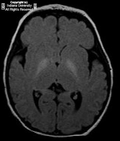
Increased signal intensity on T1 and FLAIR sequences in the bilateral globi pallidi and hippocampi.

Differential diagnosis:
- Carbon monoxide exposure
- Kernicterus
- Hepatic failure
- Creatine deficiency
- Profound hypoxic encephalopathy


Diagnosis: Kernicterus


Key points

Encephalopathy is due to deposition of toxic unconjugated bilirubin in the basal ganglia, hippocampus, substantia nigra, and brainstem nuclei. It is most often caused by hyperbilirubinemia from erythroblastosis fetalis and most commonly seen in premature infants. It is also associated with early discharge from the hospital. Physiologic and breastfeeding jaundice are benign causes of hyperbilirubinemia.
In the absence of hyperbilirubinemia, factors influencing permeability of the blood-brain barrier (e.g., acidosis, infection) and the amount of unbound (versus albumin-bound) bilirubin may play a role.
Bilirubin-induced neurologic dysfunction (BIND) refers to the clinical signs associated with bilirubin toxicity (i.e., hypotonia followed by hypertonia, opisthotonus or retrocollis, or both).
In the acute phase, neurological findings can include decreased alertness, hypotonia, and poor feeding early on. Later, hypertonia of the extensor muscles is a typical sign. Patients present clinically with retrocollis (backward arching of the neck), opisthotonus (backward arching of the back), or both. Infants who progress to this phase develop long-term neurologic deficits. Prognosis is poor once neurological complications develop. Seizures are not a usual presentation.
Chronic sequelae evolve over the first several years of life. They include extrapyramidal, auditory, and visual abnormalities, and cognitive deficits.
Noncontrast MRI is the most useful imaging modality in the acute and chronic phases. CT is not useful.
MR imaging findings in the acute phase include increased T1 signal intensity in the globus pallidus, hippocampus, substantia nigra, and dentate nucleus. T2 signal may be subtly increased.
MR imaging findings in the chronic phase include increased T2 signal in the posteromedial border of the globus pallidus, hippocampi, and occasionally the dentate nucleus. T1 weighted images are normal.
Preventative treatments include phototherapy. Exchange transfusion is indicated for patients with findings of encephalopathy and serum bilirubin levels >25mg/dL with dehydration.

giovedì 9 ottobre 2008

CSF leak


Findings

CT shows dehiscence of the cribriform plate bilaterally. Right gyrus rectus encephalocele. Nuclear medicine cisternogram shows abnormal radiotracer localization in the nasal cavities bilaterally.


Diagnosis: CSF leak


Key points

Causes include:
- Blunt head trauma
- Sequelae of skull-base surgery
Commonly functional endoscopic sinus surgery (FESS)
Transsphenoidal pituitary surgery
Translabyrinthine acoustic schwannoma
Mastoid surgery with intact tympanic membrane
- Destructive skull-base lesions, including neoplasms (both benign and malignant), and empty sella
- Developmental defects of the ethmoid, sphenoid, frontal, or petrous temporal bones with the formation of a meningocele or meningoencephalocele (with an intact tympanic membrane)
- Fracture of the petrous temporal bone or other destructive processes in which CSF in the middle ear drains to the nose in the presence of an intact tympanic membrane
- Less than 5% of all cases of CSF rhinorrhea are spontaneous

Most cases of CSF rhinorrhea begin soon after a head injury and cease spontaneously within 7-180 days.
Radionuclide cisternography is used to confirm a suspected CSF leak.
Leaks are most common in the region of the cribriform plate and ethmoid sinuses.
CSF leaks may be intermittent leading to a false negative study.
The study is performed in conjunction with ENT and possibly neurosurgery, in addition to neurointerventional radiology, who performs the lumbar puncture.
Typically Tc-99m sulfur colloid is administered into the thecal sac by lumbar puncture. Additionally cotton pledgets are placed in the superior and middle turbinates by ENT. Imaging of the spinal canal and head are obtained between 1 and 3 hours. The cotton pledgets are removed from 4 to 24 hours later and are individually placed in a well counter. Serum samples are obtained at the same time and counted for activity. Pledget to serum ratios of more than 1.5 may be interpreted as evidence of CSF leak.
CT cisternography after the administration of iodinated contrast within the thecal sac may be performed for further anatomic delineation.
Treatment is primarily surgical.

mercoledì 8 ottobre 2008

Amyloid angiopathy with subacute right temporal lobe hemorrhage


Findings

Heterogeneous right temporal lobe mass with a thin rim of peripheral enhancement and moderate surrounding parenchymal edema. There is associated susceptibility on gradient images, however no diffusion restriction. There is effacement of the posterior horn and trigone of the right lateral ventricle, as well as adjacent sulcal effacement. There are also multiple punctate/small foci of susceptibility distributed throughout the cerebral and cerebellar hemispheres centered at the corticomedullary junction. Also noted (but not shown) were foci of T2 prolongation scattered throughout the centrum semiovale, corona radiata, and subcortical white matter, consistent with microvascular disease.

Differential diagnosis:
- Hypertensive micro hemorrhages
- Ischemic stroke with micro hemorrhage
- Multiple vascular malformations
- Traumatic diffuse axonal injury
- Amyloid angiopathy
- Hemorrhagic metastasis
- CADASIL (cerebral autosomal dominant arteriopathy with subcortical infarcts and leukoencephalopathy)
- Metallic microemboli from artificial heart valves


Diagnosis: Amyloid angiopathy with subacute right temporal lobe hemorrhage


Key Points

Amyloidosis – rare systemic disease caused by extracellular deposition of ß-amyloid
10-20% localized form, inclusive of CNS
Can be idiopathic/primary or secondary/reactive (dialysis related amyloidosis)

Cerebral amyloid deposition: 3 varieties
- Cerebral amyloid angiopathy (most common)
- Amyloidoma (rare)
- Diffuse white matter involvement (rare)

Common cause of spontaneous lobar hemorrhage in elderly (1% of all strokes)
Responsible for 15-20% of ICH in patients >60 years old
27-32% of normal elderly (autopsy)
82-88% in Alzheimer's disease
Common in Down's syndrome


Imaging features

Multifocal "black dots" representing chronic micro hemorrhages.
Lobar hemorrhages of different ages.
Involves subcortical WM (gray/white junction), parietal and occipital lobes (autopsy), also frontal and temporal lobes.
Less commonly involves brainstem, deep gray matter, cerebellum, hippocampus.
Acute lobar hemorrhages tend to be large and irregular with dependent blood sedimentation.
Punctate foci of dark susceptibility on T2*GRE sequences (blooming) seen with chronic micro hemorrhages.
Focal or patchy/confluent WM disease associated in approximately 70%.

giovedì 2 ottobre 2008

Congenital cystic intracranial neoplasm


Findings

Figure 1: Prenatal ultrasound demonstrates a 4 x 5 cm intracranial lesion in the region of the right middle cranial fossa, with multiple cystic and heterogeneously echoic soft tissue areas.
Figure 2, Figure 3, and Figure 4: MR images at 32 weeks 3 days gestation. Coronal (Figure 2) and sagittal (Figure 3) single shot T2 weighted (SShT2) images and axial (Figure 4) fast field echo (FFE) image of the fetal head show a large cystic lesion with multiple internal cysts and soft tissue components in its inferior aspect in the region of the right temporal lobe. (SShT2: repetition time 15000 milliseconds, echo time 120 milliseconds, flip angle 90, number of excitations 2)
(FFE: repetition time 5.3 milliseconds, echo time 2.6 milliseconds, flip angle 60, number of excitations 2)
Figure 5: Newborn non-enhanced axial CT image shows a multicystic heterogeneous lesion within the right temporal lobe containing calcification.

Differential diagnosis of complex cystic neonatal intracranial masses:
- Teratoma
- Astrocytoma
- Hemangioma
- Cystic ependymoma


Diagnosis: Congenital cystic intracranial neoplasm


Congenital brain tumors represent only 0.5-1.9% of all pediatric brain tumors, but of these, intracranial teratomas are the most common tumor in the neonatal period. They are congenital neoplasms derived from all three embryonic layers and contain ectodermal, endodermal, and mesodermal elements and are thought to arise from embryonic cells that fail to differentiate or migrate. They originate in the 3rd to 4th weeks of fetal development, have mixed density, and are most commonly found in the pineal and suprasellar regions.

Extragonadal teratomas are thought to arise from misplaced, pluripotential, primordial germ cells. According to one theory, some of the embryonic germ cells fail to migrate normally and these ectopic germ cells become embedded in or near midline structures. The ectopic cells retain their pluripotent capabilities and may develop into teratomas or other types of germ cell neoplasms.

Intracranial teratomas can be divided into two categories with those occurring in the cerebral hemispheres and those occurring in the pineal region. Those appearing in the cerebral hemispheres are typically large and are usually discovered in the newborn period. Those in the pineal region manifest symptoms later on and are usually smaller. Intracranial teratomas are extremely rare tumors and are usually diagnosed after birth, but they have been discovered prenatally via ultrasound.

Of the intracranial prenatal neoplasms, teratomas and primitive neuroectodermal tumors (PNETs) are the most common. If a heterogeneous prenatal cystic mass is identified, a congenital cystic teratoma should be at the top of the differential diagnosis. Imaging features of teratomas show both lipid and calcification. Characteristics that help to differentiate teratomas from PNETs are the presence of lipid and the lack of blood breakdown products, which are often seen in PNETs.

When ultrasound demonstrates a brain lesion, prenatal MRI can be used to define the characteristics and extent of the tumor while also delineating the relationship of the lesion with adjacent structures. MRI is not only useful for correct diagnosis, but it also may obviate the need for immediate postnatal imaging of the newborn and findings can determine potential resectability and guide surgical planning. In this case, the prenatal MR imaging was used in prenatal diagnosis and aided in pregnancy and surgical management. While it has been shown that a fetus with an arachnoid cyst can safely undergo normal spontaneous vaginal delivery, the diagnosis of teratoma in this patient altered both the method and timing of delivery in this patient.

mercoledì 1 ottobre 2008

Lupus cerebritis


Findings

Diffuse, predominantly white matter T2/FLAIR hyperintense signal spares only the anterior temporal lobes. Focal lesions with hypointense T1 signal, low T2 signal, are heterogeneously hypointense on gradient echo, do not show restricted diffusion, and rim enhance. Ventricles, sulci, and cisterns remain normally configured and are of normal size for patient's age. No midline shift. Normal flow-voids in the major intracranial arteries.

Differential diagnosis:
- Lupus cerebritis
- Atypical infection
- Severe progressive PRES (posterior reversible encephalopathy syndrome) with high-dose steroids

Commentary: Initial study (not showed) has findings typical of lupus encephalopathy. The focal lesions in the second study are almost certainly hemorrhage. Given the rapid progression of these abnormalities, patient's history of lupus, and the development of multiple hemorrhagic foci, lupus cerebritis is most likely, with less likely differential diagnoses including severe progressive PRES with high dose steroids or atypical infection.


Diagnosis: Lupus cerebritis (presumed)


Key points

One acute subcategory of neuropsychiatric systemic lupus erythematosus (NPSLE).

Clinical
- Neurologic disorder which can present with any neurologic sign or symptom.
Focal: Seizure
Diffuse: Psychosis

Laboratory
- May be associated with high anti-ribosomal P autoantibody.
- Role of imaging
Assessment of acute focal (stroke-like) neurologic deficits.
CNS vasculitis.
Reported in up to 7% of SLE patients.


Radiology

High T2 signal in white matter.
- Sometimes in vascular distribution.
- Involves cortical and subcortical areas.
- Particularly occipital region.
- Relative sparing of periventricular white matter relative to MS.

Atrophy: Commonly found, secondary to
- Encephalopathy
- Steroid use

- Lesions may evolve in 7-10 day course
- Also possible
Hemorrhage
Subarachnoid
Intraparenchymal
Infarct
Aneurysms also reported