

Findings
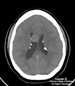
Figure 1: Axial noncontrast CT image shows an intraventricular mass near the foramen of Monro with foci of calcification, as well as several calcified subependymal nodules along the lateral ventricular surface. Hydrocephalus is also present with enlargement of the right lateral ventricle.
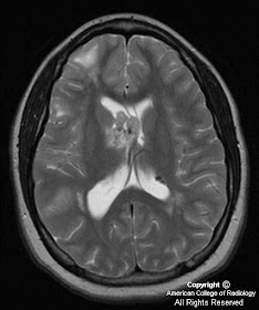
Axial T2-weighted and FLAIR MR images demonstrate a mass near the foramen of Monro with heterogenous, somewhat hyperintense signal compared to white matter. Intense homogeneous enhancement is seen on contrast-enhanced axial and coronal T1 weighted images. Subependymal nodules are seen along the lateral ventricles. Multiple foci of increased signal are seen on FLAIR images in the subcortical regions representing parenchymal tubers.
Diagnosis: Subependymal giant cell astrocytoma (SEGA)
Subependymal giant cell astrocytomas are intraventriclar neoplasms that occur in 15% of patients with tuberous sclerosis. Tuberous sclerosis (Bourneville’s disease) is a neurocutaneous phakomatosis characterized by an autosomal dominant pattern of inheritance presenting with the classical clinical triad of mental retardation, seizures, and adenoma sebaceum (although only 30% present with all three features).
The disease is characterized by hamartomatous tumors and malformations affecting multiple organ systems, the CNS being the most commonly involved, with seizure being the most frequent presenting clinical sign of the disorder. Other than the CNS manifestations, patients may present with renal angiomyolipomas, cardiac rhabdomyomas, and cystic lung disease indistinguishable from lymphangiomyomatosis.
Hamartomatous brain lesions include cortical tubers, white matter heterotopias, subependymal nodules, and the subependymal giant cell astrocytoma. Histologically, cortical tubers, white matter lesions, and subependymal nodules are identical lesions composed of disordered neurons, glia, and giant cells mostly of the astrocyte type, only differing in size and location. Subependymal nodules are usually easily identified with CT due to frequent calcification (>90%) and usually do not enhance thus helping to distinguish, but not entirely exclude a SEGA from a subependymal nodule. Cortical tubers are less likely to calcify and appear as low attenuation lesions at CT, demonstrate increased signal intensity on T2-W images, and rarely enhance. White matter lesions are seen as curvilinear or straight bands of increased T2 signal extending from the ventricles. SEGAs are characterized by slow growth and a benign biological behavior (WHO grade I), likely arising from the degeneration of subependymal nodules. On CT, SEGAs are iso-to slightly hypoattenuating intraventriuclar masses located near the foramen of Monro, with calcification and secondary hydrocephalus being common findings. On MR imaging, SEGAs exhibit hypointense signal compared to white matter on T1-weighted images, heterogenous hyperintensity on T2-weighted images, with intense homogenous enhancement (except for calcified areas). Because MR enhancement cannot always reliably distinguish between subependymal nodules and a SEGA, larger size (>1cm) and interval growth of a mass on annual follow-up CT or MR are considered better indicators of a SEGA rather than a benign subependymal nodule. Therefore, annual surveillance MR imaging is recommended in patients with tuberous sclerosis.
Nessun commento:
Posta un commento