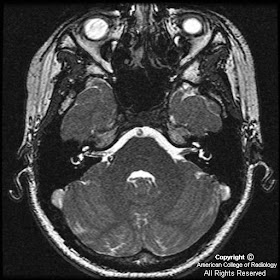


Findings
Figure 1: Nonenhanced thin-section T1-weighted axial MR image demonstrates two small lesions in the right cerebellopontine angle and the right vestibule, respectively. Both demonstrate high signal similar to subcutaneous fat.
Figure 2: T2-weighted image also demonstrates high signal in the right cerebellopontine angle lesion and the right vestibule lesion.
Figure 3: FIESTA image demonstrates a dark rim surrounding the CP angle lesion, consistent with chemical shift artifact at the boundary between the lesion and the surrounding CSF.
Figure 4: Post-contrast fat-saturated T1-weighted image shows complete suppression of signal from both lesions, confirming that they are composed of fat.
Diagnosis: Cerebellopontine angle and intravestibular lipomas
Cerebellopontine angle lipomas and vestibular lipomas are rare lesions. Any cerebellopontine angle or vestibular mass can cause a patient to present with sensorineural hearing loss, as in this case. MRI with dedicated thin images through the internal auditory canals is the preferred imaging technique for evaluation of sensorineural hearing loss.
Intracranial lipomas are thought to be congenital lesions. While most are asymptomatic, they can grow over time and cause clinically significant symptomatology. They occur most commonly in the interhemispheric, quadrigeminal/superior cerebellar, and suprasellar/interpeduncular regions, followed by the cerebellopontine angles. Intravestibular lipomas are extremely rare, with only a few cases reported in the literature. However, there is a known association between cerebellopontine angle lipomas and intravestibular lipomas.
In this case, the very high signal in both lesions on the T1-weighted images suggests fat content. Low-signal rims around the lesions on relatively T2-weighted gradient series (such as True FISP or FIESTA) is due to chemical shift at the fat-CSF boundary. Fatty content is definitively confirmed by a fat-saturated T1-weighted sequence.
Nessun commento:
Posta un commento