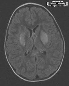


Findings
CT head: Low attenuation in the bilateral basal ganglia
MRI brain: There are small focal areas of diffusion restriction in the periventricular white matter, bilateral thalami and putamen. There is T2 prolongation affecting the basal ganglia, corpus callosum (genu and splenium), external capsules, and corona radiata.
Diagnosis: Hemolytic uremic syndrome
Key points
Most common pathogen is E. Coli O157:H7 verotoxin.
Clinical: acute renal failure, microangiopathic hemolytic anemia, thrombocytopenia, and neurologic symptoms
Neurologic complications occur in 20-50%. Symptoms include seizures, visual changes, altered consciousness and brainstem findings.
Neurologic findings most likely due to verotoxin affecting the microvascular endothelium leading to small vessel infarction and/or hemorrhage.
Patients with neurologic symptoms should undergo CT and MRI evaluation.
Treatment: fluid replacement, plasmapheresis. Avoid antibiotics which can worsen HUS.
Imaging (MRI)
Basal ganglia T2 hyper intensity is the most common finding, particularly the dorsal lateral lentiform nuclei.
Involvement of the basal ganglia is associated with a good clinical outcome.
T2 weighted hyper intensities have been reported to involved the thalamus, internal and external capsules, dorsal brainstem, corpus callosum, posterior leukoencephalopathy syndrome. Findings likely related to edema.
Diffusion restriction in the basal ganglia and thalami has been reported.
Areas of T1 hyper intensity should raise the possibility of hemorrhage.
Areas of hemorrhage are most likely to result in neurologic sequela.
Nessun commento:
Posta un commento