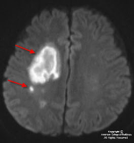


Findings
CT: Multifocal areas of hypoattenuation in the right frontal lobe which are confirmed acute infarctions on MRI with diffusion weighted imaging.
MRI: Axial MRI DWI and matching ADC maps demonstrate multifocal areas of true restricted diffusion in the right frontal lobe indicating acute infarctions from thromboemboli secondary to more proximal right internal carotid artery dissection.
CTA: Sequential Axial Neck CTA images from caudal to rostral demonstrate tapering to occlusion of the right internal carotid artery just distal to the Right common carotid artery bifurcation.
CTA neck exam frontal, oblique, and Sagittal 3D volume rendered and Sagittal MIP images demonstrate ‘flame shaped’ tapering to occlusion of the cervical right internal carotid artery just distal to the common carotid artery bifurcation, typical of dissection.
Diagnosis: Internal Carotid Artery Dissection
Carotid and vertebral artery dissection should be considered among the etiologies of brain infarct, particularly in young patients.
Symptoms typically include neck and face pain, headache, acute onset Horner’s syndrome, and ischemic symptoms that may occur initially or days to weeks after dissection.
Primarily treated with anticoagulation and aspirin.
Spontaneous carotid dissection can occur at any age but is most frequently seen in the fifth decade of life. The most common location for dissection of the internal carotid artery is the proximal extracranial segment. While brain infarct is the most feared complication, some carotid dissections may be asymptomatic from a neurologic standpoint.
Once thought to be a rare occurrence, spontaneous dissection of the internal carotid artery has become increasingly recognized as a cause of anterior circulation infarction, largely due to the advent of MR angiography. Predisposing factors include hypertension, Ehlers-Danlos disease, Marfan syndrome, fibromuscular dysplasia, migraine, oral contraceptives, and pharyngeal infections although most carotid dissections are seen in completely healthy individuals. A history of minor trauma is often elicited. The most studied association is chiropractic spinal manipulation, but carotid dissection has been described in various other minor traumas such as: yoga, ceiling painting, nose blowing, judo, coughing, sneezing, vomiting, and even ventilation associated with resuscitation or anesthesia. Blunt or penetrating major trauma to the head and neck is also a well-recognized cause of carotid dissection.
The underlying abnormality in spontaneous carotid artery dissection is thought to be an expanding hematoma within the vessel wall and, as a result, on CT angiogram an intimal flap is not always seen (unlike aortic artery dissection where contrast commonly tracks into the false lumen). Patients with carotid artery dissection can present with headaches, neck pain, acute onset Horner’s syndrome, or transient ischemic attack (TIA’s) and stroke (as in our case example). The dreaded complication of vascular dissection is thromboembolic phenomenon that may occur days to weeks after the dissection.
Imaging findings in carotid artery dissection include a tapered narrowing and occlusion of the vessel, as seen on current CTA exams with MIP images and 3D rendering. A hyperintense intramural hematoma may sometimes be seen on noncontrast axial T1 weighted imaging with fat-saturation, when blood products are in the subacute phase, representing methemoglobin. On occasion, it may also be termed the “crescent sign” because of its morphology. Signs of anterior circulation infarction can be seen on CT and MR at the time of initial presentation (as in our case).
Treatment in uncomplicated cases usually includes anticoagulation therapy and aspirin. It is important to obtain follow up MR imaging in these patients to assess for recanalization of the vascular lumen or progressive stenosis. These patients are also more prone to development of pseudoaneurysms.
Nessun commento:
Posta un commento