lunedì 30 aprile 2007

Periorbital epidermoid cyst


Findings

Small hypodense lesion is noted along the right superolateral orbital rim (Hounsfield units approximately -6), causing smooth contoured external depression of the adjacent bone. No evidence of bone destruction or abnormal periosteal reaction. The globe, extraocular muscles, and orbital soft tissues appear normal.

Differential diagnosis:
- Epidermoid cyst
- Dermoid cyst
- Subperisosteal abscess
- Eosinophilic granuloma
- Metastasis (e.g., neuroblastoma).


Diagnosis: Periorbital epidermoid cyst


Key points

Dermoid and epidermoid cysts are found in a variety of locations around the skull and midface, typically classified as true choristomas. Choristomas are tumors that are composed of tissues not normally found at their site of occurrence, instead arising from the sequestration of surface ectoderm by closure of the underlying suture lines, neural tube closure, and diverticulation of the cerebral hemispheres. Dermoid cysts contain ectoderm and skin elements, whereas epidermoid cysts contain ectoderm but no skin elements. Histologic examination is necessary to differentiate the dermoid cyst with its squamous epithelium lining containing dermal appendages from the epidermoid cyst, which has no dermal appendages. Dermoid and epidermoid cysts are most commonly seen in midline and frontotemporal locations, followed by parietal locations. Midline locations include the anterior fontanel, glabella, nasion, vertex, and subocciput. Nasal dermal sinuses and dermoid and epidermoid cysts occur at multiple locations and may be associated with external skin ostia or deep sinus tracts, which may potentially extend intracranially.
Complete surgical excision of the dermoid or epidermoid is the recommended management, as the contents of the cystic lesion are irritating and may result in lipogranulomatous inflammation if ruptured. Deep orbital cysts extending into the orbital roof, temporal fossa, or intracranially may require neurosurgical assistance for removal.


Radiology

Careful investigation is important to distinguish deep from superficial lesions. CT attenuation will vary depending on cyst content. Similarly, the signal intensity at MR imaging depends on the contents of the cyst and may range from pure fluid signal intensity in an epidermoid cyst to a more complex signal intensity in a dermoid cyst. Epidermoid cysts also typically have bright signal intensity on isotropic diffusion-weighted MR images. With nasal dermoid cysts, high-resolution surface coil MR imaging is useful in determining if there is a connecting sinus tract in the prenasal space to the foramen cecum.

giovedì 26 aprile 2007

Phthisis Bulbi secondary to endophthalmitis from Serratia meningitis


Findings

Figure 1: Axial CT with contrast in this infant with meningitis shows proptosis on the left, a large globe, scleral enhancement, and dirty retrobulbar fat consistent with endophthalmitis and retroglobar infection.
Figure 2: MRI T2-weighted sequence obtained one month later demonstrates a small shrunken calficified globe, consistent with phthisis bulbi which has progressed since the prior examination. This is phthisis bulbi as a sequela of infection.
Figure 3: FLAIR image demonstrates bright signal in the left globe as compared with the normal low signal in the right globe related to fluid.


Diagnosis: Phthisis Bulbi secondary to endophthalmitis from Serratia meningitis


There is a predisposition to endophthalmitis seen in diabetics, patients with debilitating diseases or the immmunosuppressed. Once the intraocular infection is established, exudates are deposited in the vitreous humor along with inflammatory cell infiltration of the global wall (the sclera, choroid and retina).

Phthisis bulbi is a retracted, scarred shrunken globe. It may be the end result of infection (as in our patient), noninfectious inflammation, radiation, trauma or other insult. The globe is usually one half to one third the size of the normal size due to reduced aqueous production. Furthermore, the globe has a diffuse increase in density and demonstrates an irregular contour. There are diffuse calcifications, either mottled or linear within the globe and there may be ossification of the devitalized tissue. The separate structures within the globe are often distorted and cannot be discerned. There is frequently associated atrophy of the optic nerve. Blindness and shrinkage of the globe may occur even with aggressive intraocular injection of antibiotics.

CT is the modality of choice for demonstrating the soft tissue abnormalities and possible calcification of phthisis bulbi. Enhancement of the vitreous humor may indicate that the humor is the initial site of involvement.

martedì 24 aprile 2007

Glioblastoma multiforme


Findings

Multiple enhancing masses with surrounding vasogenic edema and mass effect in the left cerebral hemisphere. There is associated sulcal effacement, gyral expansion/thickening, and mass effect with approximately 9-mm midline shift toward the right.


Diagnosis: Glioblastoma multiforme


Glioblastoma multiforme (GBM) is unfortunately both the most common and the most malignant of the glial tumors.
Of approximately 17,000 primary brain tumors diagnosed in the U.S. every year, 60% are glial tumors. Males have a slight predominance: 1.5 to 1.

The World Health Organization classification:
- Grade II: Low-grade diffuse astrocytoma
- Grade III: Anaplastic astrocytoma
- Grade IV: Glioblastoma multiforme

GBMs often arise in the subcortical white matter of the cerebral hemispheres. 31% arise in the temporal lobes. 24% arise in the parietal lobes. 23% arise in the frontal lobes and 16% arise in the occipital lobes. Sites such as the brain stem, cerebellum and spinal cord are much less common.
Classically, GBM has a thick, irregular enhancing ring of neoplastic tissue which surrounds a necrotic core.

The differential diagnosis for GBM includes:
- Abscess: Ring enhancement is usually thinner than GBM
- Metastases: Usually multiple lesions at gray-white junction
- Anaplastic astrocytoma: Usually nonenhancing white-matter mass. Enhancement may suggest degeneration to GBM
- Primary CNS lymphoma: Periventricular enhancing mass which often crosses the corpus callosum
- Tumefactive demyelination: Usually incomplete "horseshoe" enhancement which is open toward the cortex

lunedì 23 aprile 2007

Subclavian steal syndrome


Additional clinical information: Sixty-year-old man with left arm burning on use, noted to have decreased blood pressure in left upper extremity. History of prior left carotid endarterectomy.


Findings

Arteriogram demonstrates occlusion of the left subclavian artery origin with reconstitution of flow after approximately a 1-cm segment via the left vertebral artery. Findings are consistent with subclavian steal syndrome. The peripheral left common carotid artery has an approximately 40% diameter stenosis at the operative site.

Differential diagnosis - Causes of ubclavian steal syndrome:
- Atherosclerotic disease
- External compression
- Thrombosis
- Dissection
- Embolism
- Trauma


Diagnosis: Subclavian steal syndrome


Definition: Stenosis/obstruction of the subclavian artery near its origin with demonstration of ipsilateral vertebral artery flow reversal, compromising cerebral flow.
This can be either congenital or acquired secondary to atherosclerotic disease most often. Other causes of acquired steal can be secondary to trauma, embolism, tumor thrombosis, dissecting aneurysm, radiation, inflammatory arteritis, or ligation.
Incidence is 2.5% of all occlusions of extracranial arteries. Associated with additional lesions 81% of the time.
Males affected more often than females 3:1. Left-sided more often than right 3:1.

Signs and symptoms are:
- Weak or absent ipsilateral extremity pulse.
- Ipsilateral extremity has lower systolic blood pressure by >20-40 mm Hg.
- Evidence of brachial insufficiency (pain, coolness, numbness, burning, necrosis).
- Evidence of vertebrobasilar insufficiency (syncope, ataxia, vertigo, headache).

Angiographic studies show the occlusion/stenosis of the subclavian artery and can demonstrate the retrograde vertebral artery flow.
Color Doppler studies can demonstrate the reversal of the vertebral artery flow.
Treatment is surgical bypass or PTA
Occult subclavian steal syndrome is when the flow reversal can only be seen after maneuvers like ipsilateral arm exercise or inflation of blood pressure cuff.
Partial subclavian steal syndrome is when there is retrograde flow seen during systole and antegrade flow is seen during diastole.

giovedì 19 aprile 2007

Osteoporosis circumscripta


Findings

Figure 1: Axial CT image with bone windowing shows confluent areas of lysis involving the frontal bone and portions of both temporal bones with relative sparing of the cortex and thinning of the diploic space.
Figure 2: More cephalad axial CT image demonstrates involvement of the frontal bone and right parietal bone anteriorly.


Diagnosis: Osteoporosis circumscripta


Paget disease was first described by Sir James Paget in 1877 as osteitis deformans which later came to be known as Paget disease. It is the second most common bone disorder in the elderly population behind osteoporosis. The disease process has three phases, beginning with the lytic phase where exaggerated bone resorption occurs by atypical osteoclasts. This results in the second phase, or mixed phase, where morphologically normal osteoblasts move in and cause rapid increases in bone formation. The newly formed bone is disorganized, larger, weaker and more vascular. The final phase, or sclerotic phase, is when osteoblastic activity predominates with formation of thickened and enlarged sclerotic bone.

Osteoporosis circumscripta indicates involvement of the calvarium during the lytic phase of Paget disease and is characterized by asymmetric and confluent foci of calvarial lysis. The involved bone is usually thinner than the normal bone which helps to differentiate from other lesions which tend to expand the calvarium or destroy cortex.

Paget disease affects the axial skeleton, long bones and skull predominantly. The hands and feet are almost never involved. It is most commonly found in caucasian populations, mainly in the northeast and in Europe. The majority of patients are asymptomatic with the disease detected by an incidentally elevated alkaline phosphatase level or characteristic bone changes. The etiology is unknown with various hypotheses including a familial cause linked to chromosomes 6 or 18q. Infectious causes have also been theorized as biopsies from these patients demonstrate paramyxovirus antigens within osteoclasts.

mercoledì 18 aprile 2007

Spinal cord herniation


Findings

Initial non-contrast sagittal T1 and sagittal T2 weighted images demonstrate anterior (ventral) displacement of the spinal cord from T4-T6 with enlargement of the subarachnoid space dorsally.
Sagittal reconstructed image from a CT myelogram (Figure 3) performed 15 days later shows a similar finding.
Axial images from a CT myelogram demonstrate anterior displacement of the spinal cord with enlargement of the subarachnoid space dorsally (Figure 4). We also see that the subarachnoid space is continuous and fills with contrast without evidence of an arachnoid cyst, as is often mistaken for the diagnosis.


Diagnosis: Spinal cord herniation


Spinal cord herniation is a rare diagnosis to be considered in patients presenting with slow and progressive neurologic symptoms.
CT myelogram helps to confirm findings on MRI.
Pathogenesis is unclear, however is thought to be related to an initial thoracic dural defect, whether congenital or acquired.

Spinal cord herniation is a rarely encountered cause of myelopathy. The pathogenesis of thoracic cord herniation has been ascribed to a dural defect, either congenital or acquired, on the convex ventral surface of the thoracic spinal canal. The spinal cord is believed to “plug” the hole in the dura, and prolonged CSF pulsations force the cord through the dural defect. Adhesions and distortion of the cord with possible vascular compromise over time leads to progressive myelopathy. This proposed mechanism explains the absence of idiopathic spinal cord herniation occurring in the cervical and lumbar spine and posteriorly in the thoracic spine, where the cord would not be so closely applied to a potential dural defect.

The clinical presentation in the literature of thoracic cord herniation is characteristic but nonspecific. Age of presentation ranges from 22-71 years old with a mean age of 43. There is also a 2:1 female to male ratio. A majority of herniations present with a Brown-Sequard syndrome (ipsilateral hemiparesis and loss of proprioception and vibratory sensation and contralateral loss of pain and temperature sensation). A minority may present with paraparesis or bladder and bowel dysfunction. The onset of symptoms is usually slow and progressive.

Imaging studies characteristically show the cord appearing small, rotated, and displaced with an apparently dilated CSF space opposite to the direction of cord displacement. This apparently dilated CSF space represents the void left by the herniated cord. In such cases, the CSF space would be expected to follow the signal characteristics of normal CSF on MRI sequences and to fill readily with intrathecal contrast material. This could also be interpreted as an expansive communicating arachnoid cyst compressing the cord or as spinal cord adhesions from posttraumatic or postoperative fibrosis that have tethered the cord to the dura against the wall of the spinal canal. Axial images may be helpful in distinguishing a compressed cord from a herniated cord, especially if the dural defect has allowed only the lateral portion of the cord to herniate into bony notches or defects. The most common location for thoracic spinal cord herniation is between T2 and T8 over a length of 1 to 3 cm.

Treatment includes surgical release of the incarceration with repair of the dural defect. The surgical defect in the dorsal dura may require a dural graft to provide a larger dural space, as the reduced spinal cord may be swollen and edematous. In the majority of cases in the literature, the patients’ neurologic deficits resolved or improved.

venerdì 13 aprile 2007

Balo's concentric sclerosis


Findings

Figure 1: T1-weighted image shows low density in both the posterior and the left anterior periventricular region.
Figure 2: T2-weighted image shows high signal in the periventricular lesions with suggestion of concentric rings of hyperintensity and isointensity.
Figure 3: FLAIR image reveals edema in the periventricular lesions.
Figure 4 and Figure 5: The postcontrast T1-weighted images show enhancement in a concentric ring like fashion and hold the key to the diagnosis.


Diagnosis: Balo's concentric sclerosis


Balo’s concentric Sclerosis (BCS) is a rare demyelinating disorder considered to be a variant of multiple sclerosis (MS).Histologically it is characterized by alternating rings of myelin preservation or remyelination and demyelination involving the cerebral hemispheres, brain stem, spinal cord, and optic chiasm. The clinical course of BCS is considered variable with the current treatment modalities.

The MR imaging findings show hypointense and isointense concentric bands on T1 weighted images, concentric hyperintense and isointense bands on T2 weighted images. The hyperintense bands on T2 weighted images correspond to concentric bands of demyelination with gliosis and perivascular lymphocytic infiltration. The isointense bands represent the white matter which has been spared or has had remyelination. On the post contrast T1 weighted images all the lesions showed concentric rings of enhancement, indicating rings of active inflamed and spared white matter.

It has been noted that with treatment and over a period of time, the contrast enhancement decreases and so does the size of the lesions. The characteristic concentric pattern may not be observed if the MR imaging is not performed early in the disease. Our patient is still in the hospital being treated with high dose steroids.

Differential for the imaging findings would include Marburg variety of multiple sclerosis, ADEM (Acute disseminated encephalomyelitis) and lymphoma. ADEM also has multiple lesions which enhance after gadolinium administration. The age of the patient does not favor that and the patient did not have history of any viral illness. Lymphoma should have more mass effect and more enhancement. The patient did not have any history of immunosuppresion.

giovedì 5 aprile 2007

Carotid body (paraganglioma) tumor


Findings

Figure 1: Axial contrast enhanced CT image of the upper neck shows a large enhancing mass in relation to the carotid space and is displacing and compressing the vessels.
Figure 2: MRI image of the upper neck shows a large enhancing mass in relation to the carotid space and is displacing and compressing the vessels. Note the multiple small flow voids within the lesion on MRI giving the "salt & pepper" appearance.


Diagnosis: Carotid body (paraganglioma) tumor


Carotid body tumor is a rare type of head and neck tumor composed of paraganglionic cells. Paraganglionic tissue can be found in the adrenal medulla and in multiple extra-adrenal sites, including at the carotid bifurcation, the aortic arch, adjacent to the jugular vein, the tympanic nerve, the facial nerve, the vagus nerve, and others. This tissue develops embryologically from neural crest tissue. These tumors are called extra-adrenal paragangliomas and sometimes as glomus tumors or chemodectomas. The function of paraganglionic tissue in the carotid body is to function as a chemoreceptor, sensing oxygen levels as well as carbon dioxide and pH levels. In head and neck, the glomus tumors are seen at the carotid bifurcation (carotid body tumor) as seen in this case, at the jugular fossa (glomus jugulare), in the middle ear (glomus tympanicum) or along the vagus nerve (glomus vagale).

Carotid body tumors are well encapsulated and highly vascular benign tumors and can occur in a wide range of ages, from childhood to the elderly, but the average age at diagnosis is around 45. There is a slight female predominance, and people who live at high altitudes are more likely to develop carotid body tumors (thought to be due to lower oxygen levels).Most cases are sporadic but familial inheritance is also described. In addition, familial syndromes such as multiple endocrine neoplasia (MEN) can contribute to the development of multiple carotid body tumors as well as tumors at multiple other paraganglionic sites in the head and neck and in the rest of the body as well. Carotid bifurcation is the second most common site for glomus tumors in head and neck, after the middle ear(glomus tymapnicum).

Carotid body tumors like all other paragangliomas are slow-growing and may be locally invasive. They can present as a lateral cervical mass which may or may not be pulsatile. These tumors can sometimes cause cranial nerve palsies, most commonly vagal nerve palsy, and if large can even erode through the skull base or other bony structures. They are rarely malignant in that they rarely metastasize, although local recurrence is not uncommon. 10% of head and neck paragangliomas are multicentric and bilaterality is most common with carotid body tumors. Synchronous and/or metachronous tumors can develop at the contralateral carotid bifurcation as well as in other paraganglionic sites, especially if a familial syndrome is present. These types of recurrences are not usually considered metastases by most sources. Carotid body tumors and other paragangliomas can actively secrete metanephrines, most commonly norepinephrine, although this is rare (3-5% of head and neck paragangliomas). Symptoms of an actively secreting carotid body tumor would include headaches, flushing, hypertension, and palpitations.

CT of the neck is helpful for diagnosis and can identify any local lymph node enlargement or bony erosions. Additionally, MR can help determine the relationship of the tumor with respect to the carotid vessels and other neck structures and if there are multiple paragangliomas present. A classic finding of the carotid body tumor is splaying of the internal and external carotid arteries. On both enhanced CT and MR, the carotid body tumors and other paragangliomas are intensely enhancing due to their extensive vascularity. Sometimes small flow voids can be seen on nonenhanced MR, causing a “speckled” or “salt and pepper” appearance of the tumor. On dynamic enhanced CT or MR, there will be a rapid enhancement, a high peak, and rapid washout due to early arteriovenous shunting of the tumor. Conventional catheter angiography reveals similar findings. In addition, embolization of the tumor can be undertaken during the angiographic exam to reduce blood loss from this highly vascular tumor during surgery. Another study that can be performed to evaluate the carotid body tumor is an Indium-111 octreotide isotope scan which is preferentially taken up by neuroendocrine tissues and can help detect presence of multiple paragangliomas.

Surgical excision is the preferred treatment of head and neck paragangliomas in most cases although it is not without risk. The main risks of excision are injury to local cranial nerves or to the carotid artery and blood loss from these highly vascular tumors. If the tumor is found to encase the carotid extensively, then temporary balloon occlusion of the common carotid artery with EEG monitoring can help determine the adequacy of collateral circulation. If the patient is not a surgical candidate, due to extensive tumor involvement or coexisting morbidities, radiation therapy is an option, although it is considered second-line therapy.

lunedì 2 aprile 2007

Malignant otitis externa


Findings

Figure 1: Initial CT of the skull base on bone widows shows subtotal opacification of the mastoid air cells without bony destruction.
Figure 2: CT of the skull base on soft tissue windows shows an inflammatory soft tissue mass filling the external auditory canal extending into the surrounding soft tissues without involvement of the middle ear.
Figure 3: On subsequent CT of the skull base with soft tissue windows, the inflammatory soft tissue mass remains with partial destruction of the auricular cartilage. It now crosses the tympanic membrane and opacifies the middle ear with osteolysis of the anterior, bony portion of the external auditory canal.
Figure 4: Axial bone windows reveal destruction of the anterior wall of the mastoid air cells.
Figure 5: Sagittal reconstruction with bone windows reveals coalescing mastoiditis with destruction of the bony septae and resultant enlargement of the air cells. The inflammatory mass displaces the mandibular condyle anteriorly out of the articular fossa.
Figure 6: Coronal reconstruction with bone windows reveals soft tissue opacifying the middle ear with partial destruction of the tympanic portion of the temporal bone.


Diagnosis: Malignant otitis externa


Malignant otitis externa (MOE) also referred to as necrotizing otitis externa or skull base osteomyelitis can be defined as otitis externa with progression to skull base osteomyelitis. The causative organism is almost universally Pseudomonas, which invades via the fissures of Santorini, located in the cartilaginous, lateral 1/3 of the external auditory canal (EAC). Affected patients are typically immunocompromised, often diabetic and present with symptoms of otitis externa unresponsive to topical antibiotics. Examination demonstrates purulent otorrhea with granulation tissue in the EAC. Cranial nerve involvement in the setting of otitis externa is an ominous sign and suggests progression to malignant otitis externa. Leukocytosis and signs of systemic infection are not typically present.

When diagnosing MOE, imaging is mandatory. CT is the exam of choice, demonstrating osteolysis in association with mastoid opacification and marked soft tissue inflammation centering on the external auditory canal. CT is relatively insensitive for early disease; osteolysis is not seen until at least 30% of the bone has been destroyed. Nuclear scans are generally sensitive but not specific, and may play an adjunctive role. Due to the lack of marrow in the affected bones, MR is of limited use, but may be complimentary to demonstrate soft tissue involvement.

Imaging can only suggest the diagnosis of MOE, and biopsy is necessary for confirmation.

The anatomy of the temporal bone means that the disease may disrupt the TMJ or the middle ear. Involvement of the facial nerve may occur. Intracranial spread is more ominous still, leading to venous sinus thrombosis, meningitis and abscess formation.

The historical mortality for MOE was 77%; however with current treatment the mortality is closer to 10%. Systemic antibiotics are the mainstay of therapy however surgical debridement may be necessary.

Nasolacrimal duct mucocele


Findings

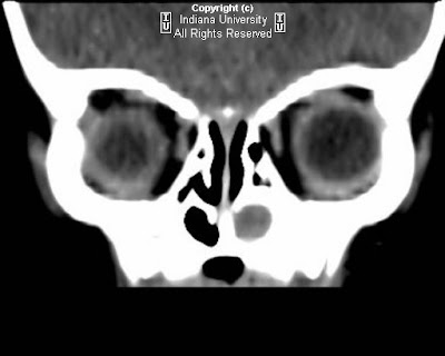
There is a rounded hypoattenuating lesion in the medial aspect of the left orbit which extends through the left nasolacrimal duct into the left nasal cavity. There is bony expansion of the left nasolacrimal duct.

Differential diagnosis:
- Mucocele
- Intraorbital abscess
- Encephalocele
- Dermoid
- Hemangioma
- Glioma


Diagnosis: Nasolacrimal duct mucocele


Congenital nasolacrimal duct mucocele is characterized by a triad of findings:
- Mass located at the medial canthus
- Bony expansion of the nasolacrimal duct
- Intranasal mass

In neonates, there is a thin mucus membrane (plica lacrimale or valve of Hasner) at the inferior aspect of the nasolacrimal duct. In 6-12% of patients, this is imperforate at birth, but shortly after birth it typically spontaneously perforates.
Failure to perforate this membrane results in outflow obstruction with subsequent mucocele formation.
Initial presentation may include a mass at the medical canthus. A secondary infection may cause physical exam findings similar to an abscess.
Epiphora may result from blockage of nasolacrimal duct drainage.
The initial presentation may also include respiratory distress secondary to nasal obstruction, as neonates are obligate nasal breathers.
Management is typically expectant, as spontaneous perforation is typical. If there is persistent failure to perforate, or there are signs of infection, the mucocele can be marsupialized endoscopically, or a dacryocystorhinostomy may be performed.
The imaging modality of choice is CT, which can show the extent of the mass lesion within the medial canthus as well as the nasal cavity, and any associated nasolacrimal duct bony expansion.