mercoledì 30 gennaio 2008

Rhombencephalosynapsis


Findings

Figure 1: Sagittal T1-weighted image demonstrates enlarged lateral ventricles with upward bowing and thinning of the corpus callosum, compatible with hydrocephalus. Slight rounding of the fastigial recess of the fourth ventricle is also identified, as well as absence of the cerebellar vermis. Included upper cervical spine shows congenital fusion of the C3 and C4 vertebral bodies.
Figure 2 and Figure 3: Coronal T2-weighted images demonstrate fusion of the cerebellar hemispheres, horizontal appearance of the cerebellar folia with gray and white matter crossing the midline. There is no intervening vermis.
Figure 4: Axial gadolinium-enhanced T1-weighted image demonstrates “keyhole” shaped fourth ventricle and absent cerebellar vallecula.
Figure 5: Axial FLAIR image demonstrates fused, horseshoe-shaped cerebellar dentate nuclei.
Figure 6: High resolution T2-weighted sagittal cisternogram is highly suggestive of aqueductal stenosis.
Figure 7: Coronal T2-weighted image shows dilated frontal horns of the lateral ventricles, which are normally separated by the septum pellucidum. Normal massa intermedia without evidence of fused thalami.


Diagnosis: Rhombencephalosynapsis


Rhombencephalosynapsis, originally described by Obersteiner in 1914, is a rare congenital malformation of the posterior fossa featuring fusion of the cerebellar hemispheres (partial or total), agenesis or severe hypogenesis of the cerebellar vermis and apposition or fusion of the dentate nuclei. Failure of dorsal induction/differentiation of the normal midline structures with disturbed cerebellar development is thought to occur following an insult between the 28th and 44th days of gestation, and may be genetic or acquired. The likely genetic defect involves the isthmic organizer, with FGF8 and LMX1A genes being considered; there have been anecdotal reports of interstitial deletion of chromosome 2q and parental consanguinity. Among predisposing maternal factors, hyperpyrexia, diabetes, and phencyclidine and alcohol use have been incriminated.

The most distinctive pathologic feature is midline fusion of the cerebellar hemispheres with folia and fissures transversely oriented (single-lobed cerebellum). There is associated agenesis or poor differentiation of the vermis, the rostral portion being the most severely affected, while the caudal vermis is better formed, usually with a well-developed flocculonodulus. The posterior fossa is smaller than normal. Superior and middle cerebellar peduncles may be fused, as well as the dentate nuclei and the inferior colliculi, sometimes leading to a characteristic diamond or “keyhole” shaped fourth ventricle (narrow, pointing posteriorly). Olivary nuclei may be hypoplastic or absent. Once the diagnosis is established, a prompt search for other supratentorial and infratentorial findings should be performed.

Hydrocephalus is the most frequently associated supratentorial anomaly and may be related to aqueductal stenosis. Other common supratentorial findings include fused thalami, fornices and cerebral peduncles, absence of the septum pellucidum, dysgenesis of the limbic system, cortical malformations and multiple suture synostoses. Associations with septo-optic dysplasia and holoprosencephaly have been suggested. Hypoplasia of the commissural system and the anterior visual pathway and agenesis of the posterior lobe of the pituitary can occasionally be seen.

Rhombencephalosynapsis can be part of the Gomez-Lopez-Hernandez syndrome (cerebellotrigeminal dermal dysplasia), which is also characterized by trigeminal anesthesia, midface hypoplasia and bilateral bands of alopecia.

Segmentation and fusion anomalies of the spine, as well as other musculoskeletal, cardiovascular, urinary tract, and respiratory abnormalities have been described in several patients.

Clinical presentation correlates with the severity of associated supratentorial anomalies, varying from mild truncal ataxia and normal intelligence to cerebral palsy. Patients may manifest hypotonia, motor and cerebellar dysfunction, seizures, strabismus and developmental delays. Compulsive self-injurious behavior is common. Variable growth hormone deficiency depends on midline supratentorial anomalies. Most reported cases are pediatric; patients usually have a short lifespan, although incidental rhombencephalosynapsis in adults has also been described. Management includes treatment of related hydrocephalus and monitoring the hypothalamic-pituitary axis.

Because of multi-planar capabilities, MR is the imaging modality of choice demonstrating transversely oriented cerebellar folia, which cross the midline without an intervening vermis, and associated “keyhole” shaped fourth ventricle, which lacks the normal cerebellar vallecula. On sagittal images, the primary fissure of the vermis is absent and the fastigial recess of the fourth ventricle may appear upwardly rounded. Fusion of the cerebellar dentate nuclei appears as horseshoe-shaped decreased signal along the posterolateral aspect of the fourth ventricle on axial T2-weighted images. On CT, the cerebellar anomaly may be difficult to detect, but may be suggested by the characteristic configuration of the fourth ventricle.


giovedì 24 gennaio 2008

Ivory vertebral body from prostate cancer metastasis


Findings

Figure 1: Lateral Rx of the neck shows dense C5 vertebral body (ivory vertebral body)

Differential diagnosis for ivory vertebral body:
- Metastases
- Lymphoma
- Paget disease
- Less common: Infection (low grade i.e. TB) or Idiopathic segmental sclerosis


Ivory vertebral body from prostate cancer metastasis


Ivory vertebral body refers to an increase in density of a vertebral body that retains its size and contours, with no change in the density and size of adjacent intervertebral disks. The added density may be diffuse and homogeneous and involve most or all of the vertebral body, giving it a white appearance as opposed to the normal or possibly osteoporotic appearance of the rest of the vertebral column

Ivory vertebral body may be caused by numerous etiologies as listed above. Osteoblastic metastases elicit a sclerotic response in which vertebral body spongiosa is replaced with a dense, amorphous bony mass. In similar fashion, lymphomatous spread may cause an osteoblastic response leading to the dense changes. On the other hand, Paget disease usually causes an expansion of the contour of the vertebral body, which sometimes takes it outside of the definition of ivory vertebral body. You may also see reactive bone formation that occurs in response to stress leading to the appearance of an ivory vertebral body. This process is called idiopathic segmental sclerosis.

In children, the finding of ivory vertebra is rare. However, when found it is usually secondary to lymphoma, most frequently Hodgkin disease. Other less frequent causes include osteosarcoma, metastatic neuroblastoma, medulloblastoma or osteoblastoma. There have been rare cases of Ewing sarcoma causing dense vertebral bodies.

In adults, by far the most common cause is osteoblastic metastases from breast or prostate cancer. Other causes include osteosarcoma, carcinoid, Paget disease, and lymphoma. A radiopaque vertebral body is most compatible with a diagnosis of metastatic disease, and in the case of an elderly male would most likely be a result of prostate carcinoma. In a female patient, the most likely diagnosis would be breast carcinoma. Metastatic disease from breast carcinoma will not infrequently present as a solitary vertebral lesion.

Paget disease can present with a uniform appearance, however more often presents with a classic “picture frame” vertebral body where the contour of the vertebra is sclerotic with a relatively lucent center. The trabecular bone is also usually thickened leading to an expansion in the anterior-posterior and lateral dimensions.

Lymphoma can sometimes lead to sclerotic lesions in the vertebra including ivory vertebral body. It usually begins in a patchy fashion and leads to a more generalized pattern and sometimes even to an ivory vertebra pattern. Often you may also see the accompanying, invading soft tissue mass.

Osteomyelitis may also lead to sclerotic changes, however it is rarely found within only a single vertebral body. There are usually erosive changes found at the disk margins.

In general, the three causes that should be considered in the differential for ivory vertebral body are: metastatic cancer, Paget disease and lymphoma.


mercoledì 23 gennaio 2008

Acute calcific tendonitis of the longus colli muscle


Findings

Figure 1: Scout image from CT scan of the head demonstrates prevertebral soft tissue swelling and loss of physiologic cervical lordosis. This finding prompted further evaluation with contrast-enhanced CT scan of the neck.
Figure 2, Figure 3, and Figure 4: Axial (Figure 2 and Figure 3) and reformatted sagittal (Figure 4) contrast-enhanced CT images of the neck demonstrate enlarged right longus colli muscle, focal amorphous calcification within it (Figure 2 and Figure 4), as well as smooth, nonenhancing prevertebral and retropharyngeal effusion (Figure 3 and Figure 4).
Figure 5 and Figure 6: Axial (Figure 5) and reformatted sagittal (Figure 6) contrast-enhanced CT images of the neck with bone settings show focal, off-midline amorphous calcification anterior to C1/C2 level, within the superior tendon of the right longus colli muscle. There is also mild reversal of the physiologic cervical lordosis.

Differential Diagnosis:
- Retropharyngeal space abscess
- Retropharyngeal edema of different etiology (internal jugular vein thrombosis, previous surgery or radiation therapy)
- Accessory ossicle inferior to the anterior arch of C1
- Infectious spondylodiscitis


Diagnosis: Acute calcific tendonitis of the longus colli muscle


Acute calcific tendonitis of the longus colli muscle, also known as acute calcific retropharyngeal (or prevertebral) tendonitis, is a clinical syndrome (originally described by Hartley in 1964) that usually affects adults in the 3rd through 6th decades of life. The presumed mechanism of disease is calcium hydroxyapatite crystal deposition within the superior oblique fibers of the longus colli muscle. Acute symptoms develop when these contained deposits rupture, triggering an acute inflammatory process that usually lasts 2 to 3 weeks. The typical clinical presentation is acute to subacute onset of neck pain, dysphagia, or odynophagia; patients may also have low-grade fever and paraspinal muscle spasm, as well as recent history of upper respiratory infection or minor trauma to the head or neck. Mild leukocytosis, elevation of C-reactive protein and erythrocyte sedimentation rate are occasionally present.

The pathognomonic radiographic findings consist of calcific prevertebral (or retropharyngeal) density on the lateral radiograph of the neck, typically at C1/C2 level (inferior to anterior arch of C1), with associated soft tissue thickening. CT is more sensitive than plain radiography for detection of characteristic amorphous calcification, which is usually off-midline and clearly separate from the vertebral body; its appearance varies from punctate to dense, prominent concretions. Retropharyngeal space edema and specific smooth retropharyngeal effusions usually extend from C1 to the C5 level. MR imaging is not typically necessary for the diagnosis, but may be useful in confirming and further defining the abnormality. T2-weighted images demonstrate hyperintense retropharyngeal space fluid with an acute inferior margin. Diffuse swelling of the longus colli muscle and associated C1/C2 bone marrow edema have also been reported. After administration of intravenous contrast, there may be diffuse prevertebral soft tissue and/or longus colli muscle enhancement. Hypointense calcifications can be detected inferior to the anterior arch of C1 within the superior longus colli tendon, however these are easy to miss on MR.

The most important differential diagnosis is a retropharyngeal space abscess, which would require aggressive antibiotic therapy and possible surgical drainage. Distinction from abscess is easy if four key observations are made: 1) the fluid smoothly expands the retropharyngeal space in all directions; 2) there is absence of an enhancing wall around this fluid, as would be seen in a true retropharyngeal abscess; 3) there is absence of associated suppurative (or presuppurative) retropharyngeal space lymph nodes with low-density centers; 4) there are pathognomonic tendinous calcifications within the longus colli.

Most cases are self-limiting and resolve within several weeks of the onset of symptoms. The calcifications may disappear when the soft tissue swelling recedes. Treatment is usually conservative and consists of analgesics and a short trial of NSAIDs. If symptoms are severe, the patients may require a short course of corticosteroids. Early diagnosis is important to avoid invasive diagnostic and therapeutic procedures. Follow-up imaging is not necessary due to the self-limiting nature of this condition.

Subarachnoid hemorrhage secondary to basilar tip aneurysm rupture


Findings

Head CT without contrast shows diffuse subarachnoid hemorrhage, most prominent around the basilar tip but also involving the right Sylvian fissure the anterior interhemispheric fissure. There is also intraventricular blood. CT Head angiography with 3D reconstruction shows a large bilobed aneurysm of the basilar tip. The aneurysm neck extends into the left P1 segment. The left superior cerebellar artery arises from the P1 segment, and therefore the aneurysm incorporates both the left superior cerebellar artery and P1 segment.

Differential diagnosis - Causes of subarachnoid hemorrhage (SAH):
- Aneurysm
- Trauma
- AVM
- Coagulopathy


Diagnosis: Subarachnoid hemorrhage secondary to basilar tip aneurysm rupture


Discussion

Intracranial aneurysms are classified into three categories:
- saccularù
- fusiform
- dissecting aneurysms.

Saccular aneurysms (i.e. berry aneurysms) are true aneurysms that occur at intracranial bifurcation points, most commonly within the circle of Willis. Although previously thought to be congenital in origin, recent studies suggest that most intracranial aneurysms develop as a result of hemodynamic-induced vascular injury.

The incidence of intracranial aneurysms is estimated to be near 1-6% of the population. These percentages increase in patients with congenital anomalies of the intracranial vasculature (fenestrations, persistent trigeminal arteries), vasculopathies (fibromuscular dysplasia), connective tissue disorders (Marfan, Ehlers-Danlos), polycystic kidney disease, coarctation of the aorta, or vascular malformations. Factors associated with increased risk of aneurysm rupture include cigarette smoking, female gender, and younger age at diagnosis.

The risk of aneurysm hemorrhage remains unknown. For many years, a bleeding risk of 1-2% per year was quoted in the literature, and this number remains reported in current investigations. However, a recent study (The International Study of Unruptured Intracranial Aneurysms) found that the risk of aneurysm hemorrhage was quite variable and dependent upon key factors: size, location, and history of prior aneurysm rupture. For example, anterior circulation aneurysms measuring less than 7 mm in a patient without a history of aneurysm hemorrhage had less than 0.05% risk of bleed per year. Unfortunately, basilar tip aneurysms larger than 10 mm and aneurysms in patients with a prior history of an aneurysm bleed were associated with a much higher risk of rupture of 0.5% per year.

Proper management of a ruptured aneurysm is critical since the risk of aneurysm rebleeding after the initial hemorrhage is very high. There is a 20-50% risk of aneurysm rebleeding during the first two weeks after the initial event. Rebleeding carries a high mortality rate of almost 85%. Patients with unruptured aneurysms and clinical symptoms such as new onset 3rd nerve palsy or visual loss should also be treated emergently since the risk of aneurysm hemorrhage in these individuals is significantly higher than incidentally discovered aneurysms in asymptomatic patients.


Radiologic overview of the diagnosis

When non-contrast head CT demonstrates subarachnoid hemorrhage in the suprasellar cistern, Sylvian fissures, and anterior interhemispheric fissure, an aneurysmal etiology should be highly suspected. Three primary modalities are utilized to image intracranial aneurysms - CTA, MRA, and catheter angiography. CTA and MRA are the modalities of choice for imaging of unruptured aneurysms. Traditionally, catheter angiography has been preferred for imaging of patients presenting with non-traumatic subarachnoid hemorrhage. However, CTA alone is gaining in popularity for these cases.

Catheter angiography: Conventional cerebral angiography has always been considered the gold standard for intracranial aneurysm imaging. It remains invaluable for identification of the aneurysm(s), presence and size of the aneurysm neck, clarification of the aneurysm's relationship to the parent vessel, evaluation of the collateral circulation, assessment for vasospasm, and determination of best treatment modality. As conventional angiography is invasive and time-consuming, it does have some disadvantages.

CTA with 3-D reconstruction: Recent studies suggest that CTA is a quick, reliable, and relatively simple diagnostic modality for imaging of intracranial aneurysms. Some reports indicate that the sensitivity of CTA for detection of aneurysms < 5mm is higher than catheter angiography. Advantages include inter-operator reliability and non-invasiveness.

MRA: MRA is typically used in combination with conventional MRI and used to screen patients with risk factors for intracranial aneurysm. MRA currently has a low sensitivity for aneurysms less than 4 mm in size.

giovedì 17 gennaio 2008

Molar extraction complicated by a masticator space infection and subdural empyema


Findings

Figure 1: Axial CT image with bone algorithm at the level of C2. There is a lucency in the socket of the extracted molar. The irregular hyperdensity within the socket represents surgical packing that was inserted, and left in place post-dental extraction.
Figure 2: Contrast enhanced axial CT image with soft tissue algorithm at the level of the occiput and C1. Irregular hypodense collections with faint rim enhancing walls are seen within the muscle bellies of the lateral pterygoid and the masseter. There is extensive pre-zygomatic subcutaneous soft tissue fat stranding suggesting an overlying cellulitis.
Figure 3: Contrast enhanced axial CT with brain algorithm at the level of the middle cerebral arteries. There is a subtle hypodense collection with leptomeningeal enhancement lateral to the anterior pole of the temporal lobe. We again note subcutaneous fat stranding and edema within the left temporalis muscle, and overlying the left aspect of the frontal and temporal bones.
Figure 4: Axial CT with bone algorithm at the level of the abnormality. This image confirms that the collection crosses the left coronal suture, therefore confirming that it is located within the subdural space.


Diagnosis: Molar extraction complicated by a masticator space infection and subdural empyema


A subdural empyema (SDE) is an intracranial collection containing purulent material located between the dura mater and the arachnoid mater. Intracranial subdural empyemas account for up to 22% of all intracranial pyogenic processes. The majority of subdural empyemas occur as a complication of paranasal sinusitis, otitis media, or mastoiditis, and uncommonly as a result of a masticator space infection. In the largest published series of 699 cases of SDE, dental infection was the fifth most common cause accounting for 0.7% of cases.

Fever, seizures and motor deficits are some of the commonly encountered clinical features of SDE. A subdural empyema is a life threatening illness with a mortality rate ranging from 6-35 %.

Intracranial extension of infection from the head and neck can occur by two primary means. Direct extension by erosion through the skull occurs with sinusitis or mastoiditis. Indirect spread, which is a more common route, occurs by retrograde thrombophlebitis via the valveless veins of the basal venous system which connect the intracranial and extracranial spaces. Masticator space infections resulting from osteomyelitis of the mandible from an uncontrolled dental infection can gain access to the subdural space via the foramen ovale or spinosum. Extension into the middle cranial fossa occurs with peri-neural extension along the mandibular division (V3) of the trigeminal nerve or peri-vascular extension along the middle meningeal artery.

Subdural empyemas (SDE) are characteristically crescentic extra-axial fluid collections extending along the cranial convexities. SDE(s) spread aggressively through the subdural space, crossing the sutures of the skull, until limited by the falx cerebri, tentorium cerebelli, base of the brain, or the foramen magnum. They demonstrate rim enhancement post contrast administration, and can lead to mass effect on the adjacent brain parenchyma. If not evacuated, intraparenchymal extension is a possible complication.

MRI has been shown to be superior to CT in demonstrating the presence, extent, and nature of a subdural fluid collection. However, CT scan remains a widely performed exam in this context due to its rapid availability, and accurate assessment of changes related to osteomyelitis.

Optimal management of a subdural empyema includes prompt surgical drainage of the subdural empyema, simultaneous drainage of the primary source of sepsis, and appropriate high dose intravenous antibiotics.

martedì 8 gennaio 2008

Left transverse sinus and superficial cortical vein thrombosis with hemorrhagic subcortical brain infarction


Findings

Head CT: Focal edema at the left temporooccipital junction.
CTA Head: Venous sinus thrombosis in the left transverse sinus.
Brain MRI: Moderate sized area of subcortical and cortical T2 prolongation at the left temporo-occipital junction, with evidence of blood product. No restricted diffusion. CTA Head: Venous sinus thrombosis in the left transverse sinus.

Differential diagnosis
- Venous thrombosis with hemorrhagic infarction
- Spontaneous intraparenchymal hemorrhage
- Low grade glioma
- AVM with hemorrhage
- Low grade late cerebritis


Diagnosis: Left transverse sinus and superficial cortical vein thrombosis with hemorrhagic subcortical brain infarction


Discussion

Occlusive venous disease is an important cause of cerebral ischemia and infarction and also intracranial hemorrhage. One readily thinks about arterial occlusion in the setting of ischemia and infarction, but venous occlusion is often forgotten and therefore can potentially go unrecognized. Additionally, venous occlusion can be a forgotten cause of intracranial hemorrhage. The identification of venous occlusive disease with or without associated hemorrhage is crucial because its treatment is considerably different than other etiologies of stroke and intracranial hemorrhage.

Venous thrombosis may occur in any or all of the following venous structures: the venous sinuses, superficial cortical veins, or the deep venous system. Typically, superficial cortical vein thrombosis is only seen in the seen in the setting of venous sinus thrombosis, and thrombosis of the deep venous system is relatively rare (albeit very serious). Approximately 1% of all strokes occur secondary to venous sinus thrombosis, and the most frequently thrombosed sinuses are the superior sagittal sinus, followed by the transverse, sigmoid and cavernous sinuses. There are numerous conditions associated with venous sinus thromboses and broadly speaking, these tend to be divided into septic or non-septic etiologies. One fourth of cases are of unknown cause. It is common for hemorrhage to be present within areas of venous infarction, whereas it is relatively uncommon to occur with arterial occlusion and infarction. In general, hemorrhagic cerebral infarctions are classified as primary or secondary with primary denoting those hemorrhages associated with intrinsic abnormalities of the blood vessel itself (elevated pressure, malformation, aneurysm, fistula, neoplasm) versus hemorrhages secondary to an ischemic event, or so-called hemorrhagic transformation. Suffice it here to say that venous occlusion and infarction is an important differential consideration in cases of intracranial hemorrhage. The treatment for venous occlusive disease and infarction is anticoagulation, even in the setting of hemorrhagic transformation, which is why the correct diagnosis is crucial to ensuring proper treatment. Clinical symptoms of venous thrombosis and infarction are variable and non-specific, even further highlighting the importance of the role of imaging in making the correct diagnosis.

Dural sinus thrombosis can be recognized on imaging as a hyperdense triangular density in the sinus on non contrasted head CT or as a filling defect in the sinus on contrasted studies (the "empty delta" sign in the sagittal sinus). On MR, acute venous sinus thrombosis can be isointense to brain on T1, making it subtle, but it is typically considerably brighter than a normal flow void. Potential pitfalls to consider include hypoplastic sinuses, normal flow void on MR, and arachnoid granulations. Cortical venous thrombosis can be demonstrated on non contrasted head CT as a hyperdense linear density over the cortical surface (the "cord sign"). Non contrasted head CT tends to be the initial imaging modality ordered, but MRI/MRV, CT angiography, or catheter angiography are the tests of choice for confirming the diagnosis.

Venous infarcts can be difficult to diagnose on imaging, but several features are important to keep in mind. The distribution of venous infarction is considerably different from arterial infarction. Venous infarctions tend to occur in subcortical locations, affecting white matter, as opposed to cortex. Therefore venous infarction should be considered when a non-arterial vascular territory has infracted, when an infarct is subcortical, and when no arterial thrombus (or if a venous thrombus) is identified. Venous infarcts may or may not have associated restricted diffusion and can have associated contrast enhancement. The differential diagnosis for venous infarcts also includes low grade glioma, encephalitis and late cerebritis.

mercoledì 2 gennaio 2008

Spinal extradural meningeal cyst


Findings

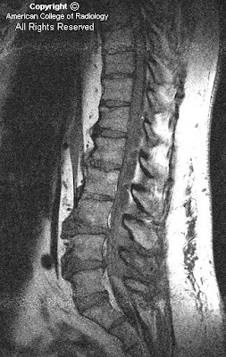
Sagittal T1 (Figure 1), T2 (Figure 2) & T2 with fat sat (Figure 3) images reveal an extradural multilobulated cystic structure which extends from the level of the T11 to L3 vertebrae. This is causing mass effect on the thecal sac. Post contrast, there was no enhancement.
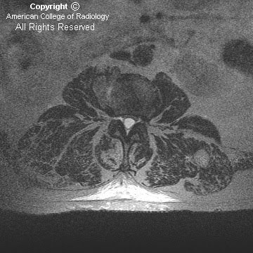
Axial T2 images (Figure 4, Figure 5, and Figure 6) once again reveal an extradural cystic structure which is causing mass effect on the thecal sac. No nerve roots are contained within the cystic structure.


Diagnosis: Spinal extradural meningeal cyst


Spinal meningeal cysts are uncommon, representing only 1-3% of all spinal masses. The pathogenesis of meningeal cysts is still unknown. Histologically, the lining of the cyst cavity may or may not be shown to be arachnoidal tissue, therefore the terms extradural arachnoid cyst and extradural meningeal cyst are used interchangeably.

Spinal meningeal cysts occur most frequently within the thoracic spine (66%), followed by the lumbar and lumbosacral spine (12%), thoracolumbar spine (12%), sacral spine (6.6%) and the cervical spine (3.3%). Most of the lesions are located posteriorly in the spinal canal. Thoracic located cysts most commonly occur in adolescents whereas sacral cysts are more commonly found in adults.

Spinal cysts mostly present through nerve compression symptoms which can be intermittent or slowly progressive. Intermittent exacerbation can occur with postural changes and Valsalva maneuvers.

MR imaging is extremely helpful in demonstrating an extradural cystic structure with CSF signal intensity. MRI can also help in identifying displacement of epidural fat and subarachnoid space, inclusion of nerve rootlets and extension into intervertebral foramina. Once the cyst is identified, CT myelography can be utilized to demonstrate a connection between the cyst and the subarachnoid space.

A classification system termed the Nabors Classification has been developed for meningeal cysts:
- Type I: lesions are extradural meningeal cysts without spinal nerve root fibers, which can be subdivided into
Type 1A extradural meningeal cysts
Type 1B sacral meningoceles
- Type II meningeal cysts are extradural and contain nerve root fibers (Tarlov’s perineural cyst)
- Type III meningeal cysts includes all intradural arachnoid cysts

Differential diagnosis for extradural arachnoid cysts include
- Intradural arachnoid cysts
- Neurenteric cysts
- Perineural cysts
- Synovial cysts
- Meningocele
- Cystic neoplasm
- Congenital and traumatic dermoid
- Inflammatory cysts or cysticercosis

Asymptomatic patients with meningeal cysts can be followed by imaging. Surgery is the treatment of choice for symptomatic patients. Immediate pain relief post surgery is common, but recurrent back pain is frequently encountered at long-term follow-up.