venerdì 30 maggio 2008
Diffuse axonal injury (DAI)
Findings
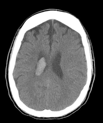
Figure 1 and Figure 3: CT images demonstrate scattered subtle hemorrhages in the interpeduncular cistern and high right frontal white matter.
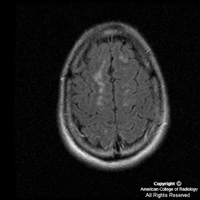
Figure 2: In the left centrum semiovale is an area of increased lucency representing edema secondary to axonal stretching.
Figure 4, 5, 6 and 7: T2 and FLAIR images demonstrate areas of high signal in the high right frontal white matter consistent with diffuse axonal injury.
Figure 8: Gradient echo image demonstrates areas of increased magnetic susceptibility in the right thalamus and right frontal white matter consistent with small areas of hemorrhage which were not identified on the CT images.
Figure 9, Figure 10, and Figure 11: Diffusion weighted images demonstrate areas of high signal and restricted diffusion in the midbrain, splenium of the corpus callosum and high right frontal white matter.
Diagnosis: Diffuse axonal injury
Diffuse axonal injury (DAI) is the result of severe closed head deceleration injury. The rotational acceleration and deceleration of the brain results in stretching of the axons. Capillaries are disrupted along with axons which produces small petechial hemorrhages. The most commonly involved sites of shear injury are the gray-white junction, corpus callosum (especially the body and splenium), brain stem, superior cerebellar peduncle and internal capsule. The body and splenium of the corpus callosum are most vulnerable because of the broadening of the falx posteriorly which allows the brain to move transiently across the midline anteriorly. Brain stem lesions, lesions within the subcortical white matter and internal capsule are associated with more severe injury. Frontal and temporal lesions at the gray-white junction are associated with more mild injury.
CT is not as sensitive as MRI in detecting lesions in DAI and initial CT examanination is normal in 50-80% of cases. Findings of DAI observed on CT are small hypodense foci which correspond to edema at the site of shearing injury. Hyperdense foci of petechial hemorrhage may be present secondary to capillary disruption. MR findings of DAI are high signal foci on T2 and FLAIR images. Hemorrhagic foci are identified as areas of low signal secondary to increased magnetic susceptibility on gradient echo images. Diffusion weighted imaging demonstrates hyperintense foci reflecting restricted diffusion.
Eighty percent of DAI lesions are microscopic and nonhemorrhagic. The traumatic axonal stretching results in swelling, microglial clusters and eventual wallerian degeneration. The most common signs and symptoms of DAI are loss of consciousness immediately following the injury with coma and persistent vegetative state. Brain stem damage (as in this case) is associated with immediate or early death. There is no treatment for diffuse axonal injury. Supportive therapy is the standard of care.
martedì 27 maggio 2008
Bilateral parotid hemangiomas
Findings
Figure 1 and Figure 2: Contrast enhanced axial CT images demonstrate bilateral, lobulated, enhancing masses in the expected region of the parotid glands. No cystic areas are seen within the masses. No normal parotid tissue is seen. Prominent enhancing vessels can be seen adjacent to these masses.
Diagnosis: Bilateral parotid hemangiomas
Hemangioma is not only the most common tumor of the parotid glands in childhood, it is also the most common tumor of infancy. The incidence of hemangioma in neonates is 1-2%. 90% of salivary gland hemangiomas occur in the parotid gland, with the remaining 10% occurring in the submandibular gland. 60% of infantile hemangiomas occur in the head and neck, and can involve virtually any space. They can be seen in association with PHACES syndrome.
On noncontrast CT imaging, parotid hemangiomas appear as lobulated, intermediate density masses without calcifications. During the proliferative phase, parotid hemangiomas typically enhance diffusely, with prominent vessels in and adjacent to the mass. As they spontaneously involute, they decrease in size and undergo fatty replacement.
The differential for these lesions includes venous and arteriovenous malformations (typically contain phleboliths), plexiform neurofibroma (look for other stigmata of neurofibromatosis), and sarcoma (tends to occur in older children and are more likely to be invasive and destructive).
In one study of 100 children with parotid hemangiomas, the female-to-male ratio was 4.5:1. Forty-five percent had a cutaneous vascular mark noted at birth. Thirty percent of the patients were asymptomatic and the lesions spontaneously involuted over time. The remaining 70% received therapy for symptomatic lesions. For most, treatment was performed to limit the size of the hemangioma and control ulceration. In the remaining patients, the indication for treatment was related to respiratory distress (26%), potential visual impairment (13%), congestive heart failure (4%), and obstruction of the auditory canal (3%).
Many hemangiomas will involute spontaneously, and watchful waiting is recommended in these cases. Symptomatic lesions or lesions causing significant cosmetic deformity can be treated. About 10-20% of hemangiomas present with indications for prompt therapy, such as expansion, destruction, obstruction, or life-threatening complications. Possible treatment options include corticosteroid or interferon injections, surgical excision, laser ablation, and embolization.
Etichette:
ACR,
Head - Neck,
Neoplasm,
Pediatric
giovedì 22 maggio 2008
Cat scratch disease
Additional clinical information: Six-year-old boy with swollen lymph glands under right-side of jaw. Concern for peritonsillar abscess.
Findings
CT shows a heterogeneously enhancing, right submandibular level I lymph node with several additional smaller, scattered enhancing subjacent lymph nodes. There is overlying infiltration of the subcutaneous fat consistent with cellulitis.
Differential Diagnosis:
- Lymphoma
- Sarcoid
- Cat scratch disease
- TB/histoplasmosis
- Mononucleosis
- Metastatic disease
- Other bacterial infectious processes
Diagnosis: Cat scratch disease
Further clinical probing subsequently revealed recent, unmonitored contact with a cat.
Discussion
Self-limiting infectious disease characterized by painful regional lymphadenopathy following the scratch of a cat (typically a kitten).
Bartonella henselae, (gram-negative bacillus) - determined to be nearly exclusively responsible for CSD.
Epidemiology:
- 22,000 cases of CSD diagnosed annually in US (1993 data).
- 70-90% of CSD cases occur in the fall and early winter months. (presumed to be due to a midsummer rise in kitten births accompanied by increased flea infestation).
- M:F 3:2
- < 21 years of age (80% of cases)
Clinical:
- >90% of patients with the disease report recent contact with a cat, usually a kitten.
- Incubation period of 3-12 days is followed by the development of one or more cutaneous papules or pustules at the inoculation site.
- Primary lesion lasts for 1-3 weeks then recedes as regional lymphadenopathy appears, generally immediately proximal to the inoculation site.
- Regional lymphadenopathy, (approx. 90%) - most notable manifestation (usually prompts medical evaluation).
- Lymphadenopathy primarily involves axillary >cervical > inguinal regions.
- Lymph nodes are often painful and spontaneously suppurate in 25-30% of cases.
- Constitutional symptoms (usually mild); include malaise, low-grade fever, anorexia, nausea, fatigue, or headache.
- Erythematous, tender papules or pustules at inoculation site.
- Tender unilateral lymphadenopathy (>90%); 50% have involvement of a single node, 30% have involvement of nodes in multiple sites, and 20% with involvement of several nodes in the same region.
- Routine laboratory tests are usually unremarkable and unlikely to aid in diagnosis. Mild leukocytosis and elevated ESR are common but nonspecific.
- Indirect fluorescent antibody (IFA) for Bartonella (84-88% sensitive and 94-96% specific), and rising immunoglobulin G (IgG) titers (titers above 1:64 supportive) provide laboratory confirmation of diagnosis. PCR from lymph node biopsy provides even more sensitive detection of disease.
- Self-limiting disease with excellent prognosis, even in patients with profound manifestations - usually resolves spontaneously over 2-5 months with rare permanent sequelae. However, immunocompromised patients may experience a dramatic and potentially life-threatening course of disease.
- Management primarily symptomatic. Antibiotics not indicated in most cases, but may be considered for severe or systemic disease.
Radiographic Overview of Diagnosis:
- Non-specific unilateral regional lymphadenopathy at/proximal to the site of inoculation.
Etichette:
AuntMinnie,
Head - Neck,
Infectious
mercoledì 14 maggio 2008
Persistent hypoglossal artery
Findings
Axial images demonstrate abnormal bifurcation of the right internal carotid artery, giving rise to a persistent hypoglossal artery, which is seen entering the hypoglossal canal.
Figure 1: Abnormal bifurcation of the right internal carotid artery.
Figure 2: Abnormal bifurcation of the right internal carotid artery.
Figure 3: Persistent right hypoglossal artery entering through the hypoglossal canal.
Figure 4: Persistent right hypoglossal artery entering through the hypoglossal canal and forming the right vertebral artery.
Sagittal images demonstrate abnormal bifurcation of the right internal carotid artery, giving rise to a persistent hypoglossal artery, which is seen entering the hypoglossal canal.
Figure 5: Abnormal bifurcation of the right internal carotid artery.)
Figure 6: Persistent right hypoglossal artery entering through the hypoglossal canal.
Figure 7: Bifurcation of ICA and persistent hypoglossal artery.
Figure 8: Persistent hypoglossal artery entering hypoglossal canal at the occipital condyle.
Figure 9: Angiogram from a different patient. Arrow points to the origin of the presistent hypoglossal artery. Please note the absence of vertebral arteries originating from the subclavian arteries.
Figure 10: Injection of left ICA opacifies a large persistent hypoglossal artery, shown to pass through the hypoglossal canal. It perfuses the entire posterior circulation.
Diagnosis: Persistent hypoglossal artery
Persistent carotid-basilar artery anastomosis occurs when there is failure of regression of the otic, hypoglossal, trigeminal and proatlantal intersegmental segments formed early during embryogenesis at day 24-28. These connections form to provide blood to the hindbrain while the posterior circulation and vertebral arteries develop.
The otic artery regresses first at the 4th week of gestation. The hypoglossal, trigeminal and proatlantal intersegmental arteries all regress by the 7th to 8th week of gestation.
Persistent trigeminal artery is the most common persistent carotid-basilar anastomosis with an incidence of 0.1-0.2% Persistent hypoglossal artery is the second most common with an incidence of 0.03-0.26%. Approximately 40 cases of persistent proatlantal intersegmental artery have been reported. Most rare is the persistent otic artery, of which there are only two well documented cases.
Persistent hypoglossal artery typically originates from the cervical portion of the ICA at the C1–C2 level and continues medially and posteriorly to enter the skull through the hypoglossal canal in conjunction with the XII nerve. With persistent carotid-basilar anastomoses the vertebral arteries are usually absent or severely hypoplastic, and there are no posterior communicating arteries. Persistent hypoglossal artery is associated with aneurysms due to the abnormal flow dynamics that often accompany this anatomical variant. They are also associated with arteriovenous malformations. It is also important to identify this anatomical variant before any endarterectomy or skull base surgery is performed as the posterior circulation is dependent on the anterior circulation.
At the 4–5mm embryonic stage bilateral longitudinal neural arteries (arrows)—one set of longitudinal neural arteries, dorsal aorta, and cervical intersegmental arteries is shown—are supplied by trigeminal artery (TA), otic artery (OA), hypoglossal artery (HA), proatlantal intersegmental artery (PA), and cervical intersegmental arteries. Internal carotid artery (ICA), external carotid artery (ECA), aortic arch (AA), dorsal aorta (DAo) and ventral aorta (VAo) are also labeled.
Etichette:
ACR,
Malformations,
Neuro,
Vascular
Brain metastasis
Additional clinical history: The patient has lung cancer and recently developed severe headaches.
Findings
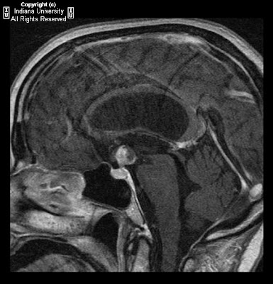
CT shows a hyperdense lesion in the suprasellar region with significant ventriculomegaly. Intraventricular air. Right frontal approach ventriculostomy tube also present. MR shows lesion is localized to pituitary stalk and hypothalamus without definite extension into body of pituitary gland. There is extension into the third ventricle causing hydrocephalus. No restricted diffusion. Lesion enhances and shows posterior hemorrhage on gradient sequences.
Differential Diagnosis:
- Pituitary adenoma
- Meningioma
- Lymphoma
- Metastatic lesion
- Thrombosed basilar tip aneurysm
- Colloid cyst
Diagnosis: Brain metastasis (CSF cytology consistent with metastatic disease)
Key Points
Metastases to the pituitary gland as a whole are rare.
Most commonly breast and lung cancer (2/3) as part of widespread metastases.
Usually to posterior lobe of the pituitary.
Most common presenting symptom is diabetes insipidus, however most are clinically silent.
Often indolent course.
Mimic pituitary adenoma, abscess, aneurysm, and any other variety of suprasellar lesions.
On CT, iso- or hyper dense mass with some enhancement. On MR, iso- or hypo intense on T1 and hyper intense on T2-weighted images with homogeneous post-gado enhancement (clearly not specific).
Metastases to the pituitary infundibulum are especially rare and also usually of lung or breast origin. They invade locally and are also indolent.
Primary tumors of the infundibulum are rare, but include pituicytomas, other astrocytomas, ependymomas, and pleomorphic xanthoastrocytomas.
Metastatic lesions to pituitary stalk have been described extending superiorly into the hypothalamus.
Metastases to the hypothalamus alone without pituitary involvement is almost never described.
mercoledì 7 maggio 2008
Wallerian degeneration
Findings
Figure 1: Noncontrast brain CT demonstrates a "dense MCA sign" in the M1 segment with loss of grey-white differentiation involving a large portion of the MCA territory.
Figure 2, Figure 3, Figure 4, and Figure 5 : T2-weighted FLAIR images obtained 60 days after the initial infarct demonstrate decrease in cytotoxic edema, evolution of intraparenchymal hemorrhage, and ex vacuo dilatation of the right lateral ventricle. Following the right corticospinal tract caudally, there is increased T2 signal to the level of the pons representing gliosis as well as atrophy of the cerebral peduncle, compatible with Wallerian degeneration.
Diagnosis: Wallerian degeneration
Wallerian degeneration is antegrade destruction of axons and myelin sheaths following damage to the proximal portion of the axon or the cell body. The most common cause of Wallerian degeneration in the brain is infarction. Other causes include hemorrhage, trauma, neoplasm, and white matter disease. Dead tissue is phagocytized with resultant gliosis throughout the affected white matter tract, which can be visualized as hyperintensity on FLAIR sequences. There is no enhancement after contrast administration. Wallerian degeneration can be demonstrated 2-6 months after the initial insult, often as an atrophied cerebral peduncle with increased T2 signal along the involved corticospinal tract.
Wallerian degeneration of the corticospinal tract (or pyramidal tract) indicates severe damage and thus marked motor function impairment. It has been shown that progressive Wallerian degeneration as demonstrated on MRI is associated with persistent moderate to severe hemiparesis.
lunedì 5 maggio 2008
Limbus vertebra
Findings
Figure 1 and Figure 2: Axial image from a CT of the lumbar spine and a sagittal reconstruction demonstrate a well-corticated, triangular bone fragment anterior to the superior end plate of L4. This is a classic finding of a limbus vertebra in a classic location. Note the well corticated nature of the bone fragment on the axial view.
Diagnosis: Limbus vertebra
Limbus vertebra is closely related to Schmorl nodes and is a relatively common finding in adults. It is a result of remote injury to a skeletally immature spine, which causes anterior intraosseous herniation of part of the nucleus pulposus through an unfused ring apophysis prior to fusion of the apophysis to the vertebral body. The apophysis fragment then remains separated from the vertebral body, and eventually develops into a well corticated, triangular fragment of bone, usually adjacent to the anterosuperior corner of a mid lumbar vertebral body. Less commonly, it can also be found near the anteroinferior aspect of a mid cervical vertebral body.
In children, the diagnosis can be trickier, since the well corticated fragment might not yet be present, and only an irregular, destructive appearing process is seen, typically involving the superior end plate of a mid lumbar vertebral body
This finding is commonly misinterpreted as a fracture, infection, or tumor when the patient presents for imaging due to back pain or following trauma, causing undue grief to the patient and the treating physician. Limbus vertebra is typically not the cause of the patient’s pain and is an incidental finding. It is important for the radiologist to be well aware of this entitiy in order to preclude further unnecessary intervention or invasive diagnostic procedures.
Skull base fibrous dysplasia and traumatic right orbital floor fracture through infraorbital canal
Findings
Small, non-displaced right orbital floor fracture extending through the infraorbital canal. Replacement of normal trabecular bone pattern of the basi-occiput, sella turcica, planum sphenoidal, and posterior nasal septum with an expansile lesion with a ground-glass matrix.
Diagnosis: Skull base fibrous dysplasia and traumatic right orbital floor fracture through infraorbital canal
Key points
Fibrous dysplasia is a fairly common benign congenital disorder caused by a defect in osteoblastic differentiation and maturation. It results in expanding bony lesions with mixture of fibrous tissue and woven bone.
Clinical presentations of FD are:
1) Monostotic (85%) – Femur, tibia, facial bones/skull base, ribs (in descending frequency)
2) Polyostotic (15%) - Skull / Facial bones (>50% cases), pelvis, long bones, ribs.
Bones most frequently involved in craniofacial FD: maxilla > mandible > frontal bone > ethmoid and sphenoid > temporal bone.
CT is the most helpful imaging tool for evaluation and, in general, demonstrates expanded bone with "ground glass" matrix.
There are actually three very general appearances that FD may be manifest (lesion appearance is thought to correlate with level of disease activity – in descending order):
- Cystic
- Pagetoid (mixture of the other two)
- Sclerotic (homogeneous "ground glass")
Lesion location of facial / skull base FD determines clinical presentation:
- Orbital FD – optic neuropathies (compression of CN II); Temporal FD – EAC sclerosis, conductive hearing loss, CN VII palsies; Sino nasal – mucocele.
- Severe facial bone involvement has a very characteristic appearance known as leontiasis ossea ("lion facies"). McCune-Albright Syndrome involves polyostotic FD, endocrine abnormalities (precocious puberty) and cutaneous hyperpigmentation (café-au-lait spots).
Sarcomatous transformation occurs rarely (< 0.5% cases, usually osteosarcoma).
Imaging points:
- CT: preferred modality, see above.
- MR: Low T1 and T2 signal usually. T2 hyper intensity may correlate with clinical-pathological activity of lesions. T1+C may be used to evaluate cranial neuropathies or suspected malignant transformation.
- Radionuclide scintigraphy: Frequently used to assess skeletal extent of polyostotic FD
Etichette:
Head - Neck,
Ophtalmic,
Other,
Signs,
Trauma
venerdì 2 maggio 2008
Moya moya disease
Findings
Axial CT shows intraventricular hemorrhage within the right lateral ventricle. MR / MRA show low intensity signal within right lateral ventricles compatible with hemorrhage. The right MCA is not visualized and there are surrounding collateral vessels (best seen on the MRA AP projection and corresponding coronal CT image). Collateral vessels are identified on the left to a lesser degree. The left internal carotid, left MCA, vertebral arteries, basilar artery, and PCA arteries are patent.
Differential diagnosis:
- Moya moya
- Carotid thromboembolic disease
- Blood dyscrasias and stroke
- Vasculitidies such as polyarteritis nodosa
- Cavernous sinus syndromes
Diagnosis: Moya moya disease
Discussion
Moya moya disease is a rare, chronic cerebrovascular disease characterized by severe stenosis or occlusion of the arteries around the circle of Willis with prominent collateral circulation. "Moyamoya" is a Japanese word meaning "puff of smoke," and describes the angiographic appearance of the abnormal vascular collateral networks that develop adjacent to the stenotic vessels. The stenotic or occlusive areas are usually bilateral but unilateral involvement does not exclude the diagnosis.
The etiology of Moya moya disease is unknown. The high incidence among the Japanese and Asian population together with a 10% familial occurrence suggest a genetic cause. Although the disease may occur by itself in a healthy individual, many disease processes have been associated with the angiographic appearance of Moya moya.
These include:
- sickle cell disease
- Neurofibromatosis type I
- Down's syndrome
- Fibromuscular dysplasia
Pathologically, bilateral concentric stenosis or occlusion is consistently found in the distal carotid arteries as well as the proximal anterior and middle cerebral arteries. The pathology is characterized by fibrocellular thickening of the intima, disruption of the internal elastic lamina, and thinning of the media without evidence of inflammation. The hallmark of Moya moya is the small collateral channels that develop within the basal or cortical regions of the brain. In addition, intracranial aneurysms may develop within the circle of Willis and cause subarachnoid hemorrhage.
Clinical manifestations from Moya moya disease may differ in children and adults. Cerebral ischemic events, either TIA or ischemic stroke, are more common in children as a consequence of the stenotic vessels while intracerebral hemorrhage usually presents in adult Moya moya due to the fragility of the abnormal vessels. In addition, epilepsy may be a presenting sign, particularly in children. Treatment of the disease is controversial and unproven.
Radiology
Cerebral angiography is the gold standard for diagnosis of Moya moya disease. Characteristic angiographic findings include stenosis or occlusion at the distal internal carotid artery with leptomeningeal, collateral networks that form intraparenchymal anastamosis. These vascular collateral networks form the "puff of smoke," for which moyamoya is named. In addition, MR angiography can demonstrate lesions of the distal carotid arteries and is shown to have a sensitivity of 98% and specificity of 100 % for the diagnosis of Moya moya. MRI is superior to CT scan for detection of the small ischemic lesions in the cortical and subcortical area that are characteristic of the disease.
Etichette:
AuntMinnie,
Neuro,
Signs,
Vascular
Iscriviti a:
Post (Atom)