lunedì 29 dicembre 2008
Schwannomatosis
Findings
Figure 1, Figure 2, and Figure 3: Sagittal T2 and post contrast sagittal T1 weighted images demonstrate numerous enhancing intradural, extramedullary masses lining the roots of the cauda equina.
Figure 4, Figure 5, and Figure 6: A heterogeneously enhancing right paraspinal mass demonstrates heterogenous signal on T2 weighted images, highly suggestive of a schwannoma. There is no adjacent osseous erosion.
Diagnosis: Schwannomatosis
Schwannomatosis is a form of neurofibromatosis that has unique diagnostic criteria but can have findings that overlap with other forms of NF that can confound diagnosis.
Spectrum of neurofibromatosis:
- Neurofibromatosis Type I: Café au lait spots, skin fold freckling, Lisch nodules, cutaneous and plexiform neurofibromas, learning disabilities, and propensity for malignancy
- Neurofibromatosis Type II: Bilateral vestibular schwannomas, various other intracranial, spinal and cutaneous schwannomas, meningiomas, ependymomas. Ocular manifestations include cataract, retinal hamartoma
- Schwannomatosis: Schwannomas in multiple locations without the presence of vestibular schwannomas
Schwannomatosis, also known as neurilemmomatosis, is a condition of multiple non-vestibular schwannomas in a patient who has no other stigmata of neurofibromatosis type 2. Schwannomas are benign nerve sheath tumors that are usually solitary and occur sporadically. A definite diagnosis of schwannomatosis can be made when a patient older than 30 years has two or more nondermal schwannomas, one of which has been pathologically confirmed. There is however, a wide spectrum of overlap between schwannomatosis and NF2 such that a definitive diagnosis may be difficult to make.
Differences between schwannomatosis and NF2 include the age of presentation. Most patients with the former will present in middle age, whereas patients with NF2 present in the second to third decade of life. Patients with schwannomatosis generally do not have a corresponding family history, unlike NF2.
Imaging characteristics of the tumors in schwannomatosis are the same as those of sporadic schwannomas. Iso/hypointensity on T1 weighted images, hyperintensity (secondary to cystic change, and occasionally hemorrhage) on T2 weighted and STIR images and avid contrast enhancement are typical findings.
Dedicated imaging of the internal auditory canals and the brain should be pursued to help clarify the diagnosis. Follow up imaging is also indicated, especially if the diagnosis is not definitive, as malignant degeneration of an NF2 lesion would manifest as rapid increase in size of a mass.
martedì 23 dicembre 2008
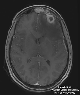
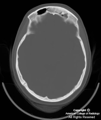
Brain abscess secondary to frontal osteoma and sinusitis
Findings
CT demonstrates a well marginated bone density lesion arising from the anterior left frontal sinus wall and protruding into the sinus lumen (Figure 1). Opacification is present in the superior left frontal sinus consistent with sinusitis (Figure 1). The posterior left frontal sinus wall is dehiscent (Figure 2).
MR demonstrates a multilocular ring enhancing process in the left anterior frontal lobe (Figure 1 and Figure 2). The center of the collection demonstrates high signal on diffusion weighted images (yellow arrows in Figure 6 and Figure 7). The overlying dura enhances as does the adjacent frontal sinus (Figure 4 and Figure 5). Vasogenic edema is present in the surrounding left frontal white matter (Figure 2 and Figure 3).
Diagnosis: Brain abscess secondary to frontal osteoma and sinusitis
Brain abscess is an infectious process which results in high morbidity and mortality if undiagnosed and untreated. In today’s era of antibiotics and refined neurosurgical technique, the major obstacle to successful treatment is delayed diagnosis. Noninvasive diagnostic imaging is critical for diagnosis and is directly responsible for improved patient outcomes in the last 10-15 years.
Brain abscess presents as a rapid developing space occupying syndrome usually beginning with a headache, followed by focal neurological deficit or seizure, and may progress to increased intracranial pressure, nausea,vomiting, and loss of consciousness. Fever is absent in 50% of cases as in this case.
Infection occurs by extension from a direct cranial focus or by hematogenous dissemination from an extracranial source with 50% incidence of each. Mastoiditis is the most common cranial source (30%) followed by sinusitis (15%). Hematogenous seeding accounts for 25%, of which endocarditis is the most common cause. No identified cause is found in 20% of cases. Rare causes include retrograde thrombophlebitis from a facial or dental infection, penetrating trauma, meningitis complication, and surgery.
The typical appearance of brain abscess on CT is a focal round lesion with hypodense center and surrounding vasogenic edema. If contrast is given, ring enhancement is seen. Abscess secondary to mastoiditis occurs in the temporal lobe and sometimes the posterior cranial fossa. Paranasal sinusitis results in frontal lobe location, and hematogenous dissemination usually results in multiple lesions following a vascular distribution.
On MRI, brain abscess demonstrates ring enhancement. Compared to other ring enhancing lesions, abscesses exhibit thinner, more regular, and more homogenously enhancing walls. Often, the cortical wall will be thinner. Classically, the lesion will have a T2 hypointense rim. A critical finding usually seen is central restricted diffusion corresponding to the infection products. Necrotic tumors seldom demonstrate restricted diffusion within the necrotic center. A dominant lesion will often demonstrate contiguous smaller satellite lesions. Another helpful finding is surrounding vasogenic edema which invariably is seen with abscess and neoplasm and rarely seen in other ring enhancing or restricted diffusion lesions such as demyelinating lesions. Lymphoma can be a difficult differential diagnosis as lymphoma usually restricts diffusion, causes vasogenic edema, ring enhances, and can have central necrosis. Clinical history of immunocompromise and location are occasionally helpful to differentiate from abscess.
This patient’s frontal sinus osteoma contributed to the frontal sinusitis and/or intracranial extension of the infection. Paranasal sinus osteomas are benign, asymptomatic bone growths that can rarely have intracranial complications including subdural empyema, abscess, CSF leak, and pneumocephalus. Osteomas demonstrate variable ossification from dense ivory type to a more ground glass appearance.
Etichette:
ACR,
Infectious,
Inflammatory,
Neoplasm,
Neuro
giovedì 18 dicembre 2008
Retinal hemorrhage in non accident trauma
Findings
Figure 1: T2*GRE axial image through the globe demonstrates nodular low signal in both retinas representing bloom artifacts from blood products suggesting retinal hemorrhage.
Figure 2: T2*GRE axial image shows low signal in the posterior parietal subdural space suggesting subdural hemorrhage.
Figure 3: Axial T1 image shows left frontal subdural fluid collection similar to CSF signal suggesting subdural hygroma.
Diagnosis: Retinal hemorrhage in non accident trauma
Child physical abuse is an unfortunately common occurrence that may manifest as any pattern of injury. Some patterns of injury (metaphyseal fractures, posterior rib fractures, subdural hemorrhages, retinal hemorrhages) are more suggestive and specific than others. Child abuse is often misdiagnosed and under-recognized by physicians and caregivers.
Non-accidental head injury (NAHI) or shaken baby syndrome (SBS), (some authors prefer the term shaken-impact syndrome), occurs in 12% of the physically abused children and is a major cause of neurological disability and death during infancy. Radiological imaging plays a crucial role in evaluating cranial injury, both for guiding medical management and the forensic aspects of abusive trauma. The neurologic injuries are more devastating than other injuries. NAHI is the leading cause of morbidity and mortality in abused children. MRI is the test of choice over CT for detecting different ages of intracranial hemorrhages, shearing injuries, and retinal hemorrhages.
Subdural hemorrhage is the most common intracranial lesion in shaken baby syndrome. Other findings are cerebral edema, subarachnoid hemorrhage, intraparenchymal hemorrhage, intraventricular hemorrhage, diffuse axonal injury, shearing injury, ischemia and brain herniation. Retinal hemorrhages may be found in 50%-100% of shaken infants, the prevalence of retinal hemorrhages in victims who die approaches 100%. Retinal hemorrhage is a characteristic and diagnostic feature of SBS. The severity of retinal hemorrhage is strongly correlated with intracranial injury in SBS. It is therefore very important to recognize retinal hemorrhage on neuroimaging.
mercoledì 17 dicembre 2008
Cerebral amyloid angiopathy (CAA)
Findings
Head CT demonstrates bilateral temporal hemorrhage (left > right) with scattered small vessel ischemic disease.
Head MRI again demonstrates the two hemorrhages seen on the CT, in addition there are multiple scattered bilateral black dots with no significant mass effect. No extracerebral fluid collections are present, and there is no abnormal enhancement. There was no diffusion restriction.
Differential diagnosis:
- Cerebral amyloid angiopathy
- Hemorrhagic metastasis
- Hypertensive micro hemorrhages
- Multiple venous malformations
Diagnosis: Cerebral amyloid angiopathy
Key points
Common cause of atraumatic parenchymal hemorrhage in elderly, particularly those with dementia.
Lobar hemorrhage > microangiopathy > focal "amyloidoma" (least common).
CAA hemorrhages frequently spare the basal ganglia. By contrast, hypertensive hemorrhages frequently involve the basal ganglia.
15-20% of atraumatic intracerebral hemorrhage in normotensive patients > 60 yrs.
27-32% of normal elderly (autopsy).
82-88% in patients with Alzheimer disease (AD).
Also common in Down syndrome.
lunedì 15 dicembre 2008
CNS schistosomiasis
Findings
Figure 1: Axial T2-weighted MR image shows vasogenic edema and mass effect involving left temporal lobe.
Figure 2: Axial contrast-enhanced T1-weighted MR image shows multiple areas of central linear enhancement surrounded by enhancing punctate nodules.
Figure 3: Coronal contrast-enhanced T1-weighted MR image confirms linear and nodular enhancement pattern in a different imaging plane.
Diagnosis: CNS schistosomiasis
CNS Schistosomiasis is caused by infestation with trematode (fluke) worms. The host is the freshwater snail that releases Schistosoma cercariae into the water. The cercariae infect humans percutaneously and transform into schistosomula. The schistosomulae migrate to the lungs and liver and then descend to the mesenteric venous system.
Characteristic MR findings demonstrate a central linear enhancement, surrounded by multiple enhancing punctate nodules 1-2 mm in diameter, clustered in a masslike structure with an arborized appearance. On T2-weighted images, the lesions are hyperintense with associated surrounding vasogenic edema and mass effect. This pattern of enhancement has not been observed in association with other CNS worm infections or disease processes. The differential diagnosis for Schistosomiasis may include cerebritis, metastatic disease, tuberculosis, sarcoidosis and possibly anaplastic astrocytoma.
At biopsy, the enhancing nodules seen in the patient were thought to represent granuloma formation with extensive surrounding inflammation and possible venous congestion. Such granulomatous lesions are believed to represent either military pseudotubercles or confluent granulomata containing fibrosis, necrosis, hemorrhage, thrombosis, demyelination, calcification, and arteritis. The morphology and placement of the spine (absent, lateral, or terminal) of the ova can be used to identify the schistosomal species. S.mansoni has a broad lateral spine (as seen in our 32-year-old patient). S. haematobium has a prominent terminal spine, and S.japonicum has a rudimentary or absent spine. It is postulated that the central linear enhancement that was observed is the result of slow blood flow and local venous obstruction caused by the anomalous migration of the adult worm in the leptomeningeal veins. Such placement might result in the concentration of ova at a focal region, forming a nodular mass.
Although the enhancement pattern observed may not be a sensitive indicator for CNS schistosomiasis, it is found to be a specific one. An early diagnosis may be made on the basis of the contrast-enhanced T1 weighted MR imaging findings alone. Early diagnosis, along with treatment, will lead to a more favorable outcome. The patient was found to have complete resolution of abnormal findings on follow-up imaging studies after undergoing treatment with Praziqunatel combined with corticosteroid therapy.
giovedì 11 dicembre 2008
Monostotic clival fibrous dysplasia
Findings
CT: Expansile, lytic lesion of the clivus is present without cortical disruption. The lesion has a ground-glass appearance. It is lytic centrally and sclerotic peripherally. Bilateral jugular tubercle and right occipital condyle involvement also present.
MRI: Heterogenous (T2 hyper intense and T1 hypo intense) expansile clival lesion. The majority of the lesion demonstrates avid contrast enhancement. The brain parenchyma is normal.
Differential diagnosis:
- Fibrous dysplasia
- Chordoma
- Chondrosarcoma
- Plasmocytoma
- Metastasis
- Lymphoma
Diagnosis: Monostotic clival fibrous dysplasia
Discussion
Monostotic clival fibrous dysplasia is a rare entity (According to Sirvanci et al., only 3 cases were reported in the literature by 2002). Overall, craniofacial fibrous dysplasia represents three percent of bone tumors. Fibrous dysplasia is a developmental disorder felt to be secondary to a genetic mutation. It is not hereditary. It results in abnormal fibroblast proliferation with replacement of normal medullary bone. This causes expansion and structural integrity compromise.
Fibrous dysplasia can be classified as monostotic and polyostotic. The polyostotic form represents 30% of cases and can present as part of Albright-McCune-Sternberg syndrome. The initial presentation in the majority of cases is in the first two decades of life. No racial or gender predilection has been described. Besides craniofacial involvement, other commonly involved areas include the ribs, femurs, tibias, and the pelvis.
Management is almost always conservative with followup imaging to evaluate for stability if warranted (typically CT). Surgical treatment may be warranted if neurological compromise or significant pain develops. Rarely (0.05% of monostotic craniofacial lesions), malignant transformation may occur.
Radiologic Overview
MRI and CT are the best imaging modalities for clival evaluation. On MRI, fibrous dysplasia appears as hypo intense on T1 weighted images. The imaging characteristics on T2 weighted images are variable from hypo intense to isointense to hyper intense. The variability on T2 weighted images is attributed to several factors including (but not limited to) cellularity, cyst formation, and presence of collagen. Avid enhancement after gadolinium administration is typically present.
On CT, a ground glass appearance is associated with fibrous dysplasia. Additionally, a sclerotic border is also present. Smaller cystic, circumscribed areas are present within the lesion. Overall, CT is superior to evaluate for subtle change and internal lesion structure. If nuclear scintigraphy is performed (not necessary), mild, nonspecific activity is usually present in the clivus.
Etichette:
AuntMinnie,
Head - Neck,
Inflammatory
mercoledì 10 dicembre 2008
Cavernous malformations
Findings
CT shows multiple well circumscribed hyperdense lesions scattered throughout the brain parenchyma without any perilesional edema or extra-axial fluid collections. No mass effect.
MRI FLAIR images show mixed signal intensity of the “popcorn” lesions seen on CT. T1 images show increased signal consistent with blood products. GRE sequences show blooming of artifact and post contrast examination shows no enhancement.
Differential diagnosis for "Popcorn ball" lesion:
- Cavernous malformation
- AVM
- Hemorrhagic neoplasm
- Calcified neoplasm (oligodendroglioma)
Differential diagnosis for multiple black dots:
- Cavernous malformation
- Old trauma (contusions, DAI)
- Hypertensive petechial hemorrhage
- Amyloid angiopathy
- Capillary telangiectasis
Diagnosis: Cavernous malformations
Key points
Benign vascular hamartoma with masses of closely apposed immature blood vessels ("caverns"), intralesional hemorrhages, no neural tissue.
CM = most common angiographically "occult" vascular malformation, approximate prevalence 0.5%.
75% occur as solitary, sporadic lesion.
10-30% multiple, familial.
Zabramski classification of CMs:
- Type 1 = subacute hemorrhage (hyper intense on T1WI; hyper- or hypo intense on T2WI)
- Type 2 = mixed signal intensity on T1-, T2WI with degrading hemorrhage of various ages (classic "popcorn ball" lesion)
- Type 3 = chronic hemorrhage (hypo- to iso on T1-, T2WI)
- Type 4 = punctate micro hemorrhages ("black dots"), poorly seen except on GRE sequences
Most common signs/symptoms:
- Seizure 50%
- Neurologic deficit 25% (may be progressive)
- 20% asymptomatic
Etichette:
AuntMinnie,
Malformations,
Neuro,
Vascular
martedì 2 dicembre 2008
Occlusive atherosclerosis of the internal carotid artery (ICA) and secondary angiographic string sign
Findings
Figure 1: The left proximal common carotid artery demonstrates an atypical high-grade waveform, and a high resistance systolic upstroke. This waveform is more characteristic of the external carotid artery, and less like the internal carotid artery.
Figure 2: There is complete absence of flow in the left ICA and the ICA on grayscale appears completely occluded.
Figure 3 and Figure 4: Lateral views show the angiographic string sign. Early arterial phase images demonstrate a 99% stenosis of the proximal ICA. A trickle of contrast material is seen in the ICA distal to the stenosis.
Differential diagnoses
- String sign secondary to atherosclerosis at the proximal ICA and distal collapse of the ICA
- Dissection of the ICA
- Subacute/chronic thrombosis of the ICA
- Carotid artery stenosis secondary to radiation
Diagnosis: Occlusive atherosclerosis of the internal carotid artery (ICA) and secondary angiographic string sign
Atherosclerosis is the most common cause of the angiographic string sign. As the plaque enlarges, the pressure threshold for continued antegrade flow distal to the stenosis is reached. The distal ICA vessel lumen collapses as a result of decreased flow leading to the production of the string sign.
Carotid duplex ultrasound with color Doppler flow imaging and CT are first line diagnostic modalities for patients with symptomatic atherosclerotic vascular disease. When an occlusion of the carotid artery is seen, confirmatory imaging may be requested because differentiation between partial occlusion and a complete occlusion has important therapeutic implications.
The North American Symptomatic Carotid Endarterectomy Trial (NASCET) demonstrated a significantly decreased risk of stroke in patients with at least 70% stensoses, undergoing endarterectomy versus medical management (9% vs. 26%, respectively).
Etichette:
ACR,
Head - Neck,
Neuro,
Signs,
Vascular
Iscriviti a:
Post (Atom)