Visualizzazione post con etichetta AuntMinnie. Mostra tutti i post

martedì 8 febbraio 2011

Subdural empyema


Findings

A large left middle cranial fossa subdural empyema is demonstrated, with a relatively thin rim of enhancement. Internally, there is a large quantity of debris. There is mass effect, with a modest midline shift and effacement of the left lateral ventricle. Inflammatory changes are demonstrated in the left temporal bone which is likely the source of the abscess. There is diffusion restriction, not marked, consistent with abscess. There is extensive dural enhancement, along with considerable surrounding edema.

Differential diagnosis:
- Subdural empyema
- Chronic subdural hematoma
- Subdural effusion
- Subdural hygroma
- Dural metastasis


Diagnosis: Large left middle cranial fossa subdural empyema; left mastoiditis


Key points

Loculated collection of pus in subdural space
Best diagnostic clue: Extra-axial collection with contrast enhancing rim
Supratentorial typical
Infratentorial (up to 10%), often associated with mastoiditis
Crescentic typical; may be lens shaped on coronal images
CT demonstrates extra-axial collection, iso-to hyper dense to CSF on noncontrasted CT; shows strong peripheral enhancement with contrast
Best imaging tool: MR with DWI to demonstrate presence, nature, extent and complications
T1W image shows:
Extra-axial collection hyper intense to CSF
Crescentic extra-axial collection
T2WI demonstrates a lesion that is Iso-to hyper intense to CSF,
FLAIR shows a crescentic fluid collection which is hyper intense to CSF, underlying brain may be hyper intense
DWI shows restricted diffusion (increased signal intensity); Differentiates subdural empyema from subdural effusions
T1WI post contrast shows:
Prominent enhancement at margin related to granulomatous tissue and inflammation
Encapsulating membranes enhance strongly, may be loculated with internal fibrous strands
May see enhancement of adjacent brain parenchyma
MRV may show venous thrombosis seen as a lack of flow
CT may miss small collections
Complications include cerebritis and brain abscess, cortical vein and dural sinus thrombosis, and cerebral edema
Subdural empyema is much more common than epidural empyema
In older children, adults: Related to paranasal sinus disease (>2/3), in infants and young children it can be a complication of bacterial meningitis
Most common signs/symptoms include fever, headaches, meningismus, sinusitis, cerebritis
Sinus or ear infection in > 75% of cases
Confused with meningitis which may lead to delayed diagnosis
Can occur at any age
Rare, yet high mortality rate.
If subdural or epidural abscess is discovered, look also for sinusitis, otomastoiditis, dural sinus thrombosis and brain abscess
Progresses rapidly, neurosurgical emergency
Surgical drainage via wide craniotomy is gold standard

lunedì 27 dicembre 2010

Lumbar disc extrusion with a wrapped disc


Findings

There is a left central disc extrusion at L5-S1 that causes mild to moderate left lateral recess narrowing and nerve root displacement without nerve root compression. At this level there is also contrast enhancement traversing the left laminectomy defect and encasing the disc extrusion, consistent with a wrapped disc. There is enhancement in the left lateral recess, suggesting post-operative fibrosis.

Differential diagnosis:
- Wrapped disc
- Peridural fibrosis
- Epidural abscess
- Epidural metastasis
- Nerve sheath tumor
- Disc pseudobulge
- Intervertebral disc protrusion
- Intervertebral disc extrusion
- Recurrent intervertebral disc herniation


Diagnosis: Lumbar disc extrusion with a wrapped disc


Key points: "Wrapped" disc


Disc herniation (protrusion, extrusion, or fragment) may be caused by trauma, repetitive or acute, and are a common source of pain and subsequent back surgery in the general population. In the acute phase, the herniated disc stimulates a fibrovascular response. A "wrapped disc" is the focal herniation (protrusion, extrusion, or fragment) that is encased in vascular scar tissue stimulated by this response and is evident by enhancement on contrast-enhanced T1-weighted images.

Asymptomatic or low back pain and/or radiculopathy are most common in the lumbar spine at L4-L5 and L5-S1. A wrapped disc is a post-surgical sequela, particularly following surgery for spinal stenosis in which the surgical procedure is more extensive, involving a laminectomy and a medial facetectomy.

Best imaging modality: MR (sequences: sagittal and axial T2WI and T1WI, as well as contrast-enhanced axial and sagittal T1WI)
Other imaging modalities: CT, myelography


Imaging findings

MR: Anterior extradural mass contiguous with the disc space extending into the spinal canal
*Contrast-enhanced T1WI: Peripheral enhancement surrounding the disc herniation or fragment with/without central canal, lateral recess, or foraminal stenosis and cord or nerve root impingement. (*most helpful MR sequence)
Non-enhanced T1WI: Isointense to parent disc
T2WI: Iso- to hyper intense to parent disc
General disc hypointensity and height loss at the level of the herniation, as well as postoperative changes (laminectomy defects, etc), degenerative facet disease, and osteophytes, are common associated findings.
CT:
Non-contrast CT: An anterior extradural soft tissue mass that may displace the nerve root / indent the thecal sac
Contrast-enhanced CT: Mild peripheral enhancement of the disc herniation/fragment
Myelography: An extradural mass that indents the thecal sac and nerve root sleeves
Imaging findings of other common differential diagnoses
Peridural fibrosis: Scar within epidural space after lumbar surgery that infiltrates epidural fat, causing homogeneous enhancement that diffusely surrounds the thecal sac and nerve root; increased in T2 signal relative to adjacent disc herniation
Epidural abscess: A distinct fluid collection in the epidural space with peripheral enhancement on post-contrast images, often associated with findings of diskitis
Epidural metastasis: Elongated (cranial-caudal orientation) enhancing mass with osseous involvement and may demonstrate paravertebral extension
Nerve sheath tumor: Avid enhancement surrounding the nerve root, some of which are in a "dumbbell" shape
Disc pseudobulge: Smooth generalized extension of the disc margin without a focal defect due to "uncovering" of disc related to spondylolisthesis
Intervertebral disc protrusion: Anterior extradural mass contiguous with disc space and triangular in shape with broader base than apex; no enhancement
Intervertebral disc extrusion: Anterior extradural mass contiguous with disc space by a "neck," in which this herniated disc material then widens in the epidural space
Recurrent intervertebral disc herniation: Extradural mass contiguous with intervertebral disc margin, demonstrating enhancement peripherally but without central enhancement
Treatment
Conservative: Anti-inflammatory and pain medications, avoid trauma
Surgical: Repeat surgery to remove herniated disc (protrusion, extrusion, fragment)

mercoledì 22 dicembre 2010

Intracranial pseudotumor (Tolosa-Hunt syndrome)


Findings

Increased CSF signal demonstrated by T2 hyper intensity within the right optic sheath. Nodular enhancement at the superior aspect of the right optic nerve at the orbital apex, which extends with prominent nodular enhancement posteriorly along the dural reflection of the right cavernous sinus. Asymmetric dilatation of the right superior ophthalmic vein.

Differential diagnosis:
- Meningitis
- Sarcoidosis
- En plaque meningioma
- Meningeal metastases
- Meningeal Non-Hodgkin's lymphoma
- Tolosa-Hunt syndrome


Diagnosis: Intracranial pseudotumor (Tolosa-Hunt syndrome)


Discussion

Intracranial pseudotumor (Tolosa-Hunt syndrome) is a part of a spectrum of "quasineoplastic" lesions that demonstrate orbital, intracranial, or pulmonary involvement, and include such other disorders as plasma cell granuloma and hypertrophic cranial pachymeningitis. It is a chronic granulomatous disease of unknown origin, which has been hypothesized to represent a low grade fibrosarcoma of inflammatory cells versus an autoimmune phenomenon. While Tolosa-Hunt syndrome is rare, orbital pseudotumor is the third most common ophthalmic disorder, and encompasses 5-8% of all orbital masses. Histologically, the lesions of Tolosa-Hunt demonstrate mixed lymphocytic and plasma cell infiltrate, with a variable degree of fibrosis. Lesions favor the cavernous sinus and basal meninges, although falcine and tentorial lesions have been described.

Patients with intracranial lesions are more frequently young adults, who present initially with chronic headaches or cranial nerve palsies. Patients with orbital involvement are more frequently middle-aged, and may present with painful proptosis and vision loss . Symptoms may be intractable, leading to severe disability. Untreated or unresponsive disease may progress to death. First-line treatment is invariably high-dose steroids, with radiotherapy or surgical resection reserved for patients with incomplete response to steroids .


Radiologic Overview of the diagnosis

The imaging hallmarks of Tolosa-Hunt syndrome are characterized by an enhancing, infiltrating meningeal mass, which favors the cavernous sinus or basal meninges, although falcine and tentorial involvement has been described . Focal meningeal thickening may range from just a few millimeters to a greater than 2 cm rind. With intracranial pseudotumor, orbital involvement is spared more than 90% of the time. Tolosa-Hunt remains a diagnosis of exclusion, once meningitis, en plaque meningioma, and meningeal metastases are ruled out.

The imaging modality of choice for imaging patients with Tolosa-Hunt remains MRI, although useful information may be gleaned from other modalities. On non-contrast enhanced CT (NECT), there are no specific findings to suggest the diagnosis; however, this modality may be of some value in differentiating the lesion from en plaque meningioma. On contrast enhanced CT (CECT), salient imaging findings include enhancing, thickened meninges or a curvilinear appearance of a single meningeal region. As aforementioned, MRI remains the primary modality for diagnosis of Tolosa-Hunt syndrome, and each sequence may provide critical information required to make the diagnosis. On T1WI, one may find focal thickening of the meninges that is isointense to gray matter. On T2WI, lesions are characterized as iso- to hypo intense regions of focal meningeal thickening, which may be more hypo intense as they become more fibrotic. FLAIR is of little help in making the diagnosis, but it is unlikely to demonstrate focal brain edema underlying the lesion . Contrast enhanced T1WI is the single most valuable sequence for evaluation of Tolosa-Hunt syndrome, and is characterized by diffusely enhancing region of meningeal thickening, which may range from a few millimeters to greater than 2 cm in some cases. Diffuse boney infiltrates may be appreciated on fat saturated contrast enhanced T1 sequence. While angiography is not considered a primary modality, severe disease may result in carotid artery narrowing, thus MRA may be a useful adjunct in the appropriate clinical setting.

The appropriate differential diagnosis of Tolosa-Hunt syndrome includes meningitis, sarcoidosis, en plaque meningioma, meningeal metastases, and meningeal Non-Hodgkin's lymphoma.

venerdì 17 dicembre 2010

Hypertensive intracranial hemorrhage


Findings

Axial CT of the head shows a large hyper dense focus with peripheral hypo density in the left frontal lobe, causing sulcal effacement at the frontal cortex but no significant midline shift. Surrounding rim of low density represents edema (image 1). Unenhanced MRI of the brain shows an the same mass-like focus in the left frontal lobe, which has an isointense center with a hyper intense rim (image 2). Post gadolinium-enhanced T1 image of the brain shows no internal enhancement of this lesion (image 3).


Differential diagnosis:
- Hypertensive intracranial hemorrhage
- Ruptured arteriovenous malformation (AVM)
- Hemorrhagic intracranial mass
- Posttraumatic cerebral contusion


Diagnosis: Hypertensive intracranial hemorrhage


Acute blood appears hyper dense of unenhanced head CT.
Without a history of trauma, intraparenchymal brain hemorrhage on head CT could represent a hemorrhagic mass, a ruptured AVM, or a hemorrhagic brain tumor (primary or metastatic).
MRI with and without contrast is the best diagnostic tool for determining if a mass lesion is present, and for evaluating the age of the intracranial hemorrhage.
Acute blood products on T1 appear hypo intense to isointense (image 2), whereas subacute and chronic hematoma are hyper intense.
Neoplasms should enhance on post contrast T1. The lack of enhancement in this patient on post contrast T1 excludes neoplasm (image 3).

giovedì 16 dicembre 2010

Sequestered disk


Findings

There is an non- enhancing ovoid mass slightly hyper intense to muscle on both T1 and T2 sequences, in the anterior epidural space at the L3 level, measuring approximatelyl 12 x 8 x 12 mm. This is not contiguous with any adjacent disks. No signal dropout on fat-saturated sequences. The mass causes severe stenosis of the left half of the spinal canal at the L3 level, compressing the left descending nerve roots. T1 and T2 hyper intensity at the endplates abutting L2-L3 disc space representing Modic Type II changes. There is intervertebral disk space height loss at L2-L3 with severe disk desiccation changes.


Differential diagnosis:
- Sequestered disk
- Extruded disk
- Failed back surgery
- Epidermoid
- Epidural abscess
- Epidural hematoma
- Lipoma


Diagnosis: Sequestered disk


A focal disk protrusion is an extension of intervertebral disc material (nucleus pulposus) beyond the vertebral margin (AP diameter < mediolateral diameter). An extruded disk is one in which the nucleus pulposus has herniated through a rent in the annulus fibrosis. The AP diameter > ML diameter, and the disk may migrate craniocaudally, but maintains attachment to the parent disk (frequently symptomatic).
When extruded disk material loses its attachment to the parent disk, it is referred to as a sequestered disk. Sequestered discs usually lodge in the anterior epidural space (AES), just anterior to the posterior longitudinal ligament, and migrate either cephalad or caudad (with equal frequency). Because there is a midline septum associated with the PLL in the AES, the fragment is usually just off midline (to the right or left). Rarely, the sequestered fragment may migrate beyond the PLL into the posterior epidural space, through the dural ( intrathecal location), or into the paraspinal muscles.
They usually resemble the parent disk on MR, with T1 hypo intense and T2 iso- / hypo intense. There may be surrounding T2 hyper intensity and a rim of enhancement from inflammatory changes.
This is a crucial diagnosis to make, as a sequestered disk is a contraindication to limited disk procedures (e.g. Percutaneous discectomy) and may result in failed back surgery.

martedì 7 dicembre 2010

Methotrexate neurotoxicity


Additional clinical history: Patient was diagnosed with acute lymphocytic leukemia 2 months previously. He is status post induction therapy with a negative bone marrow biopsy, and is currently receiving consolidation chemotherapy with methotrexate, and presents with right upper extremity weakness.


Findings

MR images of the brain demonstrate a focal area of diffusion restriction involving the left frontoparietal white matter. There is minimal associated T2/FLAIR hyperintensity. No associated enhancement. Remainder of the brain was within normal limits.
Imaging done four months later shows lesion has nearly resolved.


Diagnosis: Methotrexate neurotoxicity


Discussion

Methotrexate is a folic acid analogue. Its cytotoxic effects are carried out through inhibition of the enzyme dihydrofolate reductase, which reduces tetrahydrofolic acid levels, ultimately inhibiting cell division.

From bone marrow cell precursors to the quickly dividing cells of the intestinal tract, methotrexate exerts its effects on all dividing cells in the body. One of its rare side effects is CNS toxicity. The decreased folate levels achieved with methotrexate have implications on metabolism of adenosine, homocysteine, and biopterin. Low folate levels lead to a subsequent decrease in S-adenosyl-methionine(SAM) concentrations. This eventually leads to chronic demyelination and neurologic symptoms.

An additional side effect of MTX is the elevated levels of adenosine in the CSF. Adenosine is a vasodilator, which causes dilatation of cerebral vasculature resulting in neurotoxicity. The increased homocysteine levels caused by MTX have been shown to damage vascular endothelium and lead to subsequent strokes and thromboemboli. Methotrexate has also been found to cause cytotoxic edema, which is the most common cause of lesions that enhance on MRI DWI.

The neurotoxicity caused by MTX can be immediate, acute to subacute, or delayed. Symptoms of the disease can range from headache, nausea, vomiting, and fever, to transient or permanent focal neurologic symptoms. The immediate form occurs within a day of MTX administration and presents as a chemical meningitis. The acute to subacute form presents from days to weeks after administration of MTX, and presents with seizures or focal neurologic symptoms. The delayed form presents as leukoencephalopathy and a generalized decrease in higher cognitive function.


Radiological findings

A case series containing nine cases of MTX neurotoxicity revealed that lesions found in this disease tend to be focal and show up on DWI as well as T2 and FLAIR imaging. These abnormalities can continue to persist on imaging long after the symptoms have resolved. The DWI shows diffusion restriction with T2/FLAIR hyperintensity being less conspicuous.

In another independent case study on MTX neurotoxicity, MRI demonstrated restriction diffusion with no significant T2 or FLAIR signal abnormality. Based on a combination of these imaging findings, it was determined that cytotoxic edema was likely the cause of focal neurologic symptoms on the patient, and demyelination was a less likely cause based on the MRI findings.

A different case study had MRI findings showing subtle signal changes in the left centrum semiovale, with an obviously abnormal area of restricted diffusion, indicating the presence of increased fluid. The authors of this case also mentioned a relation between elevated choline levels in lesion areas with myelin breakdown.

The lesion in this disease is similar in appearance to ischemic stroke, but differs in distribution. The lesions in MTX neurotoxicity can show up in many different patterns, whereas ischemic strokes often follow a vascular distribution, helping differentiate the two.


Radiology

MRI:
MRI with DWI is the gold standard for diagnosis
Will show focal areas of demyelination and/or edema throughout the brain
Can be normal, even in the presence of symptoms
Must perform early to avoid unnecessary workup


CT:
Can be used to rule out other etiologies that may cause focal symptoms, but is not a sensitive test for demyelination and edema found with MTX neurotoxicity
Ultimately need MRI to make diagnosis as CT is often negative
Angiography
Not very useful as it is usually normal

mercoledì 22 settembre 2010

Melanotic Neuroectodermal Tumor of Infancy (MNTI)


Findings

Large mass involving left side of face with invasion, mass effect, and extensive bony involvement. The mass does not appear to cross the midline. Enhancing focus on MR adjacent to the superior aspect of the falx and superior sagittal sinus. FDG-avid spine lesions.


Diagnosis: Pathology-proven melanotic neuroectodermal tumor of infancy (MNTI)


Discussion

MNTI is a rare osteolytic, pigmented neoplasm that typically affects the head and neck, predominantly the maxilla, of infants. It is typically considered a benign lesion, with only a few reported cases of metastatic disease reported in the literature. It usually presents in the first year of life. It may appear as a rapidly expanding, non-ulcerated, lightly pigmented, blue or black lesion on the anterior aspect of the maxilla. It may extend intraorally, cause bone destruction and dislodgement of teeth.

MNTI is considered to be of neural crest origin, and some patients will high urinary excretion of vanillylmandelic acid (VMA). The tumor is typically non-encapsulated, showing local invasion of bone. The histologic appearance is similar to other cells of neural crest origin, demonstrating small, round blue cells, as well as containing a moderately vascular fibrous background. Part of the lesion may contain large polygonal cells arranged in sheets that contain melanin. Immunohistochemistry and electron microscopy can aid in the final diagnosis. The few reported cases of malignant disease have noted increased numbers of mitoses per high-powered field.

Conventional radiography may demonstrate a well-circumscribed or ill-defined radiolucency, with destruction of bone as the lesion progresses. CT can delineate the extent of soft tissue involvement and osteolysis. Contrast-enhanced MRI can demonstrate soft tissue tumors with nonenhancing, heterogeneous tissue density and can also demonstrate osseous involvement. There may be foci of T1-hyper/T2-hypointensity secondary to melanin.

Surgical excision with partial maxillectomy and 5 mm margins are typically curative, with 10-15% recurrence rates. There are no standards of care for malignant disease.

Initial biopsy of this patient's facial lesion demonstrated a high mitotic index. After 2 surgical excisions and recurrence, the patient developed intracranial and spinal column metastases. The patient underwent multiple cycles of radiotherapy and chemotherapy. The intracranial lesion was never biopsied due to the precarious location, however it has regressed, and there appears to be a slight interval decrease in size of the soft tissue component of the maxillary lesion.

giovedì 16 settembre 2010

Wernicke’s encephalopathy


Findings

There is increased T2 signal and diffusion restriction in the thalami bilaterally. There is no contrast enhancement.


Diagnosis: Wernicke’s encephalopathy


Discussion

Wernicke's encephalopathy occurs with vitamin B1 (thiamine) deficiency. It is associated with malnutrition and is commonly seen in alcoholics. Wernicke's encephalopathy manifests itself as memory loss, ataxia and oculomotor dysfunction. Thiamine plays a vital role as a cofactor for several enzymes involved in carbohydrate metabolism. Without thiamine, the energy requirements of neuronal cells are not met resulting in cell death and neurologic dysfunction.

Wernicke's encephalopathy is reversible and if suspected, treatment should begin promptly. Parenteral thiamine should be administered promptly. The thiamine should be given prior to any glucose infusion. The addition of glucose in a thiamine deficient patient will exacerbate the encephalopathy. Patient's with thiamine deficiency are also likely to be hypomagnesemic and parenteral magnesium sulfate should also be administered.


Radiologic overview of the diagnosis

Wernicke's encephalopathy manifests itself as increased T2 signal in the medial thalami, hypothalamus and periaqueductal grey matter. There is associated diffusion restriction in affected areas. In chronic cases, there is atrophy of the mamillary bodies. The sensitivity of MR to diagnose Wernicke's is estimated at approximately 50% so the lack of these imaging findings, do not exclude the diagnosis of Wernicke's encephalopathy. CT is even worse than MR in the diagnosis of Wernicke's and should be used to rule out an acute intracranial process. Alcoholic induced Wernicke's will manifest the same above findings, but will also show superior vermian atrophy.

In this case, there is increased T2 signal and diffusion restriction in the medial thalami bilaterally. There is no contrast enhancement. The patient has lymphoma and a chronic history of vomiting. With this history, Wernicke's encephalopathy was thought to be the cause of the patient's altered mental status. The lack of contrast enhancement makes the diagnosis of lymphoma highly unlikely.

mercoledì 15 settembre 2010

Anterior communicating artery aneurysm


Findings

The head CT without contrast shows a region of hyper density along the anterior falx along the course of the anterior cerebral artery concerning for acute blood products. CT angiogram shows an anterior communicating artery aneurysm that splays both anterior cerebral arteries and points anteriorly No definite blood products within the suprasellar cistern are seen. There is no intraventricular hemorrhage.


Diagnosis: Anterior communicating artery (ACom) aneurysm (unruptured)


Key points

Aneurysm development and rupture risk reflect complex combination of inherited susceptibility and acquired mechanically-mediated vessel wall stresses4."
Associated with connective tissue disorders such as fibromuscular dysplasia, Ehlers-Danlos syndrome (type IV), ADPCKD
Familial intracranial aneurysms: Occur in clusters of 1st degree relatives

Location: 90% arise from circle of Willis
- 90% anterior circulation (Acom and internal carotid-com most common sites)
- 10 % posterior (Basilar artery bifurcation, PICA most common)
- 1-3% misc sites distal to COW (often traumatic, mycotic, oncotic)

Risk of rupture:
- Increased risk of rupture:
- Size
- Multilobed
- Apical "bleb"
- Length: neck aspect ratio >1.6

Giant aneurysms (>2.5cm) present both the risk of rupture and symptoms related to mass effect
15-20 % multiple
Estimated risk of rupture: 1-2% year cumulative for unruptured aneurysms

Demographics:
- F>M (especially with multiple aneurysms)
- Incidence: 1/100000 (<35yo); 44/100000 (>65yo)

Treatment: Cost-effective strategy for unruptured aneurysms
- Endovascular treatment was most cost effective for anterior circulation aneurysms 7-25 mm in size
- No treatment was most cost-effective for anterior circulation aneurysms < 7mm
- Surgical treatment was most cost effective for anterior circulation aneurysms > 25m
- Endovascular coiling has been accepted as effective treatment, though controversy still exists between surgical and endovascular therapy.

Long-term (10yr) followup of 1036 coiled aneurysms required retreatment in 7% and rebleeding in 0.5%

giovedì 26 agosto 2010

Germinal matrix hemorrhage


Findings

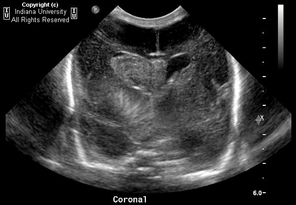
Hyperechogenic mass like hemorrhage into the right caudothalamic groove with ventricular extension, extension into the septum pellucidum, and resultant hydrocephalus; Grade 3 intraventricular germinal matrix hemorrhage.


Diagnosis: Germinal matrix hemorrhage


Key points

Risks for germinal matrix hemorrhage:
- Less than 32 weeks gestational age
- Under 1500g
- 20-25% risk in premature, low birth weight infant

Germinal matrix
- Proliferating cells; lines the periventricular spaces of the lateral ventricles and third ventricle
- Largest at 24-26 weeks, then involutes
- Begins involuting at the level of the third ventricle
- Involuted completely at 36 weeks

Mechanisms behind germinal matrix hemorrhage-
- Venous bleed
- From cardiovascular instability from lack of autoregulatory mechanism in the preterm infant
- Watershed bleed
- Nearly exclusively occurs in the first 1 wk of life, 50% on day 1
- Identifying and grading germinal matrix hemorrhage
- Blood is always as dense as choroid plexus
- Classic hemorrhage in the caudothalamic groove looks like a mass. May extend into the ventricles

Classification:
- Grade 1: Classic case caudothalamic groove looks like a mass
- Grade 2: Intraventricular extension, usually no long term sequelae
- Grade 3: Dilated ventricles
- Grade 4: Parenchyma, actually from venous hemorrhagic infarct

martedì 24 agosto 2010

Non-hemorrhagic pituitary apoplexy


Findings

A mixed signal (mildly hyper intense on T1-weighted and mixed hyper/hypo intense on T2 weighted sequences) lobular mass with thin rim-enhancement arises from the sella, extending superiorly above the diaphragm sellae (“snowman sign”), deviating the optic chiasm upwards. There is no diffusion restriction, and no susceptibility artifact (on B0 DWI sequences) to suggest hemorrhage. The clivus, and bony sella are intact. A thick rim of dural enhancement dorsal to the clivus is noted.


Diagnosis: Non-hemorrhagic pituitary apoplexy (with underlying macroadenoma)


Key points


Pituitary apoplexy (PA) (e.g. "necrosis") refers to hemorrhage or infarction of the pituitary.
Commonly there is a pre-existing macroadenoma (65-90%).
Acute clinical syndrome includes HA, visual defects/ophthalmoplegia, AMS, and variable endocrine deficiencies.
Acute imaging features show enlarged gland (T1/T2 isointense) with peripheral enhancement, with or without hemorrhage.
Subacutely – gland T1/T2 hyper intense, and chronically – appears as "empty sella" (filled with CSF) with T1 hypo / T2 hyper signal
T2*GRE sequence is very sensitive in looking for hemorrhagic products.
Sometimes there is subarachnoid hemorrhage.
Thickening/enhancement of adjacent dura (50%) and sphenoid mucosa (80%)
Differential diagnosis includes: Pituitary macroadenoma (non-hemorrhagic), craniopharyngioma, Rathke's cleft cyst, pituitary abscess, primary intrapituitary hemorrhage, or giant thrombosed intrasellar aneurysm.
Patients often suffer from long-term pituitary hormonal insufficiency.
Treatment of PA includes: Steroids, fluid/electrolyte replacement, and sometimes surgical decompression.


Discussion of disease

Pituitary adenomas are benign, slow-growing tumors which arise from the adenohypophysis, classified as either micro (< 10mm, 40 %) or macro (> 10 mm, 60%). However, occasionally leptomeningeal metastases can be seen. Patients present with indolent onset of headache, bitemporal hemianopsia (from optic chiasm compression), endocrinologic symptoms.

Pituitary apoplexy (PA), or pituitary necrosis, is usually caused by hemorrhage or infarction of the gland. There can, however, be bland necrosis (without either). The clinical syndrome is of somewhat acute onset with headache, visual defects/ophthalmoplegia, altered mental status, and variable endocrine deficiencies. Pre-existing macroadenoma is very common (65-90%). There is often associated subarachnoid hemorrhage. Thickening / enhancement of the adjacent dura and sphenoid sinus mucosa is very common. Sheehan’s syndrome is a rare peri-partum or post-partum (can be up to 15-20 yrs after index pregnancy) infarction of the pituitary with loss of anterior pituitary hormonal function. Long-term pituitary insufficiency is very common after PA. Treatment may include steroids, fluid/electrolyte replacement, or even surgical decompression.


Radiologic overview

CT (macroadenoma) – isodense to grey matter; fill and expand the sella turcica. CT is excellent for evaluating for sphenoid sinus invasion / bony destruction. Hemorrhage 10%, rarely calcify; pituitary apoplexy appears as hyper dense (acute), or as an "empty sella" if chronic, and very characteristic rim-enhancement.

MR – sellar mass without separate identifiable gland (mass is the gland) with "figure of 8" or "snowman" configuration (coronal), from constriction is caused by diaphragma sellae. Pituitary apoplexy has similar findings, with signal changes based presence/age of hemorrhage. Chronic PA will appear as an "empty sella" (T1 hypo, T2 hyper = filled with CSF). There is thickening / enhancement of adjacent dura in 50% of cases (seen in the presented case), and thickening / enhancement of sphenoid sinus mucosa 80% of the time.


lunedì 26 luglio 2010

Chronic progressive external ophthalmoplegia


Findings

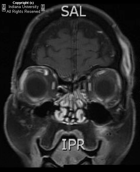
The extraocular muscles are atrophic and to some degree show fatty replacement. No abnormal enhancement is present. The globes are intact bilaterally. The intraorbital fat appears slightly increased. There is bilateral orbital proptosis with both globes anterior to the interzygomatic line.


Diagnosis: Chronic progressive external ophthalmoplegia


Key points

Chronic progressive external ophthalmoplegia (CPEO) is characterized by slowly progressive paralysis of the extraocular muscles.
Mitochondrial myopathy usually associated with skeletal muscle weakness.
Presents with bilateral, symmetrical, progressive ptosis, followed by ophthalmoparesis months to years later.

Kearns-Sayre syndrome: Related mitochondrial myopathy with CPEO, onset before age 20 years, pigmentary retinopathy, and at least one of the following: cardiac conduction defects, CSF protein of greater than 100 mg/dL, and/or cerebellar syndrome.
KSS can include mental retardation, hearing loss, seizures, short stature, delayed puberty, and various endocrine disorders.
Frequency: Rare. Males=Females.

Imaging studies: MRI, CT, and ultrasound may show thin, symmetrical extraocular muscles in CPEO, in contrast to enlarged extraocular muscles sometimes seen with Graves disease.
Those with KSS and CPEO display a wide spectrum of MRI findings, including normal brain, diffuse atrophy, and T2 prolongation in subcortical cerebral white matter, cerebellar white matter, globi pallidi, thalami, and substantia nigra.
Diagnosis: Muscle biopsy is definitive test but PCR also shown to be conclusive.