Visualizzazione post con etichetta Neoplasm. Mostra tutti i post

martedì 14 dicembre 2010

Myxopapillary ependymoma


Findings

Figure 1: Sagittal T1-weighted images reveals an isointense lobulated intradural mass at the level of the conus medullaris.
Figure 2: Sagittal T2-weighted images shows a hyperintense lobulated intradural mass extending from T11 through L2 with numerous small flow voids.
Figure 3: Sagittal T1 post-contrast images demonstrates intense enhancement of the intradural mass centered around the conus.


Diagnosis: Myxopapillary ependymoma


Myxopapillary ependymoma is a slow-growing tumor arising from the ependymal cells of the filum terminale. These tumors compromise 13% of all spinal ependymomas, and they occur almost exclusively in the conus, filum terminale, and cauda equina although extradural occurence in the sacrum and presacral region has also been reported.

The lesions tend to span two to four vertebral segments, and appear as a well-circumscribed intradural masses. In most cases the tumor is intrinsic to the conus medullaris but this is often difficult to recognize on imaging as the bulk of the mass is extramedullary. Typical MR characteristics include T1 isointensity, T2 hyperintensity, and avid enhancement on post-contrast images. As these tumors are prone to hemorrhage, a hypointensity at the tumor margin is often seen indicative of hemosiderin. Calcification and cyst formation within the mass are not infrequent.

On radiography and CT, vertebral changes can be seen which include widened interpediculate distance, thinned pedicles, posterior vertebral scalloping, and intervertebral foraminal widening due to tumor extension.

They are more common in males (M:F=2:1) with a mean age of 35 at diagnosis. Clinically, they present with back pain, paraparesis, radiculopathy, and occasionally bowel and bladder dysfunction. Because these symptoms can mimic those of disc herniation, there is often a delay in diagnosis. Treatment consists of surgical resection, and the prognosis is excellent with complete resection. Leptomeningeal seeding metastasis in myxopapillary variety is not as frequent as it is in classic spinal cord ependymomas and associated with poorer prognosis when present. Radiotheraphy after surgery improves outcome.

lunedì 29 novembre 2010

Intraventricular oligodendroglioma


Findings:

Figure 1: Non-contrast CT shows a large rounded mass in the lateral ventricles with intermediate density and large foci of calcification within it. This is causing significant hydrocephalus with enlargement of the third ventricle.


Intraventricular oligodendroglioma


Oligodentrogliomas are well differentiated, slowly growing but diffusely infiltrating cortical and subcortical tumors. Although most of them involve the frontal and frontotemporal cortex, a small proportion of them are seen in the ventricular walls (1-10%), cerebellum and exceedingly rarely within the brainstem, spinal cord and leptomeninges. No age is exempt and the peak incidence is in the 4th or 5th decades. It usually have a long standing history of symptoms and the most common being seizures and headaches.

Intraventricular oligodentrogliomas can present with hydrocephalus. Pathologically, they are well defined, grayish-pink soft unencapsulated mass and calcification is extremely common. Focal cystic degeneration and hemorrhage are frequent findings. CSF seeding is uncommon. Histopathologically, these are moderately cellular tumors with occasional mitosis. Perinuclear halos or the "fried egg artifact" is a distinctive feature of oligodentroglioma. Majority of the "intraventricular oligodentrogliomas" described in the literature are central neurocytomas and immunohistochemistry helps to distinguish them.

On CT, oligodentrogliomas usually have mixed density with nodular, clumped or linear tumoral calcification seen in majority (50-90%). Cystic degeneration is common. Intratumoral hemorrhage and edema is uncommon. The hemispheric lesions may expand, remodel or erode the calvarium. Enhancement is variable. On MR, oligodentrogliomas are typically heterogeneously hypointense or isointense to grey matter on T1 and heterogeneously hyperintense on T2 and FLAIR due to calcification, cystic change and hemorrhage. Nearly 50% show heterogeneous enhancement following contrast administration. Areas of calcification and hemorrhage demonstrate "blooming" on gradient echo MR sequences. No diffusion restriction on DWI.

Surgical resection is the primary treatment of choice. Radiation therapy and chemotherapy is reserved for recurrent tumors. Local recurrence is common and hence regular surveillance is recommended.

venerdì 26 novembre 2010

Ewing sarcoma of the occipital bone


Findings

Figure 1: Unenhanced CT shows a heterogeneous attenuation mass with cystic spaces in the posterior fossa.
Figure 2: Intense enhancement is noted after contrast administration.
Figure 3: Bone window section showing permeative destruction of the left occipital bone.
Figure 4: Axial T1 weighted image showing extra- axial mass with multiple cystic spaces.
Figure 5: Coronal T2 image showing the extra-axial origin clearly with mass effect on the cerebellum. The cystic spaces appear hyperintense.
Figure 6: Axial T1 weighted image showing intense enhancement of the mass.


Diagnosis: Ewing sarcoma of the occipital bone


Ewing sarcoma is a small round-cell tumor arising from mesenchymal cells. These tumors affect children and young adults in the age group of 5-15 years. The long bones, flat bones like the scapula and the vertebrae are the most common sites. Primary Ewing sarcoma affecting the calvarium is extremely rare, making just 1% of the cases. In the skull, the tumor more often arises from the frontal and parietal bones and less common locations include ethmoid, temporal and occipital bones.

CT scans (bone window) reveal poorly marginated permeative destructive lesion involving both inner and outer tables of the skull. The "onion peel" appearance typical of Ewing sarcoma in long bones is not seen commonly in the calvarium. The extra-dural soft tissue shows intense enhancement on contrast administration.

MR imaging provides better soft tissue delineation of these tumors. The extra dural soft tissue appears hypointense on T1 weighted images while the cystic and necrotic areas appear hyperintense on T2 weighted images. Good contrast enhancement is noted.
The differential diagnosis should include rhabdomyosarcoma, metastatic neuroblastoma and lymphomas.

Treatment is surgery followed by chemotherapy and radiation.

venerdì 19 novembre 2010

Malignant melanoma of the uvea


Findings

Thin section axial fat-suppressed fast spin-echo T2-weighted imaging
Figure 1 and Figure 2 show a V-shaped retinal detachment pointing toward the optic nerve. There is increased T1 signal posterior to the detachment suggesting hemorrhage.

Thin section axial fat-suppressed T1-weighted imaging
Figure 3 and Figure 4 show a mass arising from the nasal aspect of the right ocular globe. There is a collar button configuration to this mass, which assumes the typical configuration strongly suggesting elevation of Bruch’s membrane. The mass is near isointense to muscle on precontrast T1 and T2 weighted images. This mass extends towards the vitreous but no extension beyond the sclera is identified.

Postcontrast fat-suppressed T1-weighted imaging
Figure 5 and Figure 6: The mass is near isointense to muscle on pre-contrast T1 and T2 weighted images and demonstrates prominent contrast enhancement.


Diagnosis: Malignant melanoma of the uvea


Uveal melanoma is the most frequent primary intraocular malignant tumor in adults. It is rare in children. Its importance is that it is the main intraocular disease that can be fatal in adults.
Uveal melanoma can erupt through Bruch's Membrane. When this occurs, they develop a characteristic collar button configuration that extends through the vitreous chamber.
Uveal melanomas have differing MR characteristics, depending on the amount of melanin, which has paramagnetic properties. Melanomas containing a lot of melanin will demonstrate T1 hyperintensity, and markedly decreased T2 signal. However, in amelanotic or slightly melanotic melanomas, the typical MR pattern is isointense on T1, and slightly hypointense on T2.

Uveal melanoma is the most frequent form of intraocular malignancy in adulthood (4). As the uvea is the most vascular region of the globe, it is a common site for primary and metastatic neoplasm. Uveal melanomas comprise 70% of malignant intraocular tumors. It affects approximately 5-7 out of 1,000,000 people (3). Uveal melanoma can occur in any of the three subdivisions of the uvea: the iris, ciliary body, and choroid.

Uveal melanomas start with a flat growth profile along the choroid. With progression, they become elevated, and frequently extend through Bruch's membrane, where they can track into the subretinal space. With spread through Bruch's membrane, the melanoma can have a "mushroom shape" or "collar button" appearance that extends through the vitreous chamber.

Uveal melanomas typically appear as a solid, well-defined mass on magnetic resonance imaging. Melanin is paramagnetic, so in melanomas containing a lot of melanin, there is increased T1 signal with markedly decreased T2 signal. This signal intensity pattern is pathognomonic for uveal melanoma, as there are no other intraocular lesions with this appearance. In low-melanin or amelanotic melanomas, Magnetic Resonance imaging is less specific, but typically shows isointense signal on T1-weight images and slightly hypointense signal on T2-weighted images. Uveal melanomas typically have moderate to strong contrast enhancement following administration of gadolinium.

B-mode ultrasound typically shows a rounded, hypoechoic, highly vascular lesion. Retinal elevation and vitreous hemorrhage can also be seen, as these are complications of uveal melanomas. Uveal melanomas on unenhanced computed tomography appear sharply marginated, hyperattenuating, and elevated.

Uveal melanomas also have a propensity to metastasize hematogenously, and do so most frequently to the liver. Uveal melanoma is the most common fatal intraocular disease in the adult population.

Optimal treatment for uveal melanomas is controversial and clinical trials are ongoing. Large melanomas, typically greater than 10-mm in thickness, are usually managed with enucleation. For medium sized melanomas, 3-mm to 10-mm thick, plaque brachytherapy and external-beam radiation therapy have been accepted as alternatives to enucleation. For small lesions, less than 3-mm, routine monitoring with ultrasound is recommended as these may represent benign choroid nevi. These small lesions may also be biopsied, with a positive result placing the small melanomas into the medium melanoma treatment category.

Prognosis is dependent on many factors. Increasing tumor pigmentation is associated with a less favorable prognosis. Additionally, increasing size, infiltration through Bruch's membrane, and retinal detachment are all associated with a poorer prognosis. In metastatic disease to the liver, the mean survival has been reported to be nine months.

lunedì 8 novembre 2010

Neuroblastoma metastases


Findings

Figure 1: Axial post gadolinium T1 weighted image showing solid enhancing parenchymal lesion in the right temporal lobe with dural and leptomeningeal disease.
Figure 2: Coronal post gadolinium image showing dural enhancement along the tentorium and leptomeningeal enhancement.
Figure 3: Axial susceptibility weighted imaging revealing the hemorrhagic nature of the lesion.


Diagnosis: Neuroblastoma metastases


Neuroblastoma metastatic to the central nervous system is extremely rare, and the reported incidence varies from 1% to 16% at recurrence. Paediatric tumors that metastasise to the brain, in order of frequency, include neuroblastoma, osteosarcoma, Ewing sarcoma, rhabdomyosarcoma and Wilm tumor.

Risk factors for developing intracranial metastases include lumbar puncture at diagnosis, ages 2 to 3 years, bone marrow involvement, and MYCN gene amplification. Newer chemotherapeutic agents with better activity fail to penetrate the blood-brain barrier, thus facilitating a sanctuary for tumor cells within the central nervous system. As a result, the metastases evolve and become extensive before becoming clinically evident. Metastatic spread of tumor cells to central nervous system may occur either via hematogenous or cerebrospinal fluid routes and involve the neuroparenchyma, leptomeninges or dura.

Neuroparenchymal metastases from neuroblastoma have varied appearances. They may be cystic lesions with calcified mural nodules. The wall and mural nodules show intense enhancement with contrast. Metastases may also be solid and hemorrhagic and show homogeneous enhancement. Gradient or susceptibility weighted imaging would help in detecting hemorrhagic components. Leptomeningeal and dural metastatic involvement if present indicates poor prognosis.

lunedì 1 novembre 2010

Retinoblastoma with recurrence following enucleation


Findings

Figure 1: Contrast enhanced CT scan shows a heterogeneously enhancing intra-ocular mass in the right eye with calcification.
Figure 2: Contrast enhanced CT image shows enlargement of the right optic nerve suggestive of orbital recurrence following enucleation. Note the presence of a prosthetic globe.


Diagnosis: Retinoblastoma with recurrence following enucleation


The differential diagnosis of leukocoria in a child includes retinoblastoma, Coat’s disease, toxocariasis, retinopathy of prematurity, optic nerve drusen, persistent hyperplastic primary vitreous and phthisis bulbi. With a normal sized globe and intra-ocular calcifications the differential narrows down to retinoblastoma, Coat’s disease and toxocariasis with retinoblastoma being the most common.

Most patients with unilateral, non-metastatic retinoblastoma can be cured with enucleation alone. In patients with histologic risk factors, adjuvant chemotherapy is recommended, with the addition of orbital radiation for patients with trans-scleral involvement or tumor present at the level of the cut end of the optic nerve. Patients with metastases require intensive chemotherapy and consolidation with autologous hematopoietic stem cell rescue.

Recurrent retinoblastoma or a second primary neoplasm may be observed during follow-up. Optic nerve invasion, scleral invasion and choriodal invasion have been regarded as risk factors for recurrent disease. In general recurrent tumors have a short latent period as compared to second primary neoplasms; however this may not always be true. Recurrent retinoblastoma after enucleation can occur in the contralateral eye (type A), in the ipsilateral orbit (type B) or extend from the orbit into the brain (type C)

The most common presentation of orbital recurrence is a clinical complaint such as eyelid swelling or chemosis, suggesting that clinicians should not attribute periocular or orbital symptoms during the first year after enucleation as a sign of a benign conjunctivitis or preseptal cellulitis. A problem with the ocular prosthesis is also a frequent mode of presentation in these patients, and extrusion or displacement of a previously satisfactory prosthesis should be considered a suspicious sign for possible tumour recurrence.

Calcification maybe seen within the recurrent tumors on unenhanced CT. Most recurrent tumors appear iso to hypointense to normal temporal muscle on T1 weighted images and show moderately high signal on T2 weighted images. Contrast enhanced scans, CT or MRI, show heterogeneous enhancement of the tumor.

Cases with recurrence following enucleation should be evaluated for distant metastasis. Treatment consists of systemic chemotherapy combined with other modalities like orbital radiotherapy and stem cell rescue, depending upon the extent of metastatic disease.

venerdì 22 ottobre 2010

Adenoid cystic carcinoma with perineural spread of tumor


Findings

There is a large mass centered on the left greater sphenoid wing, extending into the left sphenoid sinus and left pterygopalatine fossa. It also extends into the left middle cranial fossa, left cavernous sinus, and left Meckel's cave. The tumor has involved V3, and foramen ovale is markedly widened on the left. There tumor has extended along the GSPN to the geniculate ganglion, and from there it has involved the tympanic and intracanalicular segments of the 7th nerve. The mass is isointense to brain on T1-weighted images and hypo- to isointense on T2-weighted images, consistent with hypercellularity. There is moderate enhancement of the mass. No macroscopic flow voids are seen to suggest a highly vascular lesion.

Figure 1: A mass lesion involving the skull base and pterygopalatine fossa on the left is shown on this axial FIESTA image. Abnormal soft tissue is seen in the left IAC as compared to fluid in the right IAC. The normal right greater wing of the sphenoid bone is shown. On the left, the greater wing of the sphenoid has been destroyed by the mass.
Figure 2: The soft tissue intensity skull base mass is again shown on the left. Infiltration of fat in the L pterygopalatine fossa is present, as compared with normal bright fatty signal in the R PPF. Asymmetrical signal is again seen in the left vs right IAC.
Figure 3: Following injection of gadolinium, moderate homogeneous enhancement of the mass lesion is shown. Also appreciated is extension into the L sphenoid sinus and displacement of the left cavernous segment of the L internal carotid artery. Meckel’s cave on the L is obliterated. Abnormal enhancement in the left IAC and abnormal thickening and enhancement of the tympanic segment of the facial nerve are also shown. Subtle linear enhancement extends posteriorly from the dominant mass along the greater superficial petrosal nerve on the left.
Figure 4: A more superior post-gadolinium image more discretely defines enhancement and enlargement of V3 on the left, posterior to the main bulk of the tumor mass. Tumor is again seen to extend posteriorly along the GSPN to the geniculate ganglion.
Figure 5: Tumor is seen to involve and expand Meckel’s cave on the left, replacing the normal CSF signal with intermediate signal intensity of a highly cellular tumor. The unaffected Meckel’s cave on the right, filled with CSF, is shown for comparison. The tumor is confined to Meckel’s cave on this image and has not extended through the dura to involve the adjacent temporal lobe.
Figure 6: A more anterior coronal T2-weighted image shows extension of the tumor into the cavernous sinus on the left, adjacent to the flow void of the internal carotid artery. Marked thickening of the third or mandibular division of the trigeminal nerve is shown on the left. The mass has markedly expanded foramen ovale on the left. This image also demonstrates atrophy and mild T2 hyperintensity of the muscles of mastication on the left, due to V3 dysfunction and resultant subacute to chronic denervation change. The masticator muscles on the right have normal bulk.
Figure 7: The enhancing tumor mass involving Meckel’s cave and the cavernous sinus on the left is again shown. This image better demonstrates the unaffected foramen ovale on the right. Also demonstrated on this image is volume loss and diffuse mild enhancement of the left temporalis muscle as compared to the right, consistent with denervation change as previously discussed). Signal drop-off due to dental hardware and inhomogeneity of the magnetic field has resulted in artifact and poor fat suppression in the right masticator space.


Diagnosis: Adenoid cystic carcinoma with perineural spread of tumor


Adenoid cystic carcinomas have a high propensity for perineural invasion and extension.
The facial and trigeminal nerves are the cranial nerves most commonly involved by perineural spread of tumor.
The facial nerve may be invaded directly by tumors of the parotid gland, or the tumor may involve the trigeminal nerve and then extend along the greater superficial petrosal nerve to reach the facial nerve.
A second “five-to-seven” connection is the auriculotemporal nerve, which also provides an important route for perineural spread of tumor.
The proximal greater superficial petrosal nerve, geniculate ganglion, and tympanic segment of the facial nerve often show normal mild enhancement due to investment by a rich vascular plexus in these regions.
Characteristics of perineural extension of tumor include abnormal enhancement and enlargement of nerves, replacement of fat in neural foramina, and widening of neural foramina. Denervation changes in innervated muscles may also be observed.


Adenoid cystic carcinoma (ACC) is the second most common malignant salivary gland tumor after mucoepidermoid carcinoma. It can arise from either the major or minor salivary glands. It is the most common malignancy of the submandibular and sublingual glands. It most commonly presents as a painless enlarging mass, though the initial presentation may be due to perineural extension of tumor (pain or paresthesia, for example) if the tumor is in a deep location. ACC is associated with a high risk of distant metastases (most commonly to the lung), and these can occur 10-20 years after initial diagnosis and treatment of the primary lesion. ACC may spread through local or direct extension of the tumor, hematogenous and lymphatic dissemination, and perineural extension.

ACC has a high propensity for perineural invasion and extension. SCCs also have a high tendency to spread perineurally, and because they are the most common head and neck cancer, one will likely encounter more cases of perineural tumor spread from SCC than from ACC in practice. Other cancers of the head and neck, including melanomas, basal cell carcinomas, and mucoepidermoid carcinomas also extend perineurally, but less commonly. Branches of the facial and trigeminal nerves are most commonly involved as they innervate the cutaneous and mucosal surfaces of the head and neck, where most tumors arise, as well as the salivary glands. The facial nerve can be invaded directly when ACC occurs in the parotid gland, or the tumor may involve the trigeminal nerve and then extend along the greater superficial petrosal nerve to reach the facial nerve. An alternate route for cranial nerve 5 to cranial nerve 7 spread (or vice versa) is the auriculotemporal nerve, which is located posterior to the neck of the mandible.

The greater superficial petrosal nerve emerges from the geniculate ganglion of cranial nerve VII carrying sensory (from the soft palate mucosa) and parasympathetic fibers. It courses anteromedially through the temporal bone and emerges through the facial hiatus. It then travels underneath Meckel’s cave and combines with the deep petrosal nerve (carrying sympathetic fibers) to form the vidian nerve. The vidian nerve travels anteriorly to the pterygopalatine ganglion, where the parasympathetic fibers synapse before being distributed to the lacrimal gland and mucosal glands of the nasal and oral cavities. This nerve pathway is vulnerable to tumor infiltration and is a common pathway for perineural extension of tumor.

Portions of the facial nerve that are invested with a rich vascular plexus may normally enhance on MRI. These normally enhancing regions are the proximal greater superficial petrosal nerve, geniculate ganglion, and tympanic segment of the facial nerve; the labyrinthine and descending mastoid segments of the facial nerve may also show mild enhancement under normal circumstances. The more distal and anterior portions of the greater superficial petrosal nerve, however, are not invested by a vascular plexus and should not enhance with contrast on MRI. Similarly, the facial nerve in the IAC does not demonstrate any enhancement under normal conditions at 1.5T. Characteristics of perineural spread to the facial nerve include thickening and abnormally intense enhancement of nerve segments, as well as replacement of fat in neural foramina by tumor. Denervation changes in the muscles of facial expression may be observed, but these are often extremely subtle due the small size of the affected muscles. Due to ACC’s high propensity for perineural spread and high tendency to recur, it is important to regularly assess for perineural extension of these tumors when MR images are being interpreted.

giovedì 14 ottobre 2010


Findings

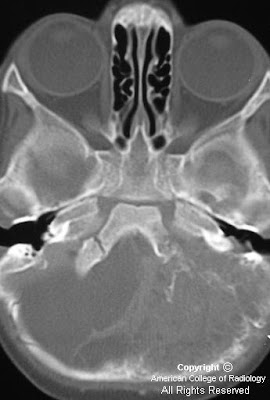
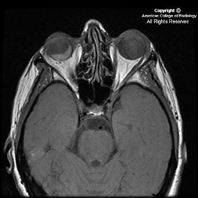
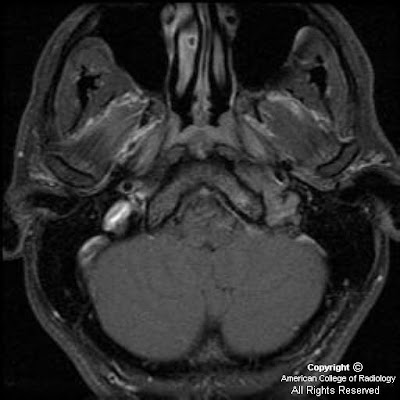
There is a poorly circumscribed jugular foramen mass with a permeative-sclerotic appearance of the involved bone with irregular margins and loss of the normal cortex with relative preservation of the bone density and architecture. There is centrifugal spread into the posterior fossa with a prominent dural tail and diffuse homogenous enhancement.


Jugular foramen meningioma


Jugular foramen meningiomas arise from the arachnoidal cap cells of the meninges which follow cranial nerves IX, X and XI into the jugular foramen. 5% of posterior fossa meningiomas arise in the jugular foramen and meningioma is the third most common jugular foramen mass after paraganglioma and schwannoma. If there are additional meningiomas or schwannomas, consider an association with neurofibromatosis II.

Meningiomas characteristically appear isointense to gray matter on both T1 and T2 weighted MR imaging but this appearance is variable. A relative T2 hypointensity is suggestive of a dense cellularity. There is an absence of high velocity flow voids within the mass which can help to distinguish from paraganglioma. There is dense, uniform contrast enhancement on MR and CT imaging which can help to identify a dural tail. Tumor matrix calcification is uncommon and internal hemorrhage is also rare.

Jugular foramen meningiomas typically infiltrate the surrounding skull base with relative preservation of the bone architecture. The margins of the jugular foramen are typically irregular with loss of the normal cortex. This combination of findings results in a permeative-sclerotic appearance and is different from the pemeative-destructive pattern of paragangliomas with erosion and infiltration without preservation of the underlying architecture or bone density. Additionally, schwannomas have a characteristic pattern of smooth enlargement without hyperostosis or permeative changes.

mercoledì 22 settembre 2010

Melanotic Neuroectodermal Tumor of Infancy (MNTI)


Findings

Large mass involving left side of face with invasion, mass effect, and extensive bony involvement. The mass does not appear to cross the midline. Enhancing focus on MR adjacent to the superior aspect of the falx and superior sagittal sinus. FDG-avid spine lesions.


Diagnosis: Pathology-proven melanotic neuroectodermal tumor of infancy (MNTI)


Discussion

MNTI is a rare osteolytic, pigmented neoplasm that typically affects the head and neck, predominantly the maxilla, of infants. It is typically considered a benign lesion, with only a few reported cases of metastatic disease reported in the literature. It usually presents in the first year of life. It may appear as a rapidly expanding, non-ulcerated, lightly pigmented, blue or black lesion on the anterior aspect of the maxilla. It may extend intraorally, cause bone destruction and dislodgement of teeth.

MNTI is considered to be of neural crest origin, and some patients will high urinary excretion of vanillylmandelic acid (VMA). The tumor is typically non-encapsulated, showing local invasion of bone. The histologic appearance is similar to other cells of neural crest origin, demonstrating small, round blue cells, as well as containing a moderately vascular fibrous background. Part of the lesion may contain large polygonal cells arranged in sheets that contain melanin. Immunohistochemistry and electron microscopy can aid in the final diagnosis. The few reported cases of malignant disease have noted increased numbers of mitoses per high-powered field.

Conventional radiography may demonstrate a well-circumscribed or ill-defined radiolucency, with destruction of bone as the lesion progresses. CT can delineate the extent of soft tissue involvement and osteolysis. Contrast-enhanced MRI can demonstrate soft tissue tumors with nonenhancing, heterogeneous tissue density and can also demonstrate osseous involvement. There may be foci of T1-hyper/T2-hypointensity secondary to melanin.

Surgical excision with partial maxillectomy and 5 mm margins are typically curative, with 10-15% recurrence rates. There are no standards of care for malignant disease.

Initial biopsy of this patient's facial lesion demonstrated a high mitotic index. After 2 surgical excisions and recurrence, the patient developed intracranial and spinal column metastases. The patient underwent multiple cycles of radiotherapy and chemotherapy. The intracranial lesion was never biopsied due to the precarious location, however it has regressed, and there appears to be a slight interval decrease in size of the soft tissue component of the maxillary lesion.

martedì 24 agosto 2010

Non-hemorrhagic pituitary apoplexy


Findings

A mixed signal (mildly hyper intense on T1-weighted and mixed hyper/hypo intense on T2 weighted sequences) lobular mass with thin rim-enhancement arises from the sella, extending superiorly above the diaphragm sellae (“snowman sign”), deviating the optic chiasm upwards. There is no diffusion restriction, and no susceptibility artifact (on B0 DWI sequences) to suggest hemorrhage. The clivus, and bony sella are intact. A thick rim of dural enhancement dorsal to the clivus is noted.


Diagnosis: Non-hemorrhagic pituitary apoplexy (with underlying macroadenoma)


Key points


Pituitary apoplexy (PA) (e.g. "necrosis") refers to hemorrhage or infarction of the pituitary.
Commonly there is a pre-existing macroadenoma (65-90%).
Acute clinical syndrome includes HA, visual defects/ophthalmoplegia, AMS, and variable endocrine deficiencies.
Acute imaging features show enlarged gland (T1/T2 isointense) with peripheral enhancement, with or without hemorrhage.
Subacutely – gland T1/T2 hyper intense, and chronically – appears as "empty sella" (filled with CSF) with T1 hypo / T2 hyper signal
T2*GRE sequence is very sensitive in looking for hemorrhagic products.
Sometimes there is subarachnoid hemorrhage.
Thickening/enhancement of adjacent dura (50%) and sphenoid mucosa (80%)
Differential diagnosis includes: Pituitary macroadenoma (non-hemorrhagic), craniopharyngioma, Rathke's cleft cyst, pituitary abscess, primary intrapituitary hemorrhage, or giant thrombosed intrasellar aneurysm.
Patients often suffer from long-term pituitary hormonal insufficiency.
Treatment of PA includes: Steroids, fluid/electrolyte replacement, and sometimes surgical decompression.


Discussion of disease

Pituitary adenomas are benign, slow-growing tumors which arise from the adenohypophysis, classified as either micro (< 10mm, 40 %) or macro (> 10 mm, 60%). However, occasionally leptomeningeal metastases can be seen. Patients present with indolent onset of headache, bitemporal hemianopsia (from optic chiasm compression), endocrinologic symptoms.

Pituitary apoplexy (PA), or pituitary necrosis, is usually caused by hemorrhage or infarction of the gland. There can, however, be bland necrosis (without either). The clinical syndrome is of somewhat acute onset with headache, visual defects/ophthalmoplegia, altered mental status, and variable endocrine deficiencies. Pre-existing macroadenoma is very common (65-90%). There is often associated subarachnoid hemorrhage. Thickening / enhancement of the adjacent dura and sphenoid sinus mucosa is very common. Sheehan’s syndrome is a rare peri-partum or post-partum (can be up to 15-20 yrs after index pregnancy) infarction of the pituitary with loss of anterior pituitary hormonal function. Long-term pituitary insufficiency is very common after PA. Treatment may include steroids, fluid/electrolyte replacement, or even surgical decompression.


Radiologic overview

CT (macroadenoma) – isodense to grey matter; fill and expand the sella turcica. CT is excellent for evaluating for sphenoid sinus invasion / bony destruction. Hemorrhage 10%, rarely calcify; pituitary apoplexy appears as hyper dense (acute), or as an "empty sella" if chronic, and very characteristic rim-enhancement.

MR – sellar mass without separate identifiable gland (mass is the gland) with "figure of 8" or "snowman" configuration (coronal), from constriction is caused by diaphragma sellae. Pituitary apoplexy has similar findings, with signal changes based presence/age of hemorrhage. Chronic PA will appear as an "empty sella" (T1 hypo, T2 hyper = filled with CSF). There is thickening / enhancement of adjacent dura in 50% of cases (seen in the presented case), and thickening / enhancement of sphenoid sinus mucosa 80% of the time.


mercoledì 18 agosto 2010

Spinal Cavernous Angiolipoma


Findings

Figure 1: Sagittal T1 image of the thoracic spine demonstrates a mixed signal soft tissue epidural mass posterior to the cord at the T4-T5 level causing posterior impression upon the thecal sac. There is also a T1-bright infiltrating lesion within the T5 vertebral body.
Figure 2 and Figure 3: Sagittal post-contrast T1 imaging shows an enhancing lesion with both bony and soft tissue components. This was localized to the right paraspinal soft tissues and there is compression of the cord. Bony involvement is seen in the vertebral bodies of T11 and L1 and in the posterior elements of multiple other thoracic levels.


Diagnosis: Spinal Cavernous Angiolipoma


Spinal angiolipomas are a rare entity that are histologically benign. These fatty tissue lesions have been quoted in the literature as representing 0.14-1.2% of all spinal axis tumors. Spinal angiolipomas are typically found in the thoracic vertebrae owing to the spine's regional variation in blood supply. The vast majority of epidural noninfiltrating angiolipomas are posterior or posterolateral in location. Patients are more commonly female and usually present in the fourth or fifth decades of life. Symptoms are slow and progressive over several months, with cord compression commonly seen at presentation. Other commonly seen symptoms include: back pain, sphincter dysfunction, progressive paraparesis, lower extremity paresthesias, and hyperreflexia. In rare cases, symptomatology may be acute due to factors such as spontaneous hemorrhage, venous thrombosis, or “steal” phenomenon.

Spinal angiolipomas are divided into two subtypes: the more common noninfiltrating type and the more aggressive (but still histologically benign) infiltrating type. An angiolipoma is termed infiltrating based on bony involvement.

Classically, spinal angiolipomas are T1 hypointense enhancing epidural lesions with or without bony involvement. The intraosseous component is usually heterogenous on T1 imaging. MR is frequently obtained due to patients symptoms of possible cord compression, and it is important to rule this entity out. Differential diagnosis would include hemangiomas, metastatic melanoma and lipid-rich metastases like liposarcoma and the clear cell variant of renal cell carcinoma. On CT, these are indistinguishable from hemangiomas. When viewed in bone windows, the typical “corduroy” appearance with linear streaks of high attenuation can be seen as well as the more stippled foci of increased density seen on axial imaging. To distinguish the two, a pathologic diagnosis is essential

The mainstay of treatment for patients with neurologic symptoms is cord decompression. When possible, surgical excision is preferred with an excellent prognosis for these patients. Heavy bleeding is typical, especially with the infiltrating type owing to the rich vascularity of these lesions. Typically, the neurosurgeon will have multiple blood products available at the time of surgery. For the case discussed above, the estimated blood loss at the time of surgery was 1800cc with 5 units of packed red blood cells administered.

martedì 10 agosto 2010

Pleomorphic Adenoma of the lacrimal gland


Findings

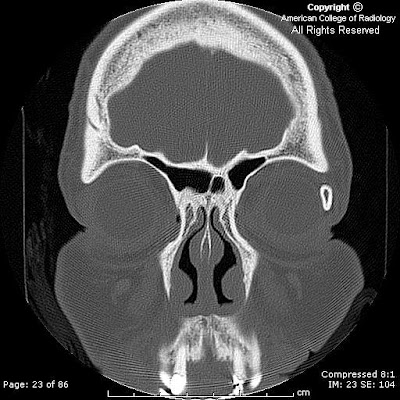
Completely ossified left lacrimal gland with no soft tissue mass, associated superficial inflammatory change or proptosis.


Diagnosis: Pleomorphic Adenoma of the lacrimal gland


Pleomorphic adenomas (benign mixed tumors) are the most common major salivary gland tumor often found in the parotid glands. When seen in minor salivary glands, the hard palate and the upper lip are the most common locations. It is, however, the most common epithelial tumor of the lacrimal gland. Approximately 50% of lacrimal masses are secondary to epithelial tumors; the other 50% are due to lymphoid and inflammatory causes. It will usually present as a palpable lacrimal fossa mass or proptosis. Other lacrimal gland tumors include: germ cell (dermoid and epidermoid), lymphoma/leukemia, metastasis, and sarcoma.
Pleomorphic adenomas are composed of epithelial and connective tissue components. Lesions may have a variable histology with growth patterns in sheets, strands, or islands of spindle and stellate cells with a myxoid configuration occasionally predominating. Cystic degeneration, squamous metaplasia, calcification or ossification may be observed in lacrimal gland pleomorphic adenomas. There may or may not be lytic remodeling or erosion of adjacent bone depending upon the chronicity of the tumor; adjacent bone abnormality does not necessarily confer malignancy. A small percentage of pleomorphic adenomas may undergo malignant transformation.