giovedì 28 gennaio 2010
Sinonasal undifferentiated carcinoma (SNUC)
Findings
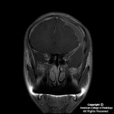
Heterogeneously enhancing mass centered at the junction of the left nasal cavity and ethmoid sinus has eroded through the cribriform plate and extends into the anterior cranial fossa. Sharp interface between the mass and the brain parenchyma, with mass-effect and vasogenic edema in the left frontal lobe. Left orbit medial wall has been eroded, and the extraconal mass has mass-effect on the left medial rectus with the fat plane between the mass and the muscle intact. Mass extends posteriorly to the margin of the left sphenoid sinus Bilateral frontal sinuses are fluid filled with the mass minimally extending into the inferior portion of the right frontal sinus. Ethmoid sinuses are completely occupied by the mass. Right maxillary and sphenoid sinuses are clear. Left maxillary sinus is almost completely fluid filled, and the left osteomeatal unit is completely obstructed by the mass.
Differential diagnosis:
- Esthesioneuroblastoma
- Squamous cell carcinoma
- Sinonasal undifferentiated carcinoma (SNUC)
- Sinonasal melanoma
- Meningioma
- Metastasis
- Lymphoma
Diagnosis: Sinonasal undifferentiated carcinoma (SNUC)
Key points
Aggressive
Rapidly growing
No histologic differentiating features
Presentation: usually older patients
Imaging appearance
Difficult to distinguish from ENB, SCCA, sinonasal adenocarcinoma
Differential diagnosis:
- Esthesioneuroblastoma (ENB)
- Squamous cell carcinoma (SCCA) of the nose
- Meningioma (specifically extracranial nasal meningioma)
- Sinonasal melanoma
- Lymphoma (specifically Non-Hodgkin lymphoma of the nose)
Esthesioneuroblastoma (ENB)
Neuroendocrine malignancy of neural crest origin
Arises from olfactory epithelial of superior nasal cavity
Presentation: adolescent or middle-aged patient with nasal obstruction with epistaxis
May bleed profusely on biopsy
Imaging appearance:
- Dumbbell mass
- Upper portion in intracranial fossa
- Lower portion in upper nasal cavity
- Waist at cribriform plate (blood-brain barrier)
- Calcifications within mass
- Cyst formation at tumor-brain interface
- Destruction of cribriform plate
- Homogenously enhance (CT or MR)
- When large, may have non-enhancing areas of necrosis
Treatment: Resection with radiotherapy
20% have nodal metastasis at presentation
Tendency to recur late
Squamous cell carcinoma (SCCA) of the nose
Malignant epithelial tumor growing from sinus surface epithelium
More common on maxillary antrum than nasal vault—only 30% primarily in nose
Presentation: Older male with sinusitis refractory to medical therapy
Exposures that increase risk:
- Nickel
- Thorotrast
- Possibly formaldehyde and asbestos
Imaging appearance:
- Typically aggressive antral mass
- Invasion and destruction of sinus walls
- Irregular margins
- Indistinguishable from esthesioneuroblastoma if begins high in nasal vault
- Enhancement: heterogenous, less than ENB, adenocarcinoma, melanoma
Treatment: Resection and XRT
With recurrence, 90% < 1 year survival
Meningioma (specifically extracranial nasal meningioma)
Presentation: Middle-aged, typically asymptomatic
Imaging appearance:
- Dural-based avidly enhancing mass
- Hyperostosis in adjacent skull base
- Peritumoral vasogenic edema
- Not associated with cyst formation at tumor-brain interface
Treatment is typically serial imaging, then resection, rarely XRT
Sinonasal melanoma
Neural crest cell malignancy arising from melanocytes in sinonasal mucosa
Presentation: Older patients
Imaging appearance:
- High T1 signal nasal cavity mass
- Can initially resemble nasal polyp on CT
Metastasis: lung, kidney, and breast
Lymphoma (specifically Non-Hodgkin lymphoma of the nose)
Malignant lymphoproliferative disorder arising from variety of immune cell types
B-cell type is most frequent in the paranasal sinuses and is less aggressive
Presentation:
- Nasal obstruction with sinusitis symptoms
- Constitutional symptoms—fever, fatigue, and weight loss
- Unilateral facial swelling
Imaging appearance:
- Dense on non-enhanced CT
- Less enhancement than ENB
- Rarely breaches skull base
Treatment:
- Primarily local XRT
- Chemotherapy if higher stage
mercoledì 27 gennaio 2010
Injured petrous internal carotid with proximal occlusion and clot from stab wound
Findings
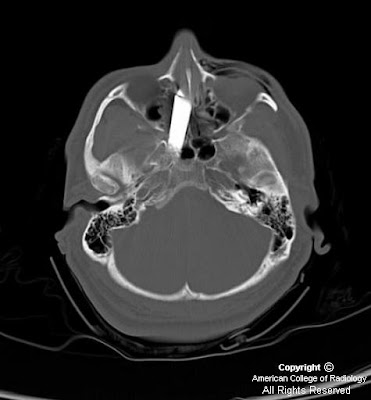
Figure 1: Transaxial CT scan of the brain. Knife entering the superolateral aspect of the left nasal cavity.
Figure 2, Figure 3, and Figure 4: Transaxial CT scan of the brain. Knife traversing the midline.
Figure 5: Transaxial CT scan of the brain. Knife traverses the carotid canal with tip at the level of the internal auditory canal.
Figure 6: Transaxial CT scan of the brain. Postoperative pneumocephalus and posttraumatic infarction in the distribution of the right middle cerebral artery. Knife has been removed.
Figure 7: Angiogram of the right internal carotid artery in an oblique projection. Knife tip in close proximity to the right internal carotid artery with little flow seen intracranially. Spasm noted at the catheter tip in the internal carotid artery.
Figure 8: Angiogram of the right internal carotid artery in an AP projection. The knife traverses the midline with the knife tip in the right carotid canal.
Diagnosis: Injured petrous internal carotid with proximal occlusion and clot from stab wound
Gunshot wounds to the head have become the leading or second leading cause of head injury in many cities in the United States; penetrating head injuries can also be the result of stab wounds, and motor vehicle or occupational accidents. Knives are the most frequent weapon used in stab wounds to the cranium although bizarre craniocerebral-perforating injuries have been reported that were caused by nails, metal poles, ice picks, keys, pencils, crochet needles, chopsticks, and power drills. A mortality rate of 17% has been reported for cranial stab wounds, mostly related to vascular injury and massive intracerebral hematomas. The orbital surfaces and squamous portions of the temporal bones are the most frequently affected since these bones are thin and easily traversed. The morbidity from stab wounds to the squamous portion of the temporal bone is more severe due to the short distance to the deep brain stem and vascular structures.
CT scans are vital in the work up of patients with penetrating head injuries since they do show the extent of intracranial injury and fracture and rule out the presence of radiopaque foreign bodies. CT angiography is limited in the presence of metallic foreign bodies because of artifact; in these cases, conventional angiography is important in ruling out vascular injury and preoperative planning. Potential signs of vascular injury include transection, pseudoaneurysm and arterial-venous fistula. Occlusion is a sign, albeit a nonspecific sign, of an underlying vascular injury. In the case of a vascular injury, injection of the contralateral artery should be done to demonstrate crossfilling via collateral circulation in the circle of Willis prior to intervention. Cerebral vasospasm is another noteworthy finding since it can portend a poor outcome.
Patients with penetrating head injuries are often taken to surgery to remove hematomas which may be causing mass effect, to remove dead brain tissue to prevent further swelling and necrosis, and to control active bleeding. Metallic foreign bodies are often removed to prevent infection, the potential for fibroglial scarring with the ensuing development of epilepsy and intracranial migration. Knives should only be removed in the OR under direct visualization. Clot was noted in the proximal ICA below the presumed level of injury; coils were placed proximal to the clot to prevent migration of the clot. In the case of an obvious direct transection, coils may also be used to prevent exsanguinations when the knife is removed. Occlusion balloons can also be used.
Etichette:
ACR,
Head - Neck,
Neuro,
Trauma,
Vascular
lunedì 25 gennaio 2010
Sialolithiasis with sialadenitis
Findings
The right parotid duct is markedly dilated. There is an obstructing calculus in the distal duct. There is inflammation in the right buccal space, the right masseter space, and in the right parotid gland. There is also a large non-obstructive stone in the right submandibular gland which was unchanged from a previous study.
Diagnosis: Sialolithiasis/sialadenitis
Discussion
Calculus disease is the most common benign condition to affect the salivary glands. The most common glands affected are the submandibular glands because secretions are more mucinous, viscous, and alkaline. Additionally, the duct drains uphill, making stasis more likely. Parotid glands are affected less frequently, and sublingual and minor salivary gland calculi are very uncommon. Patients generally have painful glands, worsened with chewing foods that precipitate salivation. Treatment is by administration of solutions that increase salivation to promote passing of the calculus. Transoral resection can be performed for calculi close to the ampulla. Resection of the gland may be used for proximal, glandular and/or recurrent sialoliths.
Radiologic overview
Imaging can be performed with plain radiographs, CT, and MRI. Most calculi are radio dense, but approximately 20% may not be visible on plain radiographs.
CT is more sensitive for detection of calcification than plain radiographs, and is also useful for detecting inflammatory masses and abscess formation.
In the case of non-radiopaque calculi, conventional sialography can be performed by cannulating the salivary duct and injecting contrast. The presence of a calculus is inferred by the presence of an intraluminal filling defect.
A new technique is MR sialography which is similar to MR cholangiopancreatography. The technique utilizes either a single shot fast spin echo T2-weighted sequence to image the duct alone, or a high resolution 3D fast spin echo T2 sequence to image the gland and duct.
Sialography is useful to demonstrate ductal strictures after passage of the stone, and in the evaluation of chronic sialadenitis from autoimmune causes.
Etichette:
AuntMinnie,
Head - Neck,
Inflammatory
venerdì 22 gennaio 2010
Neurocysticercosis (Calcified stage)
Findings
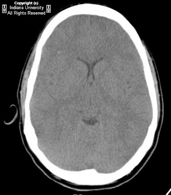
CT with contrast images showed a 7 mm calcified enhancing lesion within the posterior aspect of the left frontal lobe with a small amount of surrounding oedema. MRI images confirm the presence of a calcified ring enhancing lesion within the left posterior frontal lobe.
Differential diagnosis:
- Neurocysticercosis
- Tuberculoma
Diagnosis: Neurocysticercosis
Discussion
Cysticercosis is caused by larvae of the pork tape worm Taenia solium. Infestation occurs via the fecal-oral route. It is the most common parasitic infection involving the central nervous system in developing countries with 90% of patients present with seizures. Parenchymal cysticercosis is the most common type with lesions most commonly being peripherally distributed near the grey-white matter junction. The parasite goes through different stage of involution, each of which has different imaging features on CT and MRI.
These include the following:
- Vesical stage: CT shows hypodense non enhancing lesions. On MRI cysts follow CSF signal; T2 hyper intense scolex may be seen. No edema. Usually no enhancement.
- Colloidal stage: CT shows hypodense/isodense lesion with peripheral enhancement and perilesional edema. On MRI Cysts are hyperintense to CSF; surrounding edema, cyst wall enhances.
- Granular stage: CT shows nodular enhancing lesions. On MRI the cyst wall thickens and retracts, there is a decrease in edema, and there is nodular or ring enhancement.
- Calcified stage: When the parasite dies, nodular parenchymal calcifications are seen. These findings are best seen on CT.
Etichette:
AuntMinnie,
Infectious,
Neuro
giovedì 21 gennaio 2010
Klippel-Feil anomaly
Findings
There are multiple segmentation anomalies of the cervical spine, with fusion of C1-3.; left C6 hemivertebra.
Diagnosis: Klippel-Feil anomaly
Key points
Klippel-Feil anomaly refers to segmentation defects (congenital fusion) of the cervical spine.
Fusion of C2-3 and C5-6 most common
Frequently associated with Chiari malformation and syringohydromyelia.
Three categories:
- Group 1 = short, webbed neck, low hairline, complete lack of cervical segmentation
- Group 2 = isolated cervical segmentation defects
- Group 3 = segmentation defects affecting separate thoracic and/or lumbar level in addition of cervical involvement
Restricted motion at level of non-segmentation leads to accelerated disc degeneration.
50% of patients have at least partial dorsal splitting of the spinal cord. Defects in decussation of the corticospinal tracts can lead to "mirror movements" on physical exam.
Other CNS associations: occipital encephalocele, Dandy-Walker malformation, Duane syndrome, nasofrontal dermoid
Associated visceral anomalies: Sprengel deformity (20-30%, congenital elevation of the scapula, often with tethering omovertebral bone), cervical ribs, supranumary digits, tracheal and proximal bronchial stenosis, sickle sacrum, cleft palate, various renal anomalies.
Etichette:
AuntMinnie,
Malformations,
Neuro,
Pediatric,
Spine
venerdì 15 gennaio 2010
Lhermitte-Duclos disease
Findings
There is a small amount of increased signal on FLAIR and T2-weighted images with mild post contrast enhancement in the right cerebellar hemisphere. No mass effect is seen. Contrast images show an incidentally noted venous developmental anomaly on the right draining into the sylvian vein.
Differential diagnosis for the cerebellar lesion:
- Low-grade glioma
- Medulloblastoma
- Pilocytic astrocytoma
- Ganglioglioma
- Lhermitte-Duclos disease
- Cerebellar stroke
Diagnosis: Lhermitte-Duclos disease (dysplastic gangliocytoma of the cerebellum)
Key points
Lhermitte-Duclos disease (LDD) is a very rare benign cerebellar mass
Often associated with Cowden Syndrome–macrocephaly; benign breast, skin, and thyroid lesions; oral papillomas; GI tract hamartomas/polyps; cataracts; genitourinary neoplasia
Lesions grow very slowly or not at all
May cause obstructive hydrocephalus–presentation is usually vague neurologic symptoms related to hydrocephalus
Most commonly present at age 30-40, but can present from birth to age 60
Treatment–surgery for symptomatic patients
Rare recurrence, although complete resection is almost never possible
If confirmed LDD, no followup is needed unless patient is symptomatic
Radiologic appearance
- Well-defined cerebellar mass with striated or "gyriform" appearance
- CT–hypo dense cerebellar mass with hyper dense striations, rare enhancement
- MR
Striated mass with increased T2 and FLAIR signal +/- contrast enhancement
Bright on diffusion weighted images without signal decrease on ADC
MR Spectroscopy–decreased NAA, increased lactate, increased or normal choline
May use MRS to confirm LDD over glioma
Etichette:
AuntMinnie,
Neoplasm,
Neuro,
Systemic
giovedì 14 gennaio 2010
Arachnoid cyst with complicating intracystic and subdural hemorrhage
Findings
Figure 1 and Figure 2: Initial non-contrast (Figure 1) and contrast enhanced (Figure 2) axial head CT images. At the level of lateral ventricles an isoattenuating left sided subdural hematoma with mass effect and midline shift is demonstrated. Contrast enhanced imaging shows no abnormal enhancement within the subdural hematoma.
Figure 3 and Figure 4: More inferiorly, a hemorrhagic round extra-axial mass in anterior aspect of the left middle cranial fossa is seen. Bone window image (Figure 4) demonstrates focal thinning with endosteal scalloping of the overlying bone.
Figure 5: Diffusion weighted image through the lesion in the left middle cranial fossa shows no evidence of abnormally restricted diffusion of water. This confirms the lesion is not an epidermoid cyst as they characteristically show intense signal abnormality on DWI.
Figure 6, Figure 7, Figure 8, and Figure 9: The lesion demonstrated isointense signal on T2-weighted imaging (Figure 6). T1-weighted MR imaging (Figure 7) better demonstrates the lesion's complex nature. The lesion represents an arachnoid cyst which shows evidence of hemorrhage as demonstrated by the presence of T1 shortening. The surrounding subdural hematoma shows well demarcated prominent T1 shortening indicative of hemorrhage. Contrast enhanced axial T1-weighted MR imaging (Figure 8) shows no enhancement within the arachnoid cyst but does show enhancement of the dural membrane (arrow). The coronal contrast enhanced T1-weighted MR image (Figure 9) shows the arachnoid cyst, arachnoid membrane, and subdural hematoma.
Diagnosis: Arachnoid cyst with complicating intracystic and subdural hemorrhage
The widespread use of neuroimaging has increased incidental detection of arachnoid cysts. Arachnoid cysts constitute approximately 1% of all intracranial space-occupying lesions. Common locations include the middle cranial fossa, cerebral convexity, perisellar, retrocerebellar, cerebellopontine angle, and quadrigeminal plate cisterns. While some arachnoid cysts arise from post-inflammatory changes after trauma, intracranial hemorrhage, or infection, most are thought to be congenital. It is theorized that these cysts form due to an aberration in arachnoid development resulting in splitting or duplication of the membrane, a defect in condensation of the mesenchyme, or abnormalities of CSF flow. The exact means of arachnoid cyst genesis remains unclear.
On both CT and MRI scans arachnoid cysts are non-enhancing well-circumscribed extra-axial lesions with the same attenuation (on CT) or signal (on all MRI pulse sequences) as that of CSF. Bony erosion and remodeling, features suggestive of longstanding processes, are often associated with arachnoid cysts. These findings are seen in nearly half of cases, and are felt to be secondary to chronic fluid accumulation with transmitted CSF pulsations. Adjacent deformity or even hypoplasia of the subjacent brain may be present depending on the size and location of the cyst.
Rare complications of arachnoid cysts such as intracystic hemorrhage, subdural hematoma, or subdural hygroma may occur either after head injury or spontaneously. Rupture of the outer arachnoid cyst wall and surrounding fragile veins allows blood to accumulate within the cyst and/or subdural compartment. Intracystic hemorrhage can also be due to interruption of the highly vascular arachnoid cyst membrane or of the bridging veins traversing the cyst cavity. Fluid production by flat arachnoid cells lining the cyst walls may explain spontaneous cyst enlargement and hemorrhage if sufficient intracystic pressure is attained to breech the wall and disrupt the vasculature.
mercoledì 13 gennaio 2010
Diffuse axonal injury (DAI)
The patient was in a motor vehicle accident.
Findings
Non-contrast head CT demonstrates few punctuate hyper attenuating foci, including one near the gray-white junction in the right frontal lobe. MRI brain demonstrates multiple punctuate foci of T2/FLAIR prolongation, with corresponding restricted diffusion, within the subcortical frontal, temporal, and parietal lobes bilaterally. Most occur near the gray-white junction. The corpus callosum, deep gray matter structures, and brainstem are spared.
Differential diagnosis:
- Diffuse axonal injury.
- Multifocal nonhemorrhagic lesions:
Aging/microvascular ischemic disease
Demyelinating disease (MS, ADEM, etc.)
Marchiafava-Bignami syndrome
- Multifocal hemorrhagic lesions:
Cerebral amyloid angiopathy
Chronic hypertension
Cavernous malformation
Hemorrhagic tumors
Diagnosis: Diffuse axonal injury
Key points
Clinical Presentation
- Usually occurs in setting of high-speed MVC
- Transient loss of consciousness and retrograde amnesia common in minor traumatic brain injury
- LOC at moment of impact seen in moderate/severe TBI
Pathophysiology:
- Does not require direct impact to skull
- Occurs due to shear forces from differential acceleration/deceleration and rotational forces
- Cortex moves at different speed relative to underlying deep brain structures, creating axonal stretching.
- Non-traumatically disrupted axons are also damaged by traumatic depolarization, ion fluxes, and ultimately toxic edema.
50-80% of initial CT studies appear normal.
Hyper dense petechial hemorrhage, especially near gray-white junctions, seen in approximately20% of cases.
MRI is most-sensitive routine study for detection, especially when GRE sequences are utilized.
Typical MRI Findings:
- T1WI: Usually normal. May see hyper intense hemorrhage between 3-14 days.
- T2WI: Hyper intense foci seen at expected locations (gray-white junctions, corpus callosum, brainstem, uncommonly deep gray matter, internal/external capsules, corona radiata). Hemorrhagic lesions appear hypo intense on T2WI.
- FLAIR: Both hyper intense and hypo intense lesions at expected locations.
- DWI: Hyper intense foci of restricted diffusion.
- T2 GRE: Most sensitive sequence. Hypo intense foci due to susceptibility from blood products.
Increasing severity of traumatic force correlates with deeper brain involvement.
Staging based on Adams and Gennarelli system:
- Stage 1: Involves gray-white junctions of frontal and temporal lobes (mild)
- Stage 2: Involves corpus callosum and lobar white matter (moderate)
- Stage 3: Involves midbrain and pons (severe)
martedì 12 gennaio 2010
Optic disc coloboma
Findings
A posterior protrusion of vitreous is seen in the left globe, at the insertion of the optic nerve head. There is no hemorrhage, retrobulbar colobomatous cyst, or optic nerve atrophy. The right globe is normal.
Differential diagnosis:
- Coloboma (chorioretinal or optic disc)
- Peripapillary staphyloma
- Morning glory disc anomaly (MGDA)
- Buphthalmos (ox eye)
Diagnosis: Optic disc coloboma
Key points
Coloboma (Greek koloboun, "to mutilate") is the result of incomplete closure of the choroidal fissure.
Funnel or cone-shaped protrusion from the posterior aspect of the globe, often involving the optic nerve head insertion.
Two general types: Optic disc coloboma (ODC = excavated/everted region involves the posterior globe at the optic disc insertion only). Chorioretinal colobomas are more broad-based, extending beyond the margins of the optic disc.
May be associated with large retrobulbar colobomatous cysts (apparently encapsulated outpouchings of vitreous) which may be larger than the actual globe, may cause significant proptosis, or even globe atrophy / microphthalmia.
Majority of colobomas are sporadic and unilateral.
Associated with midline craniocerebrofacial clefting, choanal atresia, basal (particularly sphenoidal) encephalocele, corpus callosal agenesis, olfactory hypoplasia, cardiac anomalies, retardation, genital hypoplasia, and ear anomalies.
Differential diagnosis:
- Peripapillary staphyloma – more diffuse / broad based than coloboma, must feature posterior uveoscleral thinning and lack enhancement.
- Morning glory disc anomaly – fundoscopic description of ODC. Funnel or square shaped posterior defect at the optic nerve head.
- Buphthalmos ("ox eye") – 2/2 congenital glaucoma. Assoc. with Marfan's, NF-1, Sturge-Weber. Entire globe enlarged.
Etichette:
AuntMinnie,
Malformations,
Ophtalmic
lunedì 11 gennaio 2010
Wallenberg’s syndrome
Additional clinical information: Complaints of dysphagia, right facial droop, slurred speech, left lower extremity weakness following “black-out” secondary to alcohol binge. No known trauma.
Findings
The MRA shows hypoplastic right vertebral artery with areas of stenosis proximally. Long segment occlusion of right vertebral artery from mid-cervical spine to origin of the basilar artery. The MRI shows restricted diffusion involving right lateral medulla consistent with acute ischemic infarct.
Diagnosis: Wallenberg’s syndrome
Acute infarct of the posterolateral right medulla likely secondary to occlusion of the lateral medullary segment of the right PICA Also known as lateral medullary syndrome / Wallenberg's syndrome / PICA syndrome
Key points
Vertebral artery dissection secondary to trauma is an important cause
Can occur spontaneously or after minor trauma
Several case reports of Wallenberg's syndrome following chiropractic manipulation
Hypertension and vascular disease can predispose (FMD, Marfan's, Collagen Vasc Dz.)
Typically occurs in young adults (<45yo); males>females
Best diagnostic clue for acute ischemia/infarction is diffusion restriction with correlating ADC map
Restricted diffusion improves accuracy to 95%
"Time is Brain"
Intravenous Recombinant Tissue Plasminogen Activator (rTPA) therapy window within 3Hr
Intra-arterial rTPA window within 6Hr
martedì 5 gennaio 2010
Primary CNS large B-cell lymphoma
Findings
Figure 1: Axial CT show hyperdense lesion of the splenium. The CT hyperdensity is due to the hypercellularity of lymphoma.
Figure 2: Axial T1 demonstrates a low-signal mass centered in the splenium of the corpus callosum.The lesion is surrounded by a considerable amount of edema.
Figure 3: Axial T2-FLAIR: the mass is low signal on T2 images which is typical of PCNSL. There is edema surrounding the lesion.
Figure 4, Figure 5, Figure 6: T1 post gadolinium axial and coronal images demonstrate avid and homogenous enhancement of the solid mass, centered in the splenium of the corpus callosum.
DWI (Figure 7) and corresponding ADC map (Figure 8) reveal a restriction of diffusion, compatible with the hypercellularity of lymphoma.
Diagnosis: Primary CNS large B-cell lymphoma (pathologically-proven)
PCNSL is classified as an extranodal non-Hodgkin lymphoma that originates within the CNS and is confined to it at the time of diagnosis. In 90% of cases PCNLS is a non-Hodgkin B-cell lymphoma, usually of the large cell type (WHO). PCNSL is the most frequent brain tumour in immunocompromised patients, such as AIDS, and transplant recipients, although with the introduction of HAART therapy, the incidence rate in the AIDS group declined. Meanwhile PCNSL affects with increasing frequency immunocompetent patients accounting for 3% of all primary brain tumors diagnosed in the United States from 2000-2004. Median age of occurence is 63 years in immunocompetent host and 35 years in immunocompromised host.
The most characteristic presentation of PCNSL is progressive focal symptoms, followed by nonspecific mental status or personality change and cognitive function decline. Immunocompetent patients have no clear risk factors for developing PCNSL and it is slightly more common in men. Patients with AIDS developing PCNSL generally have CD4+ counts less than 30/mm3.
PCNSL is generally an aggressive disease with a median survival of only a few months after resection alone and as long as 30 months with high-dose methotrexate-based chemotherapy.
PCNSL can present as a discrete or multifocal lesion; immunocompetent patients are more likely to have a solitary lesion. The majority of lesions are supratentorial, in the cerebral hemispheres. PCNSL are typically located in the periventricular white matter. The densely packed lymphoma cells and their high nuclear-cytoplasmic ratio account for the hyperdense appearance of PCNSL on CT, the isointensity/hypointensity relative to gray matter on T2 and the restriction of diffusion as seen in DWI/ADC images. These features help in distinguishing PCNSL from most brain tumors. Most lesions enhance after administration of contrast material and in the majority of cases the pattern of enhancement is homogenous.
Gallium-67 citrate scans have been used to evaluate neoplastic and inflammatory (including infectious) diseases. There are four imaging peaks used: 93,184,296, and 388 keV. The usual dosage used is 4-6mCi (148-222 MBq), however slightly higher doses (8-10mCi) may be used for better images. Imaging in the evaluation of neoplasms is performed 48 hours after administration of the radiotracer. The mechanism of neoplastic Gallium uptake may be mediated by transferrin proteins which form complexes with the radiotracer and are taken up by the malignant cells.
Gallium scans can be recognized by their “coarse” images. There is normal physiological uptake seen in the nasopharynx, bowel, liver, lacrimal glands, and the skeleton. Gallium-avid tumors include lymphoma, hepatocellular carcinoma, and lung cancer. The resolution of a Gallium scan is on the order of one centimeter. The sensitivity in detection of Hodgkin lymphoma is approximately 85-90%. However, with non-Hodgkin lymphoma, the sensitivity is based on the histological cell type and grade. Gallium scans may also be used in post-treatment lymphoma patients where a negative scan is evidence for successful treatment.
Iscriviti a:
Post (Atom)