martedì 28 settembre 2010

Neurovascular compression syndrome of VII-VIII cranial nerves


Findings

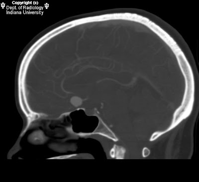
High resolution T2 images at the level of the internal auditory meatus demonstrate the facial and vestibulocochlear cranial nerves entering the internal auditory meatus. The facial nerve lies anterior while the eighth cranial nerves courses posteriorly. With the left side used for reference, a tortuous basilar artery is seen posteriorly displacing the normal course of the right vestibulocochlear/facial nerve complex. The presumed point of symptomatic compression is encountered posteriorly as the nerve complex “bends” over posterior aspect of the internal auditory meatus. Although sometimes seen in asymptomatic individuals, when findings are viewed in the appropriate clinical setting, NVCS should be raised as a diagnostic possibility.


Diagnosis: Dolichoectasia of the basilar artery causing right sided sensorineural hearing loss


Hearing loss can be characterized as either conductive or sensorineural based on clinical exam and audiometry. Conductive hearing loss involves an abnormality of the external auditory canal to the oval window and is best evaluated with high resolution CT as it is able to display the external auditory canal and middle ear structures, particularly the ossicles. Sensorineural deafness implies an abnormality of the inner ear, vestibulocochlear nerve, or its central components which are best evaluated with MRI.

Neurovascular compression syndrome (NVCS) refers to a group of disorders in which an aberrant or tortuous vessel causes nerve compression with subsequent hyperexcitation and neuropathy. Vascular compression syndrome has been described as a causative etiology for cranial nerves III, V, VII, VIII, and IX. Controversy exists, however, because of the normal intimate apposition of nerves and vasculature around the brainstem and the frequency with which it is seen in asymptomatic patients.

Vestibulocochlear NVCS is symptomatic vascular compression of cranial nerve VIII. Clinical symptoms are often non-specific including tinnitus, vertigo, and sensineural hearing loss. A recent article in the American Journal of Neuroradiology failed to reliably determine neurovascular compression as a cause of tinnitus although some authors maintain it may still be considered when presenting with so called "typewriter" tinnitus. In decreasing order of frequency, vessels indicated in NVCS include the anterior inferior cerebellar artery, posterior inferior cerebellar artery, and vertebral artery.

The following MRI classification system for neurovascular compression has been proposed to aid in surgical planning.
- Type I: Point compression where a limited segment of the nerve is in contact with the vessel.
- Type II: Longitudinal compression in which the nerve and vessel traverse parallel to each other.
- Type III: A vascular loop encircling the neve.
- Type IV: The nerve contour is deformed and/or thinned.

Definitive treatment involves retromastoid craniectomy and microvascular decompression in which a small synthetic sponge is interposed between the offending vascular structure and nerve.
giovedì 23 settembre 2010

Filar cyst


Findings

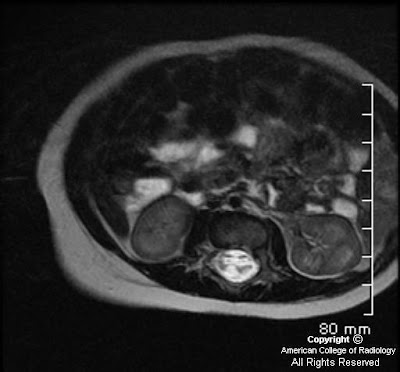
Figure 1: Axial T2-weighted image demonstrates a cystic structure in the filum terminale.
Figure 2 and Figure 3: Coronal T2-weighted images demonstrate a cystic structure in the proximal filum terminale.


Diagnosis: Filar cyst


The human spinal cord develops through three distinct stages: neurulation, canalization, and retrogressive differentiation. The conus medullaris, filum terminale, and cauda equine are mainly developed and formed in the retrogressive differentiation stage as the caudal cell mass regresses.

Cystic structures within the distal spinal cord, conus medullaris, and filum terminale are commonly seen on routine lumbosacral spine sonography in the neonates. These patients often present with abnormal laboratory values and/or external body features that are suggestive of underlying spinal dysraphism or neural tube defects. The diagnostic consideration of a cystic lesion in this region includes: syrinx, neoplasm (ependymoma or astrocytoma), persistent ventriculus terminalis in the conus medullaris, and filar cyst in the filum terminale of the cord.

Filar cysts are a relatively common entity in the neonates usually detected on screening lumbosacral sonograms. However, it has not been extensively reported in the literature. It is considered a normal variant when found as an isolated finding. The exact etiology of a filar cyst has not been reported. Literature has suggested that filar cysts are developmentally similar to the septum pellucidum and ventriculus terminalis, which can regress with age. On ultrasound, it is usually describes as an anechoic, cystic structure completely contained within the filum terminale. MRI of the lumbosacral spine can be obtained in cases that are questionable on ultrasound. Filar cyst follows the typical characteristic of a simple cyst in all sequences of an MR study. Ventriculus terminalis is a normal developmental variant described as a nonenhancing dilation of the ependyma-lined central canal at the level of the conus medullaris. Persistent ventriculus terminalis deserves special attention such that it is often used interchangeably with filar cyst in the literature, in the setting of cystic lesions seen in the distal lumbar spinal cord proximal to the conus medullaris. A cystic lesion in the absence of a solid component makes a neoplastic process less likely. A syrinx isolated to the distal spinal cord is also less common as it usually has a superior extension.

Treatment options are primarily based on patient's symptomatology. In asymptomatic neonates/infants, no further imaging is needed.

mercoledì 22 settembre 2010

Melanotic Neuroectodermal Tumor of Infancy (MNTI)


Findings

Large mass involving left side of face with invasion, mass effect, and extensive bony involvement. The mass does not appear to cross the midline. Enhancing focus on MR adjacent to the superior aspect of the falx and superior sagittal sinus. FDG-avid spine lesions.


Diagnosis: Pathology-proven melanotic neuroectodermal tumor of infancy (MNTI)


Discussion

MNTI is a rare osteolytic, pigmented neoplasm that typically affects the head and neck, predominantly the maxilla, of infants. It is typically considered a benign lesion, with only a few reported cases of metastatic disease reported in the literature. It usually presents in the first year of life. It may appear as a rapidly expanding, non-ulcerated, lightly pigmented, blue or black lesion on the anterior aspect of the maxilla. It may extend intraorally, cause bone destruction and dislodgement of teeth.

MNTI is considered to be of neural crest origin, and some patients will high urinary excretion of vanillylmandelic acid (VMA). The tumor is typically non-encapsulated, showing local invasion of bone. The histologic appearance is similar to other cells of neural crest origin, demonstrating small, round blue cells, as well as containing a moderately vascular fibrous background. Part of the lesion may contain large polygonal cells arranged in sheets that contain melanin. Immunohistochemistry and electron microscopy can aid in the final diagnosis. The few reported cases of malignant disease have noted increased numbers of mitoses per high-powered field.

Conventional radiography may demonstrate a well-circumscribed or ill-defined radiolucency, with destruction of bone as the lesion progresses. CT can delineate the extent of soft tissue involvement and osteolysis. Contrast-enhanced MRI can demonstrate soft tissue tumors with nonenhancing, heterogeneous tissue density and can also demonstrate osseous involvement. There may be foci of T1-hyper/T2-hypointensity secondary to melanin.

Surgical excision with partial maxillectomy and 5 mm margins are typically curative, with 10-15% recurrence rates. There are no standards of care for malignant disease.

Initial biopsy of this patient's facial lesion demonstrated a high mitotic index. After 2 surgical excisions and recurrence, the patient developed intracranial and spinal column metastases. The patient underwent multiple cycles of radiotherapy and chemotherapy. The intracranial lesion was never biopsied due to the precarious location, however it has regressed, and there appears to be a slight interval decrease in size of the soft tissue component of the maxillary lesion.

giovedì 16 settembre 2010

Wernicke’s encephalopathy


Findings

There is increased T2 signal and diffusion restriction in the thalami bilaterally. There is no contrast enhancement.


Diagnosis: Wernicke’s encephalopathy


Discussion

Wernicke's encephalopathy occurs with vitamin B1 (thiamine) deficiency. It is associated with malnutrition and is commonly seen in alcoholics. Wernicke's encephalopathy manifests itself as memory loss, ataxia and oculomotor dysfunction. Thiamine plays a vital role as a cofactor for several enzymes involved in carbohydrate metabolism. Without thiamine, the energy requirements of neuronal cells are not met resulting in cell death and neurologic dysfunction.

Wernicke's encephalopathy is reversible and if suspected, treatment should begin promptly. Parenteral thiamine should be administered promptly. The thiamine should be given prior to any glucose infusion. The addition of glucose in a thiamine deficient patient will exacerbate the encephalopathy. Patient's with thiamine deficiency are also likely to be hypomagnesemic and parenteral magnesium sulfate should also be administered.


Radiologic overview of the diagnosis

Wernicke's encephalopathy manifests itself as increased T2 signal in the medial thalami, hypothalamus and periaqueductal grey matter. There is associated diffusion restriction in affected areas. In chronic cases, there is atrophy of the mamillary bodies. The sensitivity of MR to diagnose Wernicke's is estimated at approximately 50% so the lack of these imaging findings, do not exclude the diagnosis of Wernicke's encephalopathy. CT is even worse than MR in the diagnosis of Wernicke's and should be used to rule out an acute intracranial process. Alcoholic induced Wernicke's will manifest the same above findings, but will also show superior vermian atrophy.

In this case, there is increased T2 signal and diffusion restriction in the medial thalami bilaterally. There is no contrast enhancement. The patient has lymphoma and a chronic history of vomiting. With this history, Wernicke's encephalopathy was thought to be the cause of the patient's altered mental status. The lack of contrast enhancement makes the diagnosis of lymphoma highly unlikely.

mercoledì 15 settembre 2010

Anterior communicating artery aneurysm


Findings

The head CT without contrast shows a region of hyper density along the anterior falx along the course of the anterior cerebral artery concerning for acute blood products. CT angiogram shows an anterior communicating artery aneurysm that splays both anterior cerebral arteries and points anteriorly No definite blood products within the suprasellar cistern are seen. There is no intraventricular hemorrhage.


Diagnosis: Anterior communicating artery (ACom) aneurysm (unruptured)


Key points

Aneurysm development and rupture risk reflect complex combination of inherited susceptibility and acquired mechanically-mediated vessel wall stresses4."
Associated with connective tissue disorders such as fibromuscular dysplasia, Ehlers-Danlos syndrome (type IV), ADPCKD
Familial intracranial aneurysms: Occur in clusters of 1st degree relatives

Location: 90% arise from circle of Willis
- 90% anterior circulation (Acom and internal carotid-com most common sites)
- 10 % posterior (Basilar artery bifurcation, PICA most common)
- 1-3% misc sites distal to COW (often traumatic, mycotic, oncotic)

Risk of rupture:
- Increased risk of rupture:
- Size
- Multilobed
- Apical "bleb"
- Length: neck aspect ratio >1.6

Giant aneurysms (>2.5cm) present both the risk of rupture and symptoms related to mass effect
15-20 % multiple
Estimated risk of rupture: 1-2% year cumulative for unruptured aneurysms

Demographics:
- F>M (especially with multiple aneurysms)
- Incidence: 1/100000 (<35yo); 44/100000 (>65yo)

Treatment: Cost-effective strategy for unruptured aneurysms
- Endovascular treatment was most cost effective for anterior circulation aneurysms 7-25 mm in size
- No treatment was most cost-effective for anterior circulation aneurysms < 7mm
- Surgical treatment was most cost effective for anterior circulation aneurysms > 25m
- Endovascular coiling has been accepted as effective treatment, though controversy still exists between surgical and endovascular therapy.

Long-term (10yr) followup of 1036 coiled aneurysms required retreatment in 7% and rebleeding in 0.5%

martedì 14 settembre 2010

Postictal Imaging Findings


46-year-old male presented to the emergency room after a first seizure. He has no significant or contributory past medical history.


Follow-up FLAIR imaging obtained approximately 5 weeks later.


Findings

Figure 1 and Figure 2: FLAIR and DWI images show abnormal increased signal in the left temporal lobe. The right temporal lobe is questionably involved. The differential diagnosis for these findings included, but is not limited to, neoplasm, infection, and postictal changes.
Figure 3: Follow-up FLAIR imaging obtained approximately 5 weeks later demonstrates resolution of these findings, consistent with postictal change.


Diagnosis: Postictal Imaging Findings


A seizure is "a sudden alteration of the CNS resulting from a paroxysmal high frequency or synchronous low frequency, high voltage electrical discharge". Imaging (CT or MRI) is indicated in cases of:
- New onset of seizure activity
- Change in pattern of previous seizure pattern
- Patients with focal neurological defects or altered mental status
- Prolonged postictal state, especially if associated with neurological defects

Following seizure activity, imaging is used to identify an underlying etiology. Differential possibilities include structural/anatomical abnormalities, space-occupying masses (primary or secondary brain neoplasms, abscesses), cerebrovascular accidents, transient ischemic attacks, hemorrhage, infectious processes (meningitis, encephalitis) venous thrombosis, and vasculitis. If performed shortly after the ictal event, CT and MR imaging may demonstrate findings that are secondary to the physiological mechanisms related to the seizure itself. These findings are most likely to occur following status epilepticus.

The mechanism and pathophysiology of these findings are unknown. Some theories are that the findings occur as a result of breakdown of the blood brain barrier. This results in transient focal brain edema, accounting for various imaging findings. Other theories propose arteriovenous shunting of blood during seizure activity that results in accumulation of toxic metabolites, ischemia, and acidosis. Most theories implicate ischemia and transient cytotoxic edema as the cause of brain changes.

Imaging findings are nonspecific and can overlap with those seen in other disease entities such as infarction/ischemia, venous thrombosis, vasculitis, infection, neoplasm, arterial thromboembolism and metabolic encephalopathy. History, presenting signs and symptoms, follow-up imaging and other relevant laboratory data can further narrow the differential diagnosis.

On CT, possible postictal imaging findings include:
- effacement of adjacent cortical sulci
- focal gyral edema
- decreased gyral attenuation
- mild to moderate gyral enhancement on contrast-enhanced images

On MRI, possible findings include:
- increased signal on T2WI (most common in the frontal and parietal lobes but also seen in the temporal and occipital lobes as well as other regions in the brain such as the hippocampus)
- corresponding hypointensity on T1WI
- abnormal contrast enhancement
- diffusion restriction and reduced ADC
- gyral swelling with effacement of adjacent sulci

Bilateral involvement is more common than unilateral. Lesions usually overlap arterial and watershed territories. In addition, lesions are usually in the cortex or subcortical white matter and spare the basal ganglia. Studies have shown leptomeningeal enhancement on post-contrast MRI. Follow-up imaging reveals complete or near-complete resolution of these findings.

It is important to recognize the various imaging findings that can be seen in the postictal period to avoid unnecessary biopsy and further workup such as angiography and biopsy. The postictal imaging appearance can be confused with other entities such as neoplasm, infection and infarction. The amount of time until resolution of these transient imaging findings is unclear. Studies have shown resolution of postictal changes in as low as 5 days. However, the majority of cases resolve over weeks - months. Patients should be re-imaged to confirm the transient nature of abnormal MR findings, usually within 4 - 6 weeks. It is important for patients to be re-imaged only after a seizure-free interval.

martedì 7 settembre 2010

Adult Onset Adrenoleukodystrophy


Findings

Figure 1 and Figure 2: Axial T2 weighted and FLAIR images demonstrate confluent symmetric hyperintensity in the peritrigonal parietoccipital deep white matter, compatible with adult onset X-ALD.


Diagnosis: Adult Onset Adrenoleukodystrophy


Adrenoleukodystrophy is an x-linked inherited disease involving the central nervous system and adrenal cortex. It results from an inability of peroxisomes to oxidize fatty acids. While seen most commonly in young males, it is also known to present in female heterozygotes. In adults, patients present with varying degrees of cognitive impairment, peripheral neuropathy, bladder and sexual dysfunction.

On imaging, "classic" adrenoleukodystrophy will demonstrate bilateral confluent hyperintense areas in the parietoccipital deep white matter on T2WI and FLAIR. If contrast is administered, it is common to see peripheral linear areas of enhancement with a “leading edge”. In AMN, T2 hyperintensity is also common in the spinal tract fibers of the pons, cerebral peduncles, and internal capsule.

Adult onset adrenoleukodystrophy has been broken down into four main subgroups. The first group is referred to as Pure AMN and is the most common subtype. Patients in this group present with thoracic spinal cord atrophy without any focal lesions and a normal brain MR. Clinically they present with spastic paraparesis and bladder dysfunction. The second group is referred to as AMN Type 1, in which MR will demonstrate T2 hyperintensity along the long tracts of the brain, such as the corticospinal, spinothalamic, and auditory pathways. AMN Type 2 demonstrates extension beyond the long tracts and crosses the corpus collosum. These patients deteriorate much more rapidly and this form most accurately fits this case presentation. The fourth type, Adult Cerebral ALD, results in severe lobar white matter involvement with marked cerebral atrophy.

It is important to consider adult onset ALD/AMN in the differential diagnosis of all adult demyelinating disorders, since it is commonly thought of as a pediatric diagnosis and patients are frequently misdiagnosed with multiple sclerosis or ALS. Early recognition can affect prognosis and delay rapidly progressive neurological deterioration.

Confirmation of the diagnosis can be obtained by various laboratory examinations. Patients will demonstrate elevated concentrations of plasma VLCF. Adrenal function testing will show diminished levels of ACTH. Genetic testing can clinch the diagnosis with a mutation of the ABCD1 locus. Treatment subsequently consists of dietary restriction, Lorenzo’s oil, and statins.

mercoledì 1 settembre 2010

Bilateral ectopia lentis


Findings

Figure 1: Axial noncontrast CT of the head at the level of the orbits demonstrates posterior dislocation of both lenses, which now rest dependently in the vitreous. The etiology in this particular patient was repetitive trauma from serial falls.


Diagnosis: Bilateral ectopia lentis


The crystalline lens of the eye is designed to refract the light entering the iris and project/focus it onto the retina. The lens itself contains no vasculature, nerves, or connective tissue. It sits behind the iris and the front of the lens is in contact with the aqueous fluid of the anterior chamber while the posterior surface of the lens is in contact with the vitreous. The lens is held in place by zonular fibers, otherwise known as suspensory ligaments. These fibers connect to the cilliary body around the circumference of the lens.

Subluxation (partial dislocation) or luxation (complete dislocation) of the crystalline lens, otherwise known as ectopia lentis, is caused by dysfunction or disruption of these zonular fibers. Trauma is the most common cause of this disorder. The absence of a traumatic history should prompt consideration of hereditary causes of zonular fiber dysfunction; predisposing conditions include Marfan syndrome, homocystinurea, tertiary syphilis, and Weil-Marchesani syndrome.

Patients will complain of monocular diplopia, markedly decreased visual acuity in the affected eye(s), and/or poor near vision.

Treatment is determined by lens position, with anterior chamber dislocation often being a surgical emergency. As the aqueous humor of the eye flows in the anterior chamber, around the iris from the cilliary body to the canal of Schlemm, this route can become acutely obstructed with anterior dislocation leading to acute glaucoma. The cornea and iris are also at risk for damage. Posterior dislocation may be treated conservatively depending on lens position, but may also lead to uveitis or glaucoma in some cases.