venerdì 27 giugno 2008
Submandibular sialadenitis
Findings
There is diffuse soft tissue and glandular edema in the left sublingual and submandibular region. There is a large calcification in the left sublingual region representing a sialolith.
Differential diagnosis:
- Submandibular sialadenitis
- Submandibular carcinoma
- Malignant lymph node
Diagnosis: Submandibular sialadenitis
Key points
Presentation
- Unilateral painful submandibular gland (SMG) swelling with eating or salivation
- Painless mass
Treatment: Removal of submandibular gland in some cases.
Radiologic Evaluation: CT with contrast.
A sialolith is a calculus found in the salivary duct
- 85% are found in the submandibular duct, Wharton's duct
- 10% are found in the parotid duct
- 5% in the sublingual duct
Acute sialadenitis:
- Unilateral enhancing enlarged SMG with dilated duct behind the calculus
- Intraglandular ductal dilatation
- SMG cellulitis and/or myositis
Chronic sialadenitis:
- SMG small
- Fatty infiltration
- Intraductal calculus
Etichette:
AuntMinnie,
Head - Neck,
Inflammatory
giovedì 26 giugno 2008
Cystic hygroma (lymphangioma)
Findings
Fetal MRI reveals a large mass extending from the anterior palate to the right supraclavicular region. It involves the anterior right neck of the fetus, as well as the right masticator space, parapharyngeal space, carotid, and parotid spaces. The airway is not well visualized, presumably due to airway compression.
Diagnosis: Cystic hygroma (lymphangioma)
Key points
Incidence is 1:5000.
Caused by absent or abnormal connections between the lymphatic and venous systems, or from sequestration of embryonic lymphatic tissue.
50% association with chromosomal abnormalities.
Often missed in the first trimester, as the most common initial finding of increased nuchal lucency is difficult to detect.
In the second trimester, is most commonly identified on ultrasound as a cystic mass with multiple thin walled septations, frequently with a posterior midline band, representing the nuchal ligament.
Important to identify anterior cervical soft tissue involvement because of increased morbidity from respiratory compromise.
Important to differentiate from posterior encephalocele, which is associated with an underlying skull defect, and cervical myelomeningocele, which is associated with an underlying vertebral defect.
MRI is usually not necessary in diagnosis, although it is helpful for surgical planning. In this case, Cesarean section with head delivery was performed at 40 weeks gestation to maintain uteroplacental circulation while tracheostomy was created because of anterior neck involvement and airway compression. Bulk resection was performed 3 weeks later.
Etichette:
AuntMinnie,
Head - Neck,
Malformations,
Pediatric,
Vascular
lunedì 23 giugno 2008
Silent sinus syndrome
Findings
Two coronal CT images (Figure 1 and Figure 2) and single axial CT image (Figure 3 ) demonstrate right maxillary sinus volume loss when compared to the left. There is inward retraction of the sinus walls (Figure 1, Figure 2, and Figure 3), increase in the size of the middle meatus (Figure 1 and Figure 2) and expansion of the retroantral fat (Figure 3). There is lateralization of the uncinate process and middle turbinate. This patient had concomitant right ethmoid air cell and frontal sinus opacification.
Diagnosis: Silent sinus syndrome
Silent sinus syndrome, or maxillary sinus atelectasis, is characterized by volume loss of the maxillary sinus after infundibular occlusion. It most often presents in the third through fifth decades of life with painless enophthalmos, facial asymmetry, and/or diplopia. Rarely do patients complain of symptoms of sinusitis.
Imaging findings of silent sinus syndrome are characteristic. There is maxillary sinus volume loss with inward retraction of the sinus walls and sinus opacification. The ethmoidal infundibulum is occluded, usually due to opposition of the uncinate process against the inferomedial orbit. The sinus volume loss accounts for the corresponding increase in ipsilateral orbital volume and size of the middle meatus.
The pathophysiology remains unclear. It is felt to be an acquired condition caused by chronic maxillary sinus obstruction and hypoventilation leading to negative intrasinus pressures. Chronic inflammation leads to osteolysis and thinning of the sinus walls which are retracted by the negative sinus pressure.
Treatment is aimed at creating an outlet for obstructed mucous via a nasal antral window or maxillary antrostomy. The goal of surgery is to prevent disease progression and further deformity.
Etichette:
ACR,
Head - Neck,
Inflammatory
mercoledì 18 giugno 2008
Normal variant consistent with parietal foramina
Findings
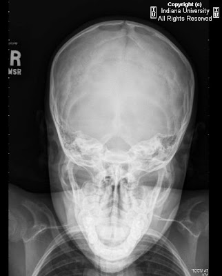
On the ultrasound, there were 2 discrete bony defects in the skull, seen in the parietal regions bilaterally, that demonstrate sharp margins. These were each approximately 2 cm in diameter. The x-rays also demonstrate these same 2 round lesions.
Diagnosis: Normal variant consistent with parietal foramina
Key points
Parietal foramina are normal variants.
Normal parietal foramina transmit the emissary veins of Santorini (there can be a depression in the outer table at the vein's exit).
Defects are insignificant except in the differential diagnosis of bony defects (from surgical intervention or trauma).
These congenital defects have a characteristic location, but may vary in size.
These are often symmetric, although they can be asymmetric and irregular.
Etichette:
AuntMinnie,
Head - Neck,
Pediatric
Oral cavity dermoid
Findings
Axial contrast enhanced CT (Figure 1 and Figure 2) demonstrate a well circumscribed cystic mass (Figure 1) in the root of tongue. The lesion is heterogeneous with focal oval areas of fat attenuation within the lesion. There is a thin wall with no significant surrounding inflammatory changes (Figure 3). The lesion is in the midline between the genioglossus muscles (Figure 4).
Diagnosis: Oral cavity dermoid and epidermoid
Epidermoid and dermoid cysts are benign lesions encountered throughout the body, with 7% occurring in the head and neck area. The orbit is the most common site in the head and neck for these congenital lesions. They rarely occur within the oral cavity, representing less than 0.01% of all oral cavity cysts.
These congenital cysts are dysembryogenetic lesions that arise from ectodermal elements entrapped during the midline fusion of the first and second branchial arches between the third and fourth weeks of intrauterine life. Acquired cysts may be derived from traumatic or iatrogenic inclusion of epithelial cells or from the occlusion of a sebaceous gland duct.
The cysts can be classified as epidermoid when the lining presents only epithelium, dermoid cysts when dermal appendages are found, and teratoid cysts when other tissue such as muscle, cartilage, and bone are present. The teratoid type is the only variety that may have a malignant change.
Anatomically, these oral cavity cystic lesions most commonly involve the floor of mouth and may occur in the root of tongue (ROT), submandibular space (SMS) or sublingual space (SLS). Dermoid cysts generally present with slow and progressive growth, and even if they are congenital, the diagnosis is usually possible in the second or third decade of life. Midline cysts of the floor of the mouth present as painless subcutaneous or submucosal lesions. When large, they can displace the tongue and result in dysphagia, dysphonia or dyspnea.
Epidermoids present on imaging as low density, unilocular, well circumscribed simple cystic lesion. Dermoid cysts are usually more heterogeneous with fatty internal material and possibly calcification.
The treatment of dermoid cysts of the floor of the mouth is extracapsular excision with an intraoral or external approach, depending on the size of the lesion and the position relative to the mylohyoid muscle. The entire cyst must be removed to prevent recurrence.
Etichette:
ACR,
Head - Neck,
Malformations
lunedì 16 giugno 2008
Glomus jugulare
Findings
Figure 1: Contrast enhanced CT shows an enhancing soft tissue mass at the jugular foramen.
Figure 2 and Figure 3: Pre and post contrast axial T1 weighted MR images demonstrate enhancement of the mass at the jugular foramen with “salt and pepper” signal drop out from vessels (Figure 3).
Figure 4: Coronal T1 post contrast MR image again demonstrates an enhancing mass at the jugular foramen with “salt and pepper” signal drop out from vessels.
Differential Diagnosis of common jugular foramen lesions:
- Glomus jugulare
- Dehiscent jugular bulb / Enlarged Jugular bulb(normal variant)
- Meningioma
- Metastasis
- Schwannoma
Diagnosis: Glomus jugulare
Glomus tumors are slow growing, encapsulated, hypervascular benign tumors which are locally invasive and originate from paraganglia cells which are neural crest progenitor cells of neuroectodermal origin. They are also referred to as paragangliomas, chromaffinomas or chemodectomas. Frequently located near nerves and vessels, their distribution can be widespread, including the periaortic area, trachea, larynx, mandible, nose, ciliary ganglion, and Fallopian canal.
Glomus tumors constitute less than 1% of all head and neck tumors. In the head and neck, they most commonly arise from within the carotid body (bifurcation), vagus nerve, middle ear, and jugular foramen. Specifically, glomus jugulare tumors originate from glomus bodies located within the wall (adventitia) of the jugular bulb. Additionally, they can be associated with either the auricular branch of the vagus nerve (Arnold nerve) or the tympanic branch of the glossopharyngeal nerve (Jacobson nerve). Arising from the jugular foramen of the temporal bone, these tumors grow within the temporal bone via pathways of least resistance, such as air cells, vascular lumens, skull base foramina, and the eustachian tube. Erosions occur frequently involving the jugular fossa and posteroinferior petrous bone. As the tumors grow they may also extend into local extracranial structures including the mastoids, occipital bone, sigmoid and petrosal sinuses. Intracranial extension with neural infiltration is also common.
Although most are sporadic, glomus tumors can be familial (autosomal dominant inheritance and incomplete penetrance). Multifocal lesions are found in 3-10% of sporadic cases and in 25-50% of familial cases. Malignant degeneration is most frequent with glomus vagale (16%) and least in jugulotympanic (4%) tumors. Metastases from glomus tumors occur in approximately 4% of cases. About 10% of head and neck paragangliomas are multicentric but bilaterality in glomus jugulare is only about 1-2%. Multiple glomus tumors can be associated with familial syndromes like multiple endocrine neoplasia (MEN) 1 and 2 , neurofibromatosis and von Hippel –Lindau disease.
Additionally, functional tumors may be found in 1-3 % of tumors producing clinically significant levels of catecholamines (norepinephrine or dopamine) with symptoms mimicking a pheochromocytoma. Rarely, an association with pheochromocytoma, parathyroid adenoma, and thyroid carcinoma may occur.
Due to their painless, slow growing nature, presentation usually manifests late in the course of disease with complaints ranging from lower cranial nerve palsies, Horner’s syndrome and hoarseness to other neuro-otologic symptoms including pulsatile tinnitus (most commonly associated symptom of glomus jugulare specifically). The glomus tumors are more frequently seen in patients with chronic hypoxia (COPD) and in those living at high altitudes. Due to the location and extent of disease which is often found at diagnosis, treatment and management is often very challenging. Patients with cervical glomus lesions typically have a more favorable prognosis. When identified in the temporal bone, however, there is often an increased rate of recurrence, residual tumor, and neurovascular compromise. Definitive treatment is surgical, however, radiation therapy may be employed for palliation for extensive tumors not amenable to surgery.
Imaging characterization of the location and extent of tumor involvement is critical in surgical planning and assessment of operative morbidity and mortality. Classic CT findings associated with these lesions include an enhancing soft-tissue mass in the carotid space, jugular foramen, or tympanic cavity. Cystic changes are not described in these tumors unlike in schwannomas from which glomus jugulare needs to be differentiated . Additionally, osseous erosion (a typical finding with the jugular type) adjacent to the tumor is best demonstrated on CT. By contrast the meningiomas, schwannomas and prominent jugular bulb tend to produce smooth expansion of bony outlines of the jugular fossa. MRI is superior to CT scan and typically demonstrates a "salt-and-pepper" appearance at standard spin-echo sequences due to signal drop out from extensive vascularity which manifests as an intense blush at angiography.
Etichette:
ACR,
Head - Neck,
Neoplasm,
Neuro,
Signs
Tethered cord
Findings
On the spinal ultrasound, the conus medullaris is abnormally positioned at the lower endplate of the L3 vertebral body level. The filum is prominent in size on ultrasound. The skin tag is confined to the subcutaneous level. The lumbar spine MRI demonstrates the conus at the same L3 level with blunting of the conus tip and thickening of the filum. Incidental note is made of a transitional vertebral body. Again, the skin tag appears confined to the subcutaneous level.
Differential Diagnosis:
- Tethered cord
- Normal variant low-lying conus
- Open or closed spinal dysraphism
- Post-surgical low-lying conus
Diagnosis: Tethered cord
Key points
Tethered cord occurs when the distal spinal cord does not completely involute and the conus improperly ascends. The stretching of the cord may result in vascular compromise.
Best demonstrated as a conus lying below the inferior endplate of the L2 vertebral body. (Normal conus medullaris tip position is at L4-5 at 16 weeks gestation, at L2-3 at birth, and then generally resides at L1-2 after 3 months of age.)
Associated with imperforate anus, diastematomyelia, filar lipoma, and filar cysts. Also, patients may demonstrate muscle atrophy, gait problems, orthopedic issues, bladder dysfunction, and may have hair patches or dermal sinus tracts.
Affects females more often than males (3:2).
On MRI, may see prolonged T1 relaxation in the cord that could represent hydromyelia or myelomalacia. Look for a thick and shortened filum terminale.
The tethered cord must meet criteria of position (below L2 at age 12 and below L3 at birth). The cord does not freely move on cine imaging, and the nerve roots may have an aberrant course. The cord may be fused to vertebral arches.
The tight filum measures more than 2mm at the L5-S1 level. May see associated cysts or lipomas on ultrasound or MRI.
On imaging, look for vertebral body abnormalities like scoliosis, spina bifida, and increased interpedicular distance.
Treated with decompressive laminectomy, removal of lipomas, untethering the spinal cord.
Etichette:
AuntMinnie,
Malformations,
Pediatric,
Spine
martedì 10 giugno 2008
Ossifying fibroma
Findings
Axial CT bone windows (Figure 1 and Figure 2) show an expansile lesion of the left maxillary sinus with dense osseous material on the periphery and low attenuation fibrous center. Coronal reformats (Figure 3) demonstrate mass effect on the left orbit. Sagittal reformats (Figure 4) show scattered foci of soft tissue density among ossified areas.
Diagnosis: Ossifying fibroma
Fibro-osseous lesions of the paranasal sinuses are a broad spectrum of bony disease with fibrous dysplasia, ossifying fibroma, and osteoma being distinct entities that lie along a continuum from the least to the most bony content. They have similar appearance and makeup; however, their clinical implications vary.
Osteoma is the most common tumor of the paranasal sinuses. The vast majority (95%) of sinonasal osteoma are found in the frontoethmoidal region. Most frequently, osteomas are asymptomatic and are discovered incidentally
Fibrous dysplasia (FD) is a slow-growing fibro-osseous lesion that can be located in the paranasal sinuses. It can be mono- or poly-ostotic. Fibrous dysplasia affects children and characteristically “burns out” during puberty. It is deforming but not destructive.
Ossifying fibroma (OF) is the most concerning of the fibro-osseous lesions in the paranasal sinuses as it can be locally destructive. It is also known as cemento-ossifying fibroma; psammomatoid ossifying fibroma, and juvenile-aggressive ossifying fibroma. It is characteristically monostotic and the most common craniofacial site is the mandible (75%). However, it is considered more aggressive when found outside the mandible.
Histologically, ossifying fibroma is an encapsulated tumor with matrix of randomly distributed mature, lamellar bone spicules mixed with fibrous stroma.
Radiographically, it is a sharply circumscribed, expansile lesion with an eggshell rim and a central low attenuation fibrous center. Ossifying fibroma is locally aggressive and can absorb tooth roots, whereas fibrous dysplasia usually encompasses the healthy roots.
Ossifying fibroma usually presents in young adults. It is generally asymptomatic and found incidentally. The juvenile variant may show aggressive, locally destructive behavior and cause compression of vital structures, severe pain or facial pain.
Because of the aggressive and locally destructive nature of ossifying fibroma, complete surgical excision is recommended.
Dural arteriovenous fistula with venous hypertension and cord edema
Findings
MRI Cervical Spine: Cord expansion involving the majority of the cervical and upper thoracic spinal cord. Abnormal increased T2-STIR signal, extending from the brainstem to the lowest portions of the visualized field-of-view in the upper thoracic cord. Prominent intradural flow-voids seen posteriorly throughout the cervical and upper thoracic cord. Spinal Angiogram: Spinal dural arteriovenous fistula arising from the left T5 radicular artery. Dilated medullary veins drain both cephalad and caudal.
Differential Diagnosis (for MRI findings):
- Normal CSF pulsations
- Spinal cord tumor
- Tortuous roots from spinal stenosis
- Spinal dural AV fistula
Diagnosis: Dural arteriovenous fistula with venous hypertension and cord edema
Key points
Presents with progressive lower extremity weakness, back pain, bowel/bladder dysfunction.
Results in spinal cord ischemia.
Progressive over many years, may lead to paraplegia.
Presents during 50 to 60 years of age.
Male more common than female.
Lesions are AV fistulas, draining into venous outflow tract.
Treatment is surgical obliteration or endovascular embolization.
Radiographic Overview
MRI:
- Enlarged, T2 hyper intense spinal cord
- Edema may spare periphery
- Abnormal vascular flow voids of dilated intradural veins
MRA/Angiogram:
- Confirms diagnosis, allows identification of exact level of shunt, localizes anterior spinal artery
Etichette:
AuntMinnie,
Malformations,
Neuro,
Spine,
Vascular
venerdì 6 giugno 2008
Cavernous malformations
Findings
Figure 1 and Figure 2: Axial noncontrast CT images demonstrate multiple foci of increased attenuation within the brain.
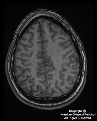
Figure 3: Axial SPGR shows two lesions, the one in the midline is heterogenous in signal. The lesion in the posterior insula on the left is more homogeneously bright.
Figure 4 and Figure 5: Axial T2 and FLAIR images demonstrate the same lesion at the level of the third ventricle. Note there is no significant parenchymal edema. The lesion in the left brain does have associated hemosiderin deposition, but centrally the blood looks more acute to subacute.
Figure 6: Axial GRE image showing multiple lesions which demonstrate “blooming” secondary to hemosiderin deposition.
Diagnosis: Cavernous malformations
Cavernous malformations are sinusoidal collections of blood vessels surrounded by a single layer of endothelium without normal intervening brain parenchyma. They occur anywhere throughout the central nervous system but are most commonly seen in the cerebral hemispheres. CMs were traditionally believed to be congenital lesions, however, CMs also develop de novo following radiation therapy, or in association with developmental venous anomalies (see Case-in-point case on 2/9/07). Both congenital and sporadic forms present with seizures, hemorrhages, focal neurological signs, headaches, or patients can be entirely asymptomatic.
On unenhanced CT, CMs are hyperdense regions due to pooling of blood within the sinusoids and exhibit focal calcification. On MR, which is the preferred modality for evaluating CMs, there is a characteristic appearance demonstrating blood products of different ages appearing as a central region of high signal intensity (representing methemoglobin), surrounded by a rim of hemosiderin that is hypodense on T2W and gradient echo imaging. Angiographically, CMs are usually occult.
Approximately 25% of patients demonstrate multiple lesions and a small percentage of these patients follow a familial pattern. Many of these lesions may be too small and only identifiable on gradient echo MR. Gradient echo imaging has increased sensitivity for magnetic field inhomogenity because it does not contain a 180 degree refocusing pulse. As a result, if multiple lesions are detected on MRI, the familial form of CM should be suspected, and close relatives should undergo evaluation. Regardless of whether CMs are of congenital or acquired origin, they follow a common clinical course. The clinical course depends on the location of the lesion, the rate of expansion of the lesion and whether a clinically overt hemorrhage has occured.
Etichette:
ACR,
Malformations,
Neuro,
Vascular
CNS primary B-cell lymphoma
Findings
CT demonstrates an intra-axial mass within the left frontal lobe, which demonstrates central hypo attenuation, subfalcine herniation with vasogenic edema, a moderate left-to-right midline shift and mass-effect. MRI shows an enhancing, heterogeneous large left frontal lobe intra-axial mass which extends to the genu of the corpus callosum and lateral ventricle ependymal surface. The mass is isointense on T1, slightly hyper intense on T2 and FLAIR. Increased signal is demonstrated on T2 and FLAIR imaging of the central portion of the mass, most consistent with edema. Diffusion weighted and ADC imaging (not shown) demonstrated some evidence of diffusion restriction. There is intense enhancement of the mass on post-contrast imaging. No acute findings on the chest or abdomen CT images.
Diagnosis: CNS primary B-cell lymphoma
Key points
Primary CNS lymphoma now represents as many as 2% of all intracranial neoplasms, 7-15% of primary brain tumors, and less than 1% of non-Hodgkin lymphomas.
CNS lymphoma affects persons at all ages, with a peak incidence in those aged 40-60 years. Almost all CNS lymphomas are non-Hodgkin B-cell tumors.
MRI is the examination of choice because of its high sensitivity and multiplanar capability. The classic appearance of CNS lymphoma is an isointense to isointense-to-hypo intense nodule or mass on nonenhanced T1-weighted MRIs and isointense-to-hyper intense on corresponding T2-weighted MRIs. On post gadolinium-enhanced T1-weighted MRIs, lymphoma tends to enhance intensely and diffusely. Usually, little or no surrounding vasogenic edema is demonstrated.
Tumor lesions can cross the midline and may appear as a butterfly tumor involving both cerebral hemispheres.
Involvement of the corpus callosum is highly suggestive of CNS lymphoma, but it also occasionally occurs with anaplastic glioma and metastatic neoplasm.
mercoledì 4 giugno 2008
Thyroid ophthalmopathy (Graves disease)
Findings
Bilateral proptosis. Diffuse, uniform thickening of all the bilateral extraocular muscles. Tapering of muscles near insertion on the globe.
Differential Diagnosis:
- Graves' orbitopathy
- Orbital pseudotumor
- Infectious myositis
- Sarcoidosis
- Lymphoma
- Metastasis
Diagnosis: Thyroid ophthalmopathy (Graves disease)
Discussion
Graves' orbitopathy or thyroid associated orbitopathy refers to autoimmune inflammatory disease of the orbits. Most affected individuals are between 30 and 50 years of age. It also has a predilection for females (80%). The majority of individuals at the time of diagnosis demonstrate laboratory values consistent with hyperthyroidism. There are two common theories explaining an offending antigen. One theory focuses on the presence of thyroid stimulating hormone receptors within the orbital tissues (similar to the thyroid). Another theory focuses on an orbital autoantigen.
The most common signs/symptoms include orbital edema, gaze restriction and proptosis. Other clinical findings include lid lag, diplopia, corneal ulceration and dryness of the eyes. In severe cases, optic nerve entrapment can ensue resulting in loss of vision. The first step in treatment involves controlling thyroid hormone dysfunction. Corticosteroids can be beneficial in limiting extraocular muscle enlargement. Lubricating agents are used to prevent corneal complications. For visual symptoms such as diplopia, orbital decompression surgery can be performed. In some cases, emergent surgery is warranted if optic nerve entrapment is suspected. Other non-emergent surgical procedures focus on eyelid lengthening to address proptosis. Cigarette smoking, male gender, uncontrolled hyperthyroidism, and diabetes are considered poor prognostic factors.
Radiologic overview of diagnosis
If the diagnosis is clinically established, imaging may not be necessary. However, imaging is used for confirmation and followup. Additionally, imaging is useful for surgical planning. CT and MRI are the imaging modalities of choice. The best diagnostic clue is exopthalmos with bilateral extraocular muscle enlargement.
On noncontrasted CT, findings to look for include isodense extraocular muscle enlargement and increased orbital fat. After contrast administration, greater than usual enhancement of the extraocular muscles is visible. Similarly, findings to look for on MRI include isointense enlargement of the extraocular muscles (T1 weighted sequences). On T2 weighted images, hyperintensity is seen with edema and hypointensity is seen with fibrosis (late stage finding). Increased signal on STIR reflects disease activity. Post contrast T1 weighted images demonstrate increased extraocular muscle enhancement. Compression of the optic nerve is more readily identified on MRI rather than CT.
The bilateral orbits are involved in over 90% of cases (even if symptoms are unilateral). The most commonly involved muscles (in descending frequency of involvement) are the inferior, medial and superior rectus muscles. The lateral rectus and oblique muscles are much less likely to be involved. Typically, isolated muscle involvement is unlikely. In the five percent of cases demonstrating isolated muscle involvement, the superior rectus is usually involved. Isolated involvement of the lateral rectus muscle should prompt the search for another inciting etiology.
Etichette:
AuntMinnie,
Inflammatory,
Ophtalmic,
Systemic
Iscriviti a:
Post (Atom)