Visualizzazione post con etichetta Inflammatory. Mostra tutti i post

venerdì 3 dicembre 2010

Basilar invagination secondary to rheumatoid arthritis


Findings

Axial and sagittal CT images demonstrate severe basilar invagination (Figure 1). The tip of the odontoid process measures 2.3 cm above Chamberlain’s line (yellow line in Figure 2). McGregor's line (red line in Figure 2) is also shown. Incidentally noted are right-sided opacified mastoid air cells (Figure 1).
Once again, severe basilar invagination is evident. On the sagittal T2 image the foramen magnum is narrowed and obliteration of the CSF space is noted at the C2-C3 level (Figure 3). On the axial T2 weighted image increased T2 signal (Figure 4) is seen within the cord at the C2-C3 level indicating edema versus myelomalacia.


Diagnosis: Basilar invagination (impression) secondary to rheumatoid arthritis.


Basilar invagination refers to a condition in which the odontoid process protrudes upward into the intracranial space. Basilar invagination may be classified as primary (congenital) or secondary (acquired). Down syndrome, Klippel-Feil syndrome and Chiari malformations are congenital causes of basilar invagination. Acquired basilar invagination, also known as basilar impression, is associated with softening of the skull base and is often due to rheumatoid arthritis, Paget disease, osteomalacia, hyperparathyroidism and osteogenesis imperfecta. Basilar invagination is probably better described as a radiologic finding rather than a diagnosis. Once the finding is identified, a cause of basilar invagination should be diligently pursued.

Plain lateral radiographs with odontoid views, although not 100% sensitive, are often the initial study used to diagnose basilar invagination. MRI is the optimal study, which also assesses the cervicomedullary junction and cervical cord. Two craniovertebral junction lines are particularly useful in defining basilar invagination. Chamberlain’s line extends between the posterior pole of the hard palate and the posterior edge of the foramen magnum (opisthion). If the dens is >3.0 mm above this line basilar invagination is present. McGregor’s line, a modification of Chamberlain’s line was developed because the opisthion could not always be seen on plain radiographs. This line extends from the posterior pole of the hard palate to the undersurface of the occiput. If the dens extends >4.5 mm above this line basilar invagination is present.

Clinical manifestations of basilar invagination include posterior skull pain, headache, signs and symptoms of brainstem and upper cervical cord compression or disturbances of CSF circulation causing obstructive hydrocephalus. The brainstem may be compressed at the level of the foramen magnum possibly resulting in compromise of the autonomic centers resulting in labile blood pressures, arrhythmias, or sudden death. Neurosurgery is recommended in patients that are symptomatic with concomitant MRI findings indicating compression. Although asymptomatic patients are often followed conservatively, many authors favor surgery even if no symptoms of cord compression are evident in rheumatoid patients.

Although often appearing together, basilar invagination or impression should not be confused with platybasia; which literally means “flattening of the base of the skull”. Platybasia, which can be seen in Klippel-Feil anomalies, cleidocranial dysplasia and achondroplasia, is present when the basal angle formed by intersecting lines from the nasion to the tuberculum sellae and from the tuberculum along the clivus to the anterior aspect of the foramen magnum (basion) is greater than 143 degrees.

mercoledì 20 ottobre 2010

Sialadenitis with an obstructing sialolith in the right submandibular gland duct


Findings

Coronal and axial post contrast CT images show a swollen right submandibular gland with dilatation of the intraglandular ducts and an obstructing stone.


Diagnosis: Sialadenitis with an obstructing sialolith in the right submandibular gland duct


The most common cause of sialadenitis of the SMG is an obstructing calculus with subsequent suppurative sialadenitis. Less common causes are suppurative sialadenitis leading to duct stenosis and chronic sialadenitis. Rare etiologies include include Sjogren syndrome, AIDS and bacterial/viral infection.

SMG accounts for 10% of sialadenitis of all major salivary glands. Other diagnostic considerations in SM space include reactive submandibular lymph node, mandibular osteomyelitis, benign mixed tumor, submandibular carcinoma and metastases.

Calculi are more common in the SMG duct. Compared to the parotid gland, the saliva in the SMG is thicker, much more mucinous and more alkaline. The SMG duct courses superiorly which makes it more prone to stasis. SMG duct is larger in diameter.

When sialadenitis is present therapy may depend on stone location. If the stone is in the anterior portion of the duct, the stone can be removed and gland salvaged. If stone is in the posterior duct, the duct and gland will likely be removed with the stone.

mercoledì 6 ottobre 2010

Labyrinthitis ossificans


Findings

Figure 1: Axial CT image through the left temporal bone shows complete ossification of the left superior semicircular canal.
Figure 3: Axial CT image at the level of the internal auditory canal shows non-erosive soft tissue attenuation lateral to the malleus and incus as well as partial ossification of the lateral semicircular canal.


Diagnosis: Labyrinthitis ossificans


Labyrinthitis ossificans (LO) is the end result of suppurative labyrinthitis where inflammation of the membranous labyrinth progresses to fibrosis and ossification. Suppurative labyrinthitis typically arises following meningitis, although other causes include direct infection from hematogenous sources or trauma.

LO is the most common cause of acquired childhood deafness and can be detected by CT as early as 2 months following an episode of meningitis. Following meningitis, approximately 6% to 30% of children develop some degree of sensorineural hearing loss which is typically bilateral.

On CT, osseous deposition is seen within the membranous labyrinth. On MRI, loss of the normal high signal on T2-weighted images from displacement of the endolymphatic fluid is suggestive of this diagnosis. It is important to the clinician to distinguish between cochlear involvement, non-cochlear involvement, or both as prognosis is determined by response to cochlear implantation. The degree of ossification is important in surgical planning and severe LO may preclude cochlear implantation.

mercoledì 30 giugno 2010

Diving Ranula


Findings

Figure 1 and Figure 2 Axial contrast-enhanced CT demonstrates a thin-walled, well-defined hypodense lesion in the right sublingual space (Figure 1) which herniates posteriorly into the submandibular space (Figure 1). This lesion also tracks into the sublingual space lateral to the right genioglossus muscle (Figure 2). It is located anterior and lateral to the right internal and external carotid arteries and effaces the submandibular gland.


Diagnosis: Diving Ranula


A diving ranula is an extravasation pseudocyst of a simple ranula in the sublingual space rupturing out into the submandibular and or parapharyngeal space. It originates from trauma or inflammation of the sublingual gland or minor salivary glands in the sublingual space. With obstruction of the gland duct, the duct dilates and eventually ruptures, allowing its secretions to leak into the surrounding soft tissue.

CT is the imaging study of choice. On CT, the lesion demonstrates water content and thin walls with subtle or no wall enhancement. If the lesion is infected, it may show thick, enhancing walls. The characteristic shape is a collapsed cystic portion in the sublingual space - “tail sign,” with its head extending into the submandibular space. The sublingual spaces are located on the floor of the mouth on either side of the midline genioglossus muscles. They are separated from the submandibular space by the mylohyoid muscle that extends from the medial inferior aspect of the mandible to the hyoid bone. Thus, the sublingual space is superomedial to the mylohyoid muscle and the submandibular space is inferolateral to the muscle. Ultrasound and MR can also be used to characterize these lesions. Ultrasound demonstrates a well-defined hypoechoic mass in the sublingual and submandibular space. MR images show the lesion with signal intensity of water, though if infected, signal intensity can vary according to the protein content or presence of hemorrhage with some wall enhancement.

The differential diagnosis would include epidermoid or dermoid cyst, lymphangioma or cystic hygroma, abscess, submandibular gland cyst (mucocele), and second branchial cleft cyst. These lesions differ in appearance on CT.

The treatment of a diving ranula is removal of the ipsilateral sublingual gland via the cervical or intraoral approach. It is not necessary to excise the pseudocyst since it puts the surrounding structures at risk for damage. Biopsy can be done to confirm the diagnosis.

martedì 25 maggio 2010

Skull base Langerhans cell histiocytosis (LCH)


Findings:

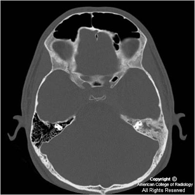
Figure 1and Figure 2: There is diffuse sclerosis of the left mastoid with near complete opacification of the mastoid air cells.
Figure 3 and Figure 4: There is diffuse sclerosis of the mastoid with a soft tissue thickening identified in the cavernous sinus. There is additional dural extension suggested along the left leaf of the tentorium.


Diagnosis: Langerhans cell histiocytosis (LCH), skull base


Langerhans cell histiocytosis (LCH) is the term used to describe the spectrum of diseases characterized by proliferation of specialized bone marrow-derived dendritic cells, called Langerhans cells (LC) with bone and/or soft tissue involvement. Although the etiology and pathogenesis of LCH is poorly understood, studies suggest that it is a reactive disease caused by immune dysregulation. The disorders once collectively called Histiocytosis X (eosinophilic granuloma, Hand-Schüller-Christian disease, and Letterer-Siwe disease) are now part of this category. LCH is defined in terms of whether it involves a single site, multiple sites, a single organ, or multiple organs. Prognosis and treatment are closely linked to the extent of disease at presentation and whether or not “risk” organs (ie: liver, spleen, lung, bone marrow) are involved. In general, patients with LCH localized to the bone have a favorable prognosis whereas patients with multisystem involvement incur a poor prognosis.

Langerhans cell histiocytosis of the bone is a relatively rare disorder with an incidence of one case per 2 million persons per year. The skull is the most commonly involved bony site in the pediatric age group. Otologic involvement usually occurs during the course of multisystemic disease, and may be the only symptom in up to 25% of patients. Because the otologic symptoms are similar to otomastoiditis, diagnosis of LCH of the skull is often delayed. The most frequent otologic symptom is otorrhea resistant to medical treatment. Other findings include mastoid swelling, aural polyps, periauricular eczema, sagging of the canal wall skin from erosion of the posterior bony external auditory canal, conductive hearing loss, and otalgia. Neurosensorial deafness, vertigo from involvement of bony labyrinth, and paralysis of cranial nerves (particularly VII and VIII) have been described. The classic clinical profile is a young man with otalgia, otorrhea, and post-auricular mass. If the diagnosis is suspected, a full examination and work-up is warranted to exclude the possibility of multisystemic or multiostotic disease.

The best imaging modality to diagnose LCH of the skull base is dedicated temporal bone CT. The skull base may show focal or diffuse bone destructive change and may present as: 1) sharply-defined “punched out” appearance, 2) irregular sclerotic lesions or 3) fragments of bone within soft tissue component. Other locations that are affected include the petrous apex, mandible, maxilla, and vertebral body. Ossicular and otic capsule destruction are common. Bilateral disease occurs in up to 30% of patients. The radiologic differential diagnosis for LCH of the skull base includes mastoiditis, cholesteatoma, cholesterol granuloma, and rhabdomyosarcoma. Rhabdomyosarcoma may exactly mimic unilateral LCH- biopsy may be required to differentiate the two entities.

Contrast-enhanced examination may help differentiate inflammatory mastoid lesions from LCH. Findings include a heterogeneously enhancing mass in the temporal bone, including mastoid complex, middle ear, extracalvarial, intracranial, and extra-dural compartments. The skull base tends to show a variable amount of enhancing soft tissue. On MR, T1 weighted images reveal iso- to hypointense mastoid, middle ear or skull base mass. T2 weighted images reveal hyper- to isointense soft tissue mass. Contrast-enhanced T1 weighted images show a heterogeneously-enhancing mastoid, middle ear, or skull base mass. MR may be obtained to delineate boundaries of soft tissue component which generally displays poorly defined borders.

The prognosis of LCH localized to the temporal bone is excellent, with a cure rate of 90%. The soft tissue component resolves initially, followed by reossification of the bone lesion. The treatment of choice for LCH localized to the mastoid-middle ear consists of surgical curettage or mastoidectomy. Low dose radiotherapy is used for larger, destructive lesions. Systemic disease is treated with chemotherapy in combination with radiation therapy.

lunedì 24 maggio 2010

Multiple bilateral infarcts secondary to IV drug abuse-related cerebral vasculitis


Additional clinical history: Woman in acute heroin withdrawal who also has bilateral lower extremity weakness.


Findings:

Brain MRI shows restricted diffusion within the bilateral parietal and occipital lobes as well as over the motor cortex of the frontal lobes bilaterally, left greater than right. T2 prolongation is noted in all the locations where restricted diffusion is seen. Coronal and sagittal reformatted CTA images show areas of subtle beaded appearance of arteries, with focal narrowing and dilatation.


Diagnosis: Multiple bilateral infarcts secondary to IV drug abuse-related cerebral vasculitis


Key points

Increased T2 signal lesions with restricted diffusion have a broad differential
Clinical history, distribution (vascular territories involved, unilateral vs. bilateral), number (single vs. multiple), the presence or absence of mass effect, and the presence or absence of a peripheral ring or central necrosis are key diagnostic clues.
Abscesses may be single or multiple, may be unilateral or bilateral, often have a peripheral ring, and often display mass effect.
Tumors often have mass effect, often have a peripheral ring, may be single or multiple, may be unilateral or bilateral, and may show central necrosis.
Hemorrhage is often in the subdural or subarachnoid space, but may be intraparenchymal, especially if there is history of head trauma or associated fracture.
In acute infarct, there is no mass effect, no peripheral ring, and no central necrosis and the lesions are usually in a single vascular distribution and are usually- but not always- unilateral.
Examples of bilateral acute infarcts, as seen in this patient (note multiple bilateral lesions without mass effect, peripheral ring or central necrosis), are watershed infarcts occurring in typical watershed zones and infarcts occurring secondary to cardiac emboli or vasculitis, both of which are not distributed with respect to single vascular territories (as in this patient).
Besides IV drug abuse (as in this patient), cerebral vasculitis with secondary infarct can be seen in bacterial meningitis, tuberculous meningitis, viral, mycotic, syphilitic or post radiation arteritis, cell mediated arteritis, collagen vascular disease, sarcoid, Wegener's granulomatosis, and Moyamoya disease.

giovedì 22 aprile 2010

Pars flaccida cholesteatoma


Findings

There is an 8mm mass in Prussak’s space with erosion of the malleus and scutum.

Differential diagnosis:
- Pars flaccida cholesteatoma
- Cholesterol granuloma
- Paraganglioma
- Pars tensa cholesteatoma


Diagnosis: Pars flaccida cholesteatoma


Key points

A pars flaccida cholesteatoma often occurs when a patient has chronic middle ear inflammation and/or TM perforation. The cholesteatoma forms when there is an accumulation of stratified epithelial cells in Prussak's space. The cholesteatoma can be seen in all age groups, but tend to be more aggressive in children. Patients can present with aural discharge, conductive hearing loss, and otalgia. Early treatment with surgery can preserve hearing.


Radiologic overview of the diagnosis

A pars flaccida cholesteatoma appears as a mass in Prussak's space with erosion of the scutum and/or adjacent ossicle. Ossicle erosion is seen ~70% of the cases. There is no enhancement of the cholesteatoma itself, though surrounding granulation tissue may enhance.

High resolution temporal bone CT is the best modality to evaluate a suspected cholesteatoma. A pars tensa cholesteatoma is far less common and involves the sinus tympanum. A cholesterol granuloma appears blue on otoscopy and may have similar bony erosions as that of a cholesteatoma. A paraganglioma appears as a cherry red mass on otoscopy and usually does not erode bone.

In this case, there is an 8mm mass in Prussak's space with erosion of the malleus and scutum.

venerdì 26 febbraio 2010

Sarcoidosis involving the lacrimal glands


Findings

Axial (Figure 1) and coronal (Figure 2) non contrast CT images, as well as an axial T2 MRI image (Figure 3), demonstrate symmetrically enlarged lacrimal glands which protrude anterior to the lacrimal fossa bony contours. Homogeneous enhancement is identified on the selected contrast enhanced axial CT image (Figure 4). There is no evidence of adjacent bony orbit invasion. An AP chest radiograph (Figure 6) demonstrates predominantly right hilar lymphadenopathy with “eggshell” calcification and bilateral reticulonodular opacities within the lung parenchyma.


Diagnosis: Sarcoidosis involving the lacrimal glands


20 to 25 percent of patients with systemic sarcoidosis develop ophthalmic manifestations usually between the third and fifth decades. The most common finding is inflammation of the uveal tract. Less commonly, patients will present with inflammation of the optic nerve or orbital involvement including pseudotumor like intraorbital masses, extraocular muscle enlargement, or lacrimal gland infiltration and hypertrophy. Isolated orbital disease is uncommon and usually limited to the lacrimal glands.

Lacrimal gland involvement occurs in approximately 15-28% of patients; usually as painless bilateral gland swelling evident on physical examination. In many cases, lacrimal gland involvement may occur long before lung and other organs are affected, thereby aiding in the early diagnosis of systemic sarcoidosis.

Imaging studies such as orbital CT and MRI are an integral part of early diagnosis. CT findings include symmetric enlargement of the lacrimal glands with diffuse homogeneous post contrast enhancement. There may be associated medial displacement of the optic globes as well as proptosis. Pertinent negative findings include the absence of adjacent bony orbital invasion and destruction. MRI is optimal to evaluate for additional orbital involvement such as optic nerve infiltration which manifests as a thickened and enhancing intraorbital nerve. An MRI of the brain is also recommended to evaluate for extension into the intracranial optic pathways and to exclude findings of coexisting neurosarcoidosis.

While diagnostic imaging tests may reveal findings highly suggestive of lacrimal gland sarcoidosis; definitive diagnosis requires biopsy of the glandular tissue and histopathologic assessment. Noncaseating granulomas characterized by clustered epithelial cells, central giant cells and abundant surrounding lymphocytes are characteristic.

The mainstay of therapy is high dose systemic steroids, usually oral prednisolone for approximately two weeks, followed by gradual tapering after the inflammation appears controlled. Some patients may require maintenance doses for several weeks to months.

lunedì 22 febbraio 2010

Baastrup's disease


Findings

There is diffuse disk desiccation and multilevel narrowing of the lumbar spine disc spaces with multilevel disc bulges or protrusion. There is close approximation of the spinous processes of the lower lumbar spine with sclerosis (low signal) and flattening of the adjacent spinous processes. There are focal fluid collections seen as high signal on the T2 sequence between the opposing spinous processes in the expected locations of the interspinous ligaments. These correspond with adventitious bursae and surrounding inflammatory changes. There are cystic changes of the L4 spinous process at the pseudarthrosis.


Diagnosis: Baastrup's disease


Key points

The clinical syndrome of pain in the back when standing erect which is relieved by bending forward was described in 1929. In 1933, Christian Baastrup, a Danish radiologist described in detail the clinical and radiological features of the syndrome. It manifests clinically as localized midline lumbar tenderness and pain on spinal extension that can be relieved by spinal flexion, local anesthetic injection and excision of part of the involved spinous processes.

Baastrups's disease is characterized on plain films by close approximation of spinous processes ("kissing spines") with associated sclerosis, enlargement and squaring off/ flattening of the involved spines. MR may document the development of adventitious bursae between the spines, seen as high signal fluid between the processes. This condition usually arises from chronic postural hyperlordosis and regional loss of discal spacings. Hypertrophy of the tips of the spinous processes may occur in the elderly persons especially in those with an occupational history of long periods of back flexion.

Synonyms: Arthrosis interspinosa, diarthrosis interspinosa, kissing osteophytes, kissing spine, kissing spinous disease, osteoarthrosis processus spinosi vertebrarum lumbalum, osteoarthrosis interspinalis

Rasmussen encephalitis


Findings

Figure 1, Figure 2, and Figure 3: Atrophy of the left cerebral hemisphere and numerous punctate calcifications involving predominately the cortex of the left hemisphere.
Figure 4: Left sided perihemispheric areas of increased signal representing areas of wallerian degeneration.
Figure 5: Unihemispheric volume loss involving the left hemisphere.


Diagnosis: Rasmussen encephalitis


Rasmussen encephalitis is a rare progressive CNS disorder characterized by chronic inflammation, most commonly involving a single hemisphere of the brain. Although the exact cause of the disease remains unknown, there are several theories regarding its etiology. One theory suggests that the disease has an autoimmune component where the body produces antibodies which activate the brain’s glutamate receptors. Other theories suggest that the encephalitis is related to primary viral infection, or to secondary immune mediation in response to a viral infection.

The disease occurs most commonly in children under the age of 10. The disease progression is characterized by intractable seizures and progressive neurologic deterioration including loss of motor skills and speech. Patient presentation can be characterized into three phases. The first phase (prodrome stage), is characterized by mild hemiparesis, and low frequency seizure activity, which generally lasts for 7-8 months. The second phase (acute stage) is characterized by acute increase in seizures, progressive hemiplegia, and brain atrophy. Often, this occurs in the form of epilepsia partialis continua, a phenomenon considered the status epilepticus for simple partial motor seizures. During the third phase (residual stage), patients develop permanent hemiparesis, and continue to have many seizures (although less than in the acute stage).

On imaging, Rasmussen encephalitis appears as unilateral cortical atrophy on CT and MR. There may be cortical swelling initially, with atrophy that ensues. The distribution is variable, although in some cases the entire cerebral hemisphere may be affected. No enhancement is usually seen. On PET/SPECT, there is decreased cerebral perfusion and hypometabolism of the affected hemisphere. There may be crossed cerebellar diaschisis although this is nonspecific. EEG is recommended to characterize the seizures.

The treatment involves hemispherectomy since anti-epileptics are usually not effective at controlling the seizures. Alternative treatments include plasmapharesis to remove the antibodies against glutamate receptors. Prognosis is poor with most patients inevitably developing hemiplegia with or without treatment.

lunedì 25 gennaio 2010

Sialolithiasis with sialadenitis


Findings

The right parotid duct is markedly dilated. There is an obstructing calculus in the distal duct. There is inflammation in the right buccal space, the right masseter space, and in the right parotid gland. There is also a large non-obstructive stone in the right submandibular gland which was unchanged from a previous study.


Diagnosis: Sialolithiasis/sialadenitis


Discussion

Calculus disease is the most common benign condition to affect the salivary glands. The most common glands affected are the submandibular glands because secretions are more mucinous, viscous, and alkaline. Additionally, the duct drains uphill, making stasis more likely. Parotid glands are affected less frequently, and sublingual and minor salivary gland calculi are very uncommon. Patients generally have painful glands, worsened with chewing foods that precipitate salivation. Treatment is by administration of solutions that increase salivation to promote passing of the calculus. Transoral resection can be performed for calculi close to the ampulla. Resection of the gland may be used for proximal, glandular and/or recurrent sialoliths.


Radiologic overview

Imaging can be performed with plain radiographs, CT, and MRI. Most calculi are radio dense, but approximately 20% may not be visible on plain radiographs.

CT is more sensitive for detection of calcification than plain radiographs, and is also useful for detecting inflammatory masses and abscess formation.
In the case of non-radiopaque calculi, conventional sialography can be performed by cannulating the salivary duct and injecting contrast. The presence of a calculus is inferred by the presence of an intraluminal filling defect.
A new technique is MR sialography which is similar to MR cholangiopancreatography. The technique utilizes either a single shot fast spin echo T2-weighted sequence to image the duct alone, or a high resolution 3D fast spin echo T2 sequence to image the gland and duct.
Sialography is useful to demonstrate ductal strictures after passage of the stone, and in the evaluation of chronic sialadenitis from autoimmune causes.

mercoledì 30 dicembre 2009

Labyrinthine ossificans


Findings

On the left, there is severe ossification and encroachment of the cochlea with extension to the vestibule and semicircular canals, which are also severely involved. The cochlear aqueduct is slightly stenotic. On the right, there is peripheral osseous hyper density of the cochlea with mild encroachment.

Differential diagnosis:
- Labyrinthine ossificans
- Cochlear aplasia
- Intravestibular schwannoma
- Cochlear otosclerosis
- Labyrinthine schwannoma


Diagnosis: Labyrinthine ossificans


Key points

Most common cause of acquired childhood deafness
Most common clinical presentation – bilateral sensorineural hearing loss 2-18 months after meningitis
May also occur after other infectious, inflammatory, trauma, or surgery
After meningitis or hematogenous infection – bilateral
After otitis media – unilateral
Rarely – severe vertigo
Suppurative membranous labyrinthitis starts inflammatory cascade which leads to fibrosis and eventually ossification
Dedicated IAC/temporal bone CT best for detection
Mild – mild increased haziness in the fluid spaces of the membranous labyrinth and prominent modiolus
Severe – complete obliteration of the membranous labyrinth with bony replacement of the fluid spaces
On MR, low signal bone encroaches (mild) or obliterates (severe), high signal fluid spaces of the membranous labyrinth
Importance – must identify cochlear labyrinthitis ossificans before cochlear implant so that it may be surgically treated
Treatment – cochlear implant, or labyrinthectomy for severe vertigo

giovedì 12 novembre 2009

Submandibular sialadenitis


Findings

The left submandibular gland is hypervascular, inflamed and markedly enlarged (Figure 1 and Figure 2). There is dilatation of the submandibular duct leading to a calculus within the distal aspect of the duct (Figure 3 and Figure 4). There are no drainable fluid collections. There is injection and stranding of the overlying dermis (Figure 5). The right submandibular gland is unremarkable (Figure 2).


Diagnosis: Submandibular sialadenitis


Acute sialadenitis may be secondary to a bacterial/viral infection or an obstructing lesion such as a calculus or tumor at the floor of the mouth.
Associated conditions include HIV, sarcoidosis, Sjogren syndrome, dehydration, diabetes mellitus and immunocompromised/postoperative patients.
Imaging is often helpful to delineate the location of the calculus and the presence/absence of subsequent complications (abscess formation, osteomyelitis, etc).

Inflammation of the submandibular gland accounts for approximately 10-15% of cases of sialadenitis involving any of the major salivary glands. Risk factors for submandibular sialadenitis include immunocompromised/postoperative patients, debilitation, elderly patients, dehydration, diabetes mellitus, hypothyroidism, hypercalcemia, radiation/chemotherapy, eating disorders (bulimia, anorexia nervosa), and other concomitant medical problems (malignancy, head and neck infections). Associated conditions also include HIV, sarcoidosis, Sjogren syndrome, tuberculosis, mumps, and cat scratch disease. It is rare in pediatric patients.

Most commonly, acute sialadenitis is a result of a bacterial infection (common organisms include Staphylococcus aureus, Streptococcus, Haemophilus influenzae, and Pseudomonas). Less commonly, the infection may be related to a virus such as mumps, coxsackie virus, herpes and influenza. On the other hand, chronic sialadenitis is usually a result of salivary stasis, ductal stenosis, calculi or other obstructive lesions such as a tumor at the floor of the mouth. Sialolithiasis is most common in the submandibular gland, accounting for approximately 80% of cases. The majority of calculi are radio-opaque, vary in size, and can be single or multiple. Often, the calculus obstructs a duct, resulting in secondary inflammation of the affected salivary gland which then becomes suppurative.

Although plain film radiography can depict the majority of calculi, CT is often first-line imaging. The affected submandibular gland is enlarged, hypervascular and there may be associated cellulitis/myositis. Calculi are easily identified and described as being either distal (towards the ductal opening) or proximal (towards the submandibular hilum). Chronic sialdenitis manifests as a small, fatty gland.

Complications of sialadenitis include abscess formation, bacteremia/septicemia, osteomyelitis, cranial nerve involvement (facial nerve paralysis), and respiratory complications. Management of acute sialadenitis includes both medical (conservative) and surgical options. Antibiotics, analgesics, sialogogues, warm compresses, glandular massage and intravenous fluids are the mainstay of medical management. Surgical options include duct cannulation with subsequent removal of the calculus and complete gland excision.