Visualizzazione post con etichetta Other. Mostra tutti i post

lunedì 7 marzo 2011

Chloroma of epidural space


Additional clinical history: Acute leukemia.


Findings

Large anterior epidural mass extending from posterior clinoid to the cervicothoracic junction measuring approx. 6x3x1.5 cm with mass effect on the anterior pons, medulla, and upper cervical cord. No post-contrast imaging obtained.

Differential diagnosis: Epidural mass
- Metastatic disease
- Lymphoma
- Leukemia/chloroma
- Chordoma
- Osteomyelitis/epidural abscess
- Epidural hematoma
- Primary tumor such as neurofibroma/schwannoma


Diagnosis: Chloroma of epidural space


Key points

AKA granulocytic sarcoma, extramedullary myeloblastoma.
Most commonly occurs in the setting of AML.
Can also occur in setting of chronic myelogenous leukemia and other myeloproliferative disorders.
These tumors can involve any part of the body, either concurrently or sequentially.

Imaging characteristics:
- NECT: Isodense or hyper dense to brain or muscle
- MR: Hypo intense or Iso intense on T1-weighted MR images, heterogeneously Iso intense or hyper intense on T2-weighted MR images
- MR+C: Enhance homogeneously after injection of contrast medium

Paraspinal and intraspinal lesions are also thought to arise from perivenous arachnoid spread of leukemic cells. Uncommonly, spinal involvement by granulocytic sarcoma may cause compression of the spinal cord, cauda equina, or nerve roots

venerdì 31 dicembre 2010

Benign perimesencephalic SAH


Findings

Figure 1, Figure 2, and Figure 3: Axial CT images of the brain demonstrate SAH in the premedullary, prepontine, suprasellar, and interpeduncular cisterns.
Other figures (not shown): Representative images from a 4-vessel cerebral angiogram demonstrate no evidence of aneurysm or vascular malformation.


Diagnosis: Benign perimesencephalic SAH


Trauma and aneurysm are the two most common causes of SAH. At least 80% of cases of atraumatic SAH are caused by rupture of an intracranial aneurysm. When SAH is present, many clinicians request CT or MR angiography in order to quickly and non-invasively diagnose aneurysm. If an aneurysm is not detected with one of these modalities, conventional cerebral angiography (the gold standard for exclusion of aneurysm) is necessary. If the initial angiogram is negative, a second cerebral angiogram, typically performed 1-3 weeks after the first, is mandatory. This is because occasionally an aneurysm will be missed on the initial angiogram due to spasm or partial/complete thrombosis. The diagnosis of non-aneurysmal SAH can be applied to patients who have two consecutive negative technically adequate 4-vessel cerebral angiograms. Additionally, many clinicians request MRI of the spine to exclude the possibility of spinal AVM as a source for SAH.

The classic variety of non-aneurysmal SAH is known as benign perimesencephalic SAH or pretruncal nonaneurysmal SAH. As the name implies, the hemorrhage is situated around the midbrain and anterior to the brainstem in the ambient, interpeduncular, and prepontine cisterns. The term “benign” refers to the fact that after recovery from the initial episode, there is no increased risk of repeat hemorrhage. Cerebral vasospasm is less likely in these patients, but does occur. Hydrocephalus also remains a possibility during the acute phase. Although not clearly understood, one proposed mechanism of benign perimesencephalic SAH is rupture of the venous plexus anterior to the pons (the anterior pontomesencephalic plexus). This is postulated to occur as a result of increased venous pressure from strenuous activities such as exercise. Intramural hematoma of the basilar artery and rupture of a basilar perforating artery have also been suggested as alternate hypotheses.

Although benign perimesencephalic SAH has been known as a distinct clinical entity for some time, patients may present with non-aneurysmal SAH in an atypical distribution (non-perimesencephalic). In some of these patients, the total volume of hemorrhage is increased such that blood is present throughout the basal cisterns and extends over the cerebral convexities. In other patients, the hemorrhage is confined to the convexities, quadrigeminal cistern, or other atypical locations. In today’s case, Patient #1 presented with the classic variety of benign perimesencephalic SAH. Patient #2 presented with atypical non-aneurysmal SAH. Both patients recovered, and have had no repeat episodes of hemorrhage to date.

Possible causes of SAH:
- Trauma
- Aneurysm
- AVM
- Vasculitis
- Dural AV fistula
- Extension from intraparenchymal hemorrhage
- Dural venous sinus thrombosis
- Infection
- Neoplasm
- Idiopathic

venerdì 24 dicembre 2010

Pseudotumor cerebri - Idiopathic Intracranial Hypertension (IIH)


Findings

T2W axial MRI (Figure 1) shows signs of increased ICP, but only increased fluid within the optic nerve sheaths, flattening of the posterior orbit, and a partially empty sella.
The 3D TOF MRV Towne and RPO projections (Figure 2 and Figure 3) show bilateral, right greater than left, focal transverse-sigmoid venous sinus junction narrowing’s. It is not a normal MRV given the pt’s history, with more explanation in the discussion. There is no aneurysm or collection of collateral blood vessels seen in these images.

The AP and lateral (Figure 4 and Figure 5) venous phase carotid arteriogram shows long segment stenosis at transverse-sigmoid venous sinus junction distal to the vein of Labbé. Pre procedure venography showed a venous pressure gradient across this lesion of 17 mmH2O with 37 mmH2O on transverse sinus side and 15 mmH2O on internal jugular vein side.

AP and lateral (Figure 6 and Figure 7) venous phase carotid arteriogram shows long segment stenosis at transverse-sigmoid venous sinus junction with a balloon crossing the gradient lesion.


Diagnosis: Pseudotumor cerebri - Idiopathic Intracranial Hypertension (IIH)


Pseudotumor cerebri is defined by typical clinical symptoms which occur in the setting of elevated “idiopathic” ICP and a normal composition of CSF. Classic clinical symptoms include diffuse recalcitrant headaches, vision changes (including vision loss), and hearing changes (e.g., tinnitus), and the disease is typically seen in obese women who are 20-50 years of age. Papilledema is the most common physical exam finding, but visual loss and sixth nerve palsy are also seen. Other symptoms include disabling headaches and blindness. LP opening pressure is greater than 25 cm H2O. Brain computed tomography (CT) and magnetic resonance imaging (MRI) are typically normal, however, the following suggestive non-pathognomonic findings are frequently present:

– Cerebral venous sinus stenoses
– Flattening of the bilateral posterior sclera
– Partially or fully empty sella; enlargement of the chiasmatic recess of the 3rd ventricle
– Distension of perioptic nerve subarachnoid space
– Intraocular protrusion of the optic nerve head
– Orbital optic nerve vertical tortuosity

Treatment for pseudotumor cerebri typically includes medical management with acetazolamide and pain control for headaches. Furosemide and corticosteroids have been used, as well. Surgical interventions to treat pseudotumor cerebri include lumboperitoneal shunt (LPS) and ventriculoperitoneal shunt (VPS), which often produce immediate results, however, eventual return of pseudotumor symptoms occur in approximately 50% within three years. Optic nerve sheath fenestration is also used to treat vision changes, with variable headache relief. Dominant transverse/sigmoid venous sinus angioplasty and stenting are relatively new methods for the treatment of pseudotumor cerebri for those who have significant dural sinus stenosis. Given that 80% of intracranial vascular compliance is provided from the venous vasculature, reduction of pressure in the sinuses reduces CSF pressure. Better results are achieved in patients with documented high pressure gradients, and greater efficacy is seen with regard to arrest of visual loss (>90%) than with headache relief (~50%). Long-term results are lacking. however.

In this case, cerebral angiography demonstrated bilateral high-grade transverse/sigmoid sinus stenoses distal to vein of Labbe insertions. Selective catheterization of the right transverse sinus revealed an estimated 80% narrowing to a luminal diameter of 1mm, and a pressure gradient across the stenosis of 13 mmHg (normal <5 mmHg). The contralateral sinus was smaller, but distally stenotic. A stent was placed across the right sided stenosis.
The patient was placed on antiplatelet medication to preserve stent patency immediately after the procedure. She had no headaches after the procedure and demonstrated objective visual improvement at her one- and six-week follow-up examinations.

mercoledì 22 dicembre 2010

Intracranial pseudotumor (Tolosa-Hunt syndrome)


Findings

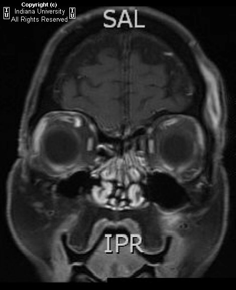
Increased CSF signal demonstrated by T2 hyper intensity within the right optic sheath. Nodular enhancement at the superior aspect of the right optic nerve at the orbital apex, which extends with prominent nodular enhancement posteriorly along the dural reflection of the right cavernous sinus. Asymmetric dilatation of the right superior ophthalmic vein.

Differential diagnosis:
- Meningitis
- Sarcoidosis
- En plaque meningioma
- Meningeal metastases
- Meningeal Non-Hodgkin's lymphoma
- Tolosa-Hunt syndrome


Diagnosis: Intracranial pseudotumor (Tolosa-Hunt syndrome)


Discussion

Intracranial pseudotumor (Tolosa-Hunt syndrome) is a part of a spectrum of "quasineoplastic" lesions that demonstrate orbital, intracranial, or pulmonary involvement, and include such other disorders as plasma cell granuloma and hypertrophic cranial pachymeningitis. It is a chronic granulomatous disease of unknown origin, which has been hypothesized to represent a low grade fibrosarcoma of inflammatory cells versus an autoimmune phenomenon. While Tolosa-Hunt syndrome is rare, orbital pseudotumor is the third most common ophthalmic disorder, and encompasses 5-8% of all orbital masses. Histologically, the lesions of Tolosa-Hunt demonstrate mixed lymphocytic and plasma cell infiltrate, with a variable degree of fibrosis. Lesions favor the cavernous sinus and basal meninges, although falcine and tentorial lesions have been described.

Patients with intracranial lesions are more frequently young adults, who present initially with chronic headaches or cranial nerve palsies. Patients with orbital involvement are more frequently middle-aged, and may present with painful proptosis and vision loss . Symptoms may be intractable, leading to severe disability. Untreated or unresponsive disease may progress to death. First-line treatment is invariably high-dose steroids, with radiotherapy or surgical resection reserved for patients with incomplete response to steroids .


Radiologic Overview of the diagnosis

The imaging hallmarks of Tolosa-Hunt syndrome are characterized by an enhancing, infiltrating meningeal mass, which favors the cavernous sinus or basal meninges, although falcine and tentorial involvement has been described . Focal meningeal thickening may range from just a few millimeters to a greater than 2 cm rind. With intracranial pseudotumor, orbital involvement is spared more than 90% of the time. Tolosa-Hunt remains a diagnosis of exclusion, once meningitis, en plaque meningioma, and meningeal metastases are ruled out.

The imaging modality of choice for imaging patients with Tolosa-Hunt remains MRI, although useful information may be gleaned from other modalities. On non-contrast enhanced CT (NECT), there are no specific findings to suggest the diagnosis; however, this modality may be of some value in differentiating the lesion from en plaque meningioma. On contrast enhanced CT (CECT), salient imaging findings include enhancing, thickened meninges or a curvilinear appearance of a single meningeal region. As aforementioned, MRI remains the primary modality for diagnosis of Tolosa-Hunt syndrome, and each sequence may provide critical information required to make the diagnosis. On T1WI, one may find focal thickening of the meninges that is isointense to gray matter. On T2WI, lesions are characterized as iso- to hypo intense regions of focal meningeal thickening, which may be more hypo intense as they become more fibrotic. FLAIR is of little help in making the diagnosis, but it is unlikely to demonstrate focal brain edema underlying the lesion . Contrast enhanced T1WI is the single most valuable sequence for evaluation of Tolosa-Hunt syndrome, and is characterized by diffusely enhancing region of meningeal thickening, which may range from a few millimeters to greater than 2 cm in some cases. Diffuse boney infiltrates may be appreciated on fat saturated contrast enhanced T1 sequence. While angiography is not considered a primary modality, severe disease may result in carotid artery narrowing, thus MRA may be a useful adjunct in the appropriate clinical setting.

The appropriate differential diagnosis of Tolosa-Hunt syndrome includes meningitis, sarcoidosis, en plaque meningioma, meningeal metastases, and meningeal Non-Hodgkin's lymphoma.

venerdì 12 novembre 2010

Idiopathic Thoracic Cord Herniation


Findings

These MR images demonstrate focal anterior displacement of the spinal in the mid thoracic spine. The cord (Images 1,2 and 3) appears to be either tethered anteriorly or compressed from the posterior aspect. The intradural space behind the cord is widened and has signal characteristics identical to CSF (Images 1,2 and 3).


Diagnosis: Idiopathic Thoracic Cord Herniation


Spinal cord herniation occurs when the cord herniates through a defect in the dura mater. These dural defects are typically located anteriorly or laterally, and occur most often in the mid-thoracic region. They may be idiopathic, post-traumatic or iatrogenic related to prior spinal surgery. Some have suggested that a herniated and calcified disk may cause thinning, erosion, or rupture of the dura, which may also be secondary to congenital weakening of the ventral dural fibers. The presence of free flow of cerebral spinal fluid dorsal to the herniated cord is key to differentiating a spinal cord herniation from an arachnoid cyst. Spinal cord herniation occurs most commonly in the middle-aged. Symptoms of myelopathy including chronic leg pain, gait disturbance, incontinence, and leg weakness are commonly seen and may slowly worsen over time if left untreated. The most common clinical feature reported is the Brown-Séquard syndrome consisting of hemiplegia and contralateral temperature sensation deficits and pain.

Typical imaging findings are focal anterior displacement of the spinal cord with expansion of the dorsal subarachnoid space. The preferred imaging modality in the setting of myelopathy is MRI, which is often sufficient for making the correct diagnosis. Myelography with CT may be required in ambiguous cases and to demonstrate the exact location of the dural defect. With cord herniation, myelography reveals uninterrupted flow of contrast and the absence of a filling defect posterior to the herniated cord segment. An arachnoid cyst will present during myelography as an early filling defect posterior to the displaced cord. Contrast may fill the cyst with time, so rapid acquisition of CT-myelograpgy after the initial myelographic images is essential. Phase contrast cine MR imaging may provide similar CSF flow information, in addition to restricted cord motion.

Treatment consists of surgically reducing the herniation by repositioning the protruding spinal cord back into the thecal sac followed by the repair of the defect in the dural mater in order to prevent recurring herniation. After surgery, symptoms typically improve and may completely resolve, even when longstanding. Patients whose symptoms are milder and non-progressive may be eligible for less invasive therapy or conservative management with monitoring.

martedì 14 settembre 2010

Postictal Imaging Findings


46-year-old male presented to the emergency room after a first seizure. He has no significant or contributory past medical history.


Follow-up FLAIR imaging obtained approximately 5 weeks later.


Findings

Figure 1 and Figure 2: FLAIR and DWI images show abnormal increased signal in the left temporal lobe. The right temporal lobe is questionably involved. The differential diagnosis for these findings included, but is not limited to, neoplasm, infection, and postictal changes.
Figure 3: Follow-up FLAIR imaging obtained approximately 5 weeks later demonstrates resolution of these findings, consistent with postictal change.


Diagnosis: Postictal Imaging Findings


A seizure is "a sudden alteration of the CNS resulting from a paroxysmal high frequency or synchronous low frequency, high voltage electrical discharge". Imaging (CT or MRI) is indicated in cases of:
- New onset of seizure activity
- Change in pattern of previous seizure pattern
- Patients with focal neurological defects or altered mental status
- Prolonged postictal state, especially if associated with neurological defects

Following seizure activity, imaging is used to identify an underlying etiology. Differential possibilities include structural/anatomical abnormalities, space-occupying masses (primary or secondary brain neoplasms, abscesses), cerebrovascular accidents, transient ischemic attacks, hemorrhage, infectious processes (meningitis, encephalitis) venous thrombosis, and vasculitis. If performed shortly after the ictal event, CT and MR imaging may demonstrate findings that are secondary to the physiological mechanisms related to the seizure itself. These findings are most likely to occur following status epilepticus.

The mechanism and pathophysiology of these findings are unknown. Some theories are that the findings occur as a result of breakdown of the blood brain barrier. This results in transient focal brain edema, accounting for various imaging findings. Other theories propose arteriovenous shunting of blood during seizure activity that results in accumulation of toxic metabolites, ischemia, and acidosis. Most theories implicate ischemia and transient cytotoxic edema as the cause of brain changes.

Imaging findings are nonspecific and can overlap with those seen in other disease entities such as infarction/ischemia, venous thrombosis, vasculitis, infection, neoplasm, arterial thromboembolism and metabolic encephalopathy. History, presenting signs and symptoms, follow-up imaging and other relevant laboratory data can further narrow the differential diagnosis.

On CT, possible postictal imaging findings include:
- effacement of adjacent cortical sulci
- focal gyral edema
- decreased gyral attenuation
- mild to moderate gyral enhancement on contrast-enhanced images

On MRI, possible findings include:
- increased signal on T2WI (most common in the frontal and parietal lobes but also seen in the temporal and occipital lobes as well as other regions in the brain such as the hippocampus)
- corresponding hypointensity on T1WI
- abnormal contrast enhancement
- diffusion restriction and reduced ADC
- gyral swelling with effacement of adjacent sulci

Bilateral involvement is more common than unilateral. Lesions usually overlap arterial and watershed territories. In addition, lesions are usually in the cortex or subcortical white matter and spare the basal ganglia. Studies have shown leptomeningeal enhancement on post-contrast MRI. Follow-up imaging reveals complete or near-complete resolution of these findings.

It is important to recognize the various imaging findings that can be seen in the postictal period to avoid unnecessary biopsy and further workup such as angiography and biopsy. The postictal imaging appearance can be confused with other entities such as neoplasm, infection and infarction. The amount of time until resolution of these transient imaging findings is unclear. Studies have shown resolution of postictal changes in as low as 5 days. However, the majority of cases resolve over weeks - months. Patients should be re-imaged to confirm the transient nature of abnormal MR findings, usually within 4 - 6 weeks. It is important for patients to be re-imaged only after a seizure-free interval.

venerdì 16 luglio 2010

Mesial temporal sclerosis with infarct of the parahippocampal gyrus


Findings

There is expansion and abnormal FLAIR and T2 signal within the right hippocampal formation and parahippocampal gyrus. There is diffusion restriction of the parahippocampal gyrus.


Diagnosis: Mesial temporal sclerosis with infarct of the parahippocampal gyrus


Discussion

Mesial temporal sclerosis (MTS) is a poorly understood phenomenon involving atrophy and sclerosis of the hippocampus and adjacent structures, namely, the amygdala, parahippocampal gyrus, and uncus. MTS may be acquired in the setting of prolonged febrile seizures, status epilepticus, or cerebral ischemia. Histopathology demonstrates neuronal loss and fibrillary gliosis. There has been controversy regarding whether MTS is the cause or the result of temporal lobe epilepsy. However, there is a clear connection since the majority of temporal lobe resection specimens done for temporal lobe epilepsy demonstrate MTS. Approximately 15% of temporal lobe resection specimens exhibit both MTS and another lesion such as cortical dysplasia. Approximately 25% of patients are successful with medical therapy. The patient presented here had longstanding seizures and had a recent seizure which resulted in acute infarction of the parahippocampal gyrus. This may result from seizure related hypoxemia.


Radiologic overview

MR demonstrates increased T2 signal as a result of neuronal loss and gliosis. Increased FLAIR signal is also seen but caution is necessary as limbic structures all demonstrate slight hyper intensity on FLAIR. Magnetic resonance spectroscopy can be used to evaluate MTS since interictal N- acetyl aspartate (NAA) is reduced in the ipsilateral temporal lobe compared with the uninvolved temporal lobe. Lactate and lipid peaks may be increased if scanned within 24 hours of seizure. Nuclear medicine studies demonstrate reduced activity if injected interictally and increased activity if injected ictally.

martedì 30 marzo 2010

Fahr disease


Findings

Figure 1 and Figure 2: Select axial non-contrast CT images demonstrate dense symmetric calcification in the basal ganglia (Figure 1) and dentate nuclei (Figure 2). Cortical atrophy is also seen.
Figure 3: Axial T1-weighted image demonstrates symmetric cerebellar hyperintense foci corresponding to calcification seen on CT.


Diagnosis: Fahr disease (Idiopathic familial basal ganglia calcification)


Fahr disease (Idiopathic familial basal ganglia calcification, bilateral striopallidodentate calcification, Familial cerebrovascular ferrocalcinosis) is a rare disorder characterized by idiopathic basal ganglia calcification with associated cognitive and neurobehavioral manifestations.

Calcification is found primarily in the globus pallidus, but the putamen, caudate, thalamus, cerebellum (especially dentate nucleus), corona radiata, and subcortical white matter can also be affected. There are no detectable abnormalities of calcium or phosphate metabolism. Patients develop progressive parkinsonism, dystonia, and neuropsychiatric disturbance.

There is a bimodal pattern of onset. Those affected in early adulthood may be asymptomatic in the first two decades, despite the presence of basal ganglia calcification. The disease presents with schizophreniform psychosis. A second peak of onset is seen in late middle age, when patients present with subcortical dementia or Parkinsonian symptoms, which are permanent and progressive. Paroxysmal dystonic choreoathetosis and seizures are common. Eventually patients develop symmetrical spastic paralysis progressing to a decerebrate state.

The disease process involves the deposition of calcium in the walls of the capillaries and larger arteries and veins. Other elements, including magnesium, zinc, aluminum, and iron have also been found deposited in the vessels. No definitive treatment is available.

In studying a three-generation family with an autosomal dominant form of the disease, Geschwind et al, established that the chromosomal locus, IBGC1, lies on chromosome 14, and found that this form of the disease demonstrates genetic anticipation. Autosomal recessive inheritance has also been documented. The disease demonstrates variable expressivity.

CT images demonstrate bilateral, symmetric calcification in the globus pallidus, cerebellum, and white matter. On T1-weighted MR images calcifications are hyperintense, while on T2-weighted and FLAIR images, calcification may be hypo- or hyperintense. T2 hyperintense regions in the white matter, which do not correspond to calcification can also be seen. This finding may reflect progressive inflammation.

Differential diagnosis for inherited and acquired basal ganglia calcification
Postinflammatory causes:
- Tuberculosis
- Toxoplasmosis
- Cystercercosis
- Congenital HIV

Endocrine causes:
- Hyperparathyroidism
- Hypoparathyroidism
- Pseudohypoparathyroidism
- Hypothyroidism

Congenital causes:
- Tuberous sclerosis
- Down syndrome
- MELAS
- Neurofibromatosis

Toxic causes:
- Exposure to carbon monoxide
- Chemotherapy
- Radiation therapy
- Lead intoxication

In addition, incidental basal ganglia calcification is seen frequently on CT imaging in patients over the age of 50.

giovedì 18 febbraio 2010

Benign enlargement of the subarachnoid spaces in infancy


Findings

The head CT shows there is prominence of bilateral frontal extra-axial CSF spaces without evidence of mass effect on the adjacent cerebral parenchyma. The lateral ventricles are also mildly prominent.

Differential diagnosis:
- Benign enlargement of the subarachnoid spaces in infancy (BESSI)
- Cerebral atrophy
- Non-accidental trauma
- Acquired external obstructive hydrocephalus


Diagnosis: Benign enlargement of the subarachnoid spaces in infancy (presumed)


Key points

BESSI – enlarged extra-axial CSF spaces with little to no ventricular dilation in an infant with an enlarging head
Previously used terms for this condition – external hydrocephalus, extraventricular obstructive hydrocephalus, benign subdural collections of infancy
Key to diagnosis is enlarged head circumference and resolution without treatment
Resolves by 2 years; normal outcome
Etiology unclear but may be related to immature CSF drainage pathways
Familial cases have been reported


Radiology

Widening of the bifrontal and anterior interhemispheric CSF spaces (>5mm). No flattening of adjacent gyri. Usually normal sulci posteriorly (unlike in atrophy)
Enlarged basal cisterns
Mild ventriculomegaly in the majority of cases
Symmetric
Cortical veins traverse the fluid when visualized with CT, MR or US (displaced to the cortical surface with subdural collections)
No blood products on MRI
Normal intraventricular CSF flow with phase contrast MRI

lunedì 8 febbraio 2010

Pneumoparotid


Findings

Figure 1: Noncontrast head CT in brain window demonstrates no other abnormality.
Figure 2: Noncontrast head CT demonstrates small foci of gas within the right parotid gland. The parotid gland is otherwise normal without any definite inflammatory changes.


Diagnosis: Pneumoparotid


Pneumoparotid refers to air within the parotid gland without any demonstrable inflammation or infection. This is caused by reflux of intra-oral air into the parotid gland through Stenson’s duct. It is associated with any process that significantly increases intra-oral pressure. Intra-oral pressure must increase enough to overcome the small, slit like orifice of Stensen’s duct with surrounding redundant mucosal folds that normally prevent reflux of salvia and air into the duct and the parotid gland.

Iatrogenic causes, underlying medical conditions, occupational hazards and self-induced mechanisms have been reported. Iatrogenic causes include: dental instrumentation, general anesthesia with endotracheal intubation, spirometry. Pneumoparotid has been descibed with conditions associated with chronic cough including COPD, cystic fibrosis and allergic rhinitis. Additionally, wind instrument players, SCUBA divers and glass blowers can develop this condition. Finally, pneumoparotid has been reported to be self-induced to simulate mumps to avoid school or military duty and in children who obsessively puff their cheeks in response to psychological stress.

If intra-oral pressure increases adequately, often in the setting of chronic or recurrent cases, rupture of air through parotid acini and dissection into surrounding soft tissues including the retropharyngeal space, facial and neck soft tissues as well as pneumomediastinum can occur.

Pneumoparotid can be an incidental finding as in the current case or associated with unilateral or bilateral parotid swelling. The swelling is generally painless, however occasionally can be tender with associated mild warmth and erythema. Crepitus and air bubbles at Stensen’s duct with palpation may be observed at physical examination. Symptoms usually resolve spontaneously in a few days. Occasionally this process can be recurrent and lead to superimposed infection and/or inflammation secondary to reflux of oral bacteria and some authors recommend treatment with prophylactic antibiotics. Treatment also involves avoidance of further increases in intra-oral pressure. Surgery is indicated only in chronic and recurrent cases.

martedì 22 dicembre 2009

Extramedullary hematopoiesis


Findings

There are at least three non-enhancing T1 mildly hypo intense, T2 hypo intense masses in the posterior epidural space of the mid thoracic spinal canal causing mass effect on the adjacent spinal cord. There is also abnormal T1 and T2 hypo intensity of the vertebral body marrow.

Differential diagnosis:
- Extramedullary hematopoiesis
- Epidural hematoma
- Epidural abscess
- Neurogenic tumor
- Lymphoma
- Metastases


Diagnosis: Extramedullary hematopoiesis


Key points

Extramedullary hematopoiesis (EH) is a compensatory response to deficient bone marrow blood cell production.
Either in response to continued RBC destruction (e.g. sickle cell disease, thalassemia, spherocytosis) or inability of normal RBC precursor to produce cells (e.g. iron deficiency, pernicious anemia, myelofibrosis, leukemia, lymphoma, diffuse osseous mets).
Liver and spleen most common sites followed by the spine. Other common sites include thymus, cardiac, adrenals, kidneys, lymph nodes, and GI tract.
Spinal involvement most common with thalassemia.
Epidural EH thought to arise from primitive rests in the dura mater or epidural space. Another theory attributes it to direct extension from vertebral bone marrow.
Typical imaging appearance is of a lobular multi-segmental mass in the mid thoracic posterior epidural space that is T1 isointense and T2 hypo intense to the cord with variable enhancement.
Cord and nerve root compression can occur.
Treatment: EH very radiosensitive with prompt response; surgery considered when severe symptoms or failed radiotherapy. Hydroxyurea and blood transfusions may also be used.

martedì 8 dicembre 2009

Right cerebellar encephalomalacia


Findings

There is increased signal in the right cerebellar hemisphere on T2 weighted images. On FLAIR images, there is decreased signal arising from the right cerebellar hemisphere secondary to fluid attenuation. There is also apparent diffusion restriction in the left cerebellar hemisphere on DWI and ADC map images. In fact the L side is the normal side.


Diagnosis: Right cerebellar encephalomalacia


Discussion

Encephalomalacia is usually a consequence of aging and/or brain insult, and in this case, the patient had a prior right cerebellar infarct. The brain parenchyma becomes atrophic and becomes replaced by CSF. Encephalomalacia usually does not cause acute symptoms and was likely not the cause of this patient's symptoms.


Radiologic overview

Diffusion weighted imaging is the most sensitive MR sequence to detect acute stroke. Changes in diffusion weighted images can be seen as early as 30 minutes after insult. At the cellular level, there is random movement and diffusion of water through cellular membranes. In tissues with greater water mobility, there is increased signal loss on diffusion weighted images. In stroke, there is a disruption of cellular membrane ion pumps, which leads to an influx of water into the intracellular space due to osmosis. Intracellular water does not move as freely as extracellular water and this decreased movement leads to increased signal on diffusion weighted images. An increased signal on diffusion weighted images alone is not sufficient to diagnose acute ischemia however, as tumor, infection and trauma can increase intra- and extra-cellular water leading to increased signal on DWI. The apparent diffusion coefficient map diminishes the increased signal associated with increased extracellular water and takes into account only the mobility of water. As the movement of water decreases in ischemic cells, the water diffusion coefficient decreases. This manifests as signal loss on ADC map images. Extracellular fluid on ADC map images will remain bright. Thus, acute ischemia is identified by increased signal on DWI and associated dark signal in the same region on ADC map. The use of DWI and ADC map images is sensitive (88-100%) and specific (86-100%) for acute stroke.

In this patient, there was a sequence of DWI images that demonstrated increased signal in the left cerebellar hemisphere. However, on ADC map images, there was no associated loss of signal. The adjacent right cerebellar encephalomalacia (and bright signal due to the high extracellular water e.g. CSF), gave the illusion of signal loss in the left cerebellar hemisphere. On T2 and FLAIR images, acute ischemia shows up as increased signal. In this case, there was no increased T2 signal throughout the brain parenchyma. The encephalomalacia in the right cerebellar hemisphere shows up bright on T2 weighted images due to CSF replacement of brain parenchyma.

mercoledì 21 ottobre 2009

Hirayama disease


Findings

Figure 1 and Figure 2: Neutral position cervical spine MR images demonstrate mild focal atrophy at C5-C6. Low-lying cerebellar tonsils are also present, although this finding is unrelated to Hirayama disease.
Figure 3: Flexion cervical spine MR image demonstrates increase in the posterior epidural space and a crescent shaped epidural mass at C3-C6.
Figure 4: Extension cervical spine MR demonstrates narrowing of posterior epidural space.


Diagnosis: Hirayama disease


Hirayama disease is a rare condition that primarily affects males aged 15-25 of Southeast Asian decent, although it also can be seen in patients of other heritage. It presents with an insidious onset of asymmetric hand and wrist weakness, muscular atrophy, cold paresis, and exertional fasciculations. The disease progresses for approximately 5 years and then stabilizes. This is a lower motor neuron disease with both acute and chronic denervation identified on EMG in the C7-T1 myotomes. There are no upper motor neuron and generally no objective sensory deficits, although paresthesias can occur.

The specific cause of Hirayma disease is unknown. The prevailing theories are based on the presumption of differential growth of the dura and the osseous spinal canal during puberty. The dura is most strongly anchored near the foramen magnum and the coccyx. Usually, there is enough dural slack to accommodate changes in the length of the spinal canal in cervical flexion. In patients with Hirayama disease, however, it is believed that a relatively short dural sac becomes taut in flexion, pulling the dural sac and spinal cord anterior. This results in cord atrophy, possibly from repeated mechanical trauma or from circulatory insufficiency. Correlative pathological data is limited, but ischemic changes in the anterior horn cells at C7-T1 have been described in autopsy data. Selective involvement of the anterior horn likely results from increased sensitivity of this area compared to white matter to ischemia.

Early in the disease, a cervical collar can be used to mitigate progression. Patients that do not respond to cervical collar treatment and demonstrate compression of the cervical cord against the subjacent vertebral body can be treated with duraplasty and posterior spinal fusion.


Imaging

There are several characteristic radiological features of Hirayma disease. In a neutral position, a cervical spine MR often demonstrates atrophy of the lower cervical spinal cord in the anteroposterior direction, which can be subtle. Some authors have reported that cord atrophy is often asymmetric, predominantly affecting the side corresponding the weakest limb. There can be loss of attachment of the posterior dura to the vertebral lamina or occasionally T2 signal change in the atrophic cord.

On flexion views, the dural sac moves anteriorly, and a crescent shape mass forms posterior to the cord. Interestingly, while the forward movement can be pronounced in the progressive stage of the disease, this finding can be subtle or absent in older, affected patients. Myelography can also demonstrate the forward shift of the cervical spinal cord with flexion, although this examination is technically difficult. The crescent-shaped mass is believed to be due to internal vertebral venous congestion, since it disappears in the neutral position, can have flow voids, and reportedly has demonstrated flow on cinematographic MR. This crescentic mass typically demonstrates high signal on both T1 and T2 weighted images.