Visualizzazione post con etichetta Vascular. Mostra tutti i post

martedì 1 marzo 2011

Basilar dolichoectasia determining a vascular loop compression syndrome


Findings

Figure 1, Figure 2, Figure 3, Figure 4, and Figure 5: Axial high resolution T2 fiesta images show a dilated and tortuous basilar artery which extends into the left cerebellopontine angle. The visualized inner ear structures are normal.
Figure 6: The basilar artery appears to contact the left trigeminal nerve at the root-exit zone.


Diagnosis: Basilar dolichoectasia


Trigeminal neuralgia is a clinical syndrome composed of paroxysmal facial pain usually confined to the maxillary (V2) and/or mandibular (V3) branches of the trigeminal nerve. Occasionally the opthalmic division (V1) is also affected. This syndrome is more common in patients over the age of 65, with no gender specificity.

VLCS is a recognized cause of trigeminal neuralgia. The offending vessel courses into the anterior cerebellopontine cistern with subsequent irritation of the 5th cranial nerve at the preganglionic root entry zone (REnZ). Additional causes of trigeminal neuralgia include anuersysms, AVMs, and tumors of the cerebello-pontine angle. Demyelinating disorders such as multiple sclerosis are also described as a potential cause.

Thin section high resolution T2 MRI of the CPA/IAC allows the best visualization of the vascular loop. These images also show the anatomic course of the 5th cranial nerve from the root entry zone into meckel’s cave. The imaging protocol should include whole brain T2/FLAIR to exclude additional etiologies such as multiple sclerosis. Axial and coronal T1 of the brainstem with gadolinium enhancement is also helpful to look for cranial neuritis, perineural tumor, and cisternal tumor such as an epidermoid, schwanomma, or meningioma.

mercoledì 23 febbraio 2011

Lateral medullary syndrome (Wallenberg syndrome)


Findings

Axial FLAIR (Figure 1 and Figure 2) and T2-weighted (Figure 3 and Figure 4) images demonstrate mild signal hyperintensity in region of the left lateral and posterior medulla PICA territory.
Axial DWI (Figure 5 and Figure 6) and matching ADC maps (Figure 7 and Figure 8) demonstrate true restricted diffusion in the left lateral and posterior medulla PICA suggestive of cytotoxic edema fort an acute infarction.
3D TOF posterior circulation MIP projection (Figure 9) demonstrates absence of a normal left PICA. It's possibile to see the right PICA for comparison, arising from the distal right intracranial vertebral artery. There is also a mild narrowing of the basilar artery. It's possibile also to appreciate bith the superior cerebellar arteries.


Diagnosis: Lateral medullary syndrome (Wallenberg syndrome)


Adolf Wallenberg (November 10, 1862-1949) was a German internist and neurologist who first described the clinical manifestations (1895) and the autopsy findings (1901) in occlusions of the arteria cerebelli posterior inferior (Wallenberg syndrome).

Lateral medullary syndrome is characterized by sensory deficits affecting the trunk and extremities on the opposite side of the infarct and sensory, and motor deficits affecting the face and cranial nerves on the same side with the infarct. Other clinical symptoms and findings include ataxia, facial pain, vertigo, nystagmus, diplopia, Horner syndrome, and dysphagia. The cause of this syndrome is secondary to occlusion of the PICA near its origin. Similar symptoms may be produced by vertebral artery occlusion near the origin of the PICA.

Afflicted persons can have dysphagia resulting from involvement of the nucleus ambiguus and slurred speech (dysphonia and dysarthria). Damage to the spinal trigeminal nucleus causes absence of pain on the ipsilateral side of the face as well as an absent corneal reflex. The spinothalamic tract can be damaged, resulting in loss of pain and temperature sensation to the opposite side of the body. Damage to the cerebellum can cause ataxia. Damage to the hypothalamospinal fibers disrupts sympathetic nervous system relay and gives symptoms analogous to Horner syndrome (ptosis, anhidrosis, and miosis).

In older patients, the most common cause of posterior circulation ischemia is thromboembolic disease resulting from accelerated atheromatous disease or embolic disease from a cardiac source. In young patients with posterior fossa ischemia, in addition to embolic disease, the diagnosis of arterial dissection should also be considered.
Wallenberg syndrome synonyms: dorsolateral medullary syndrome, lateral bulbar syndrome, lateral medullary infarction syndrome, and PICA syndrome.

venerdì 31 dicembre 2010

Benign perimesencephalic SAH


Findings

Figure 1, Figure 2, and Figure 3: Axial CT images of the brain demonstrate SAH in the premedullary, prepontine, suprasellar, and interpeduncular cisterns.
Other figures (not shown): Representative images from a 4-vessel cerebral angiogram demonstrate no evidence of aneurysm or vascular malformation.


Diagnosis: Benign perimesencephalic SAH


Trauma and aneurysm are the two most common causes of SAH. At least 80% of cases of atraumatic SAH are caused by rupture of an intracranial aneurysm. When SAH is present, many clinicians request CT or MR angiography in order to quickly and non-invasively diagnose aneurysm. If an aneurysm is not detected with one of these modalities, conventional cerebral angiography (the gold standard for exclusion of aneurysm) is necessary. If the initial angiogram is negative, a second cerebral angiogram, typically performed 1-3 weeks after the first, is mandatory. This is because occasionally an aneurysm will be missed on the initial angiogram due to spasm or partial/complete thrombosis. The diagnosis of non-aneurysmal SAH can be applied to patients who have two consecutive negative technically adequate 4-vessel cerebral angiograms. Additionally, many clinicians request MRI of the spine to exclude the possibility of spinal AVM as a source for SAH.

The classic variety of non-aneurysmal SAH is known as benign perimesencephalic SAH or pretruncal nonaneurysmal SAH. As the name implies, the hemorrhage is situated around the midbrain and anterior to the brainstem in the ambient, interpeduncular, and prepontine cisterns. The term “benign” refers to the fact that after recovery from the initial episode, there is no increased risk of repeat hemorrhage. Cerebral vasospasm is less likely in these patients, but does occur. Hydrocephalus also remains a possibility during the acute phase. Although not clearly understood, one proposed mechanism of benign perimesencephalic SAH is rupture of the venous plexus anterior to the pons (the anterior pontomesencephalic plexus). This is postulated to occur as a result of increased venous pressure from strenuous activities such as exercise. Intramural hematoma of the basilar artery and rupture of a basilar perforating artery have also been suggested as alternate hypotheses.

Although benign perimesencephalic SAH has been known as a distinct clinical entity for some time, patients may present with non-aneurysmal SAH in an atypical distribution (non-perimesencephalic). In some of these patients, the total volume of hemorrhage is increased such that blood is present throughout the basal cisterns and extends over the cerebral convexities. In other patients, the hemorrhage is confined to the convexities, quadrigeminal cistern, or other atypical locations. In today’s case, Patient #1 presented with the classic variety of benign perimesencephalic SAH. Patient #2 presented with atypical non-aneurysmal SAH. Both patients recovered, and have had no repeat episodes of hemorrhage to date.

Possible causes of SAH:
- Trauma
- Aneurysm
- AVM
- Vasculitis
- Dural AV fistula
- Extension from intraparenchymal hemorrhage
- Dural venous sinus thrombosis
- Infection
- Neoplasm
- Idiopathic

venerdì 24 dicembre 2010

Pseudotumor cerebri - Idiopathic Intracranial Hypertension (IIH)


Findings

T2W axial MRI (Figure 1) shows signs of increased ICP, but only increased fluid within the optic nerve sheaths, flattening of the posterior orbit, and a partially empty sella.
The 3D TOF MRV Towne and RPO projections (Figure 2 and Figure 3) show bilateral, right greater than left, focal transverse-sigmoid venous sinus junction narrowing’s. It is not a normal MRV given the pt’s history, with more explanation in the discussion. There is no aneurysm or collection of collateral blood vessels seen in these images.

The AP and lateral (Figure 4 and Figure 5) venous phase carotid arteriogram shows long segment stenosis at transverse-sigmoid venous sinus junction distal to the vein of Labbé. Pre procedure venography showed a venous pressure gradient across this lesion of 17 mmH2O with 37 mmH2O on transverse sinus side and 15 mmH2O on internal jugular vein side.

AP and lateral (Figure 6 and Figure 7) venous phase carotid arteriogram shows long segment stenosis at transverse-sigmoid venous sinus junction with a balloon crossing the gradient lesion.


Diagnosis: Pseudotumor cerebri - Idiopathic Intracranial Hypertension (IIH)


Pseudotumor cerebri is defined by typical clinical symptoms which occur in the setting of elevated “idiopathic” ICP and a normal composition of CSF. Classic clinical symptoms include diffuse recalcitrant headaches, vision changes (including vision loss), and hearing changes (e.g., tinnitus), and the disease is typically seen in obese women who are 20-50 years of age. Papilledema is the most common physical exam finding, but visual loss and sixth nerve palsy are also seen. Other symptoms include disabling headaches and blindness. LP opening pressure is greater than 25 cm H2O. Brain computed tomography (CT) and magnetic resonance imaging (MRI) are typically normal, however, the following suggestive non-pathognomonic findings are frequently present:

– Cerebral venous sinus stenoses
– Flattening of the bilateral posterior sclera
– Partially or fully empty sella; enlargement of the chiasmatic recess of the 3rd ventricle
– Distension of perioptic nerve subarachnoid space
– Intraocular protrusion of the optic nerve head
– Orbital optic nerve vertical tortuosity

Treatment for pseudotumor cerebri typically includes medical management with acetazolamide and pain control for headaches. Furosemide and corticosteroids have been used, as well. Surgical interventions to treat pseudotumor cerebri include lumboperitoneal shunt (LPS) and ventriculoperitoneal shunt (VPS), which often produce immediate results, however, eventual return of pseudotumor symptoms occur in approximately 50% within three years. Optic nerve sheath fenestration is also used to treat vision changes, with variable headache relief. Dominant transverse/sigmoid venous sinus angioplasty and stenting are relatively new methods for the treatment of pseudotumor cerebri for those who have significant dural sinus stenosis. Given that 80% of intracranial vascular compliance is provided from the venous vasculature, reduction of pressure in the sinuses reduces CSF pressure. Better results are achieved in patients with documented high pressure gradients, and greater efficacy is seen with regard to arrest of visual loss (>90%) than with headache relief (~50%). Long-term results are lacking. however.

In this case, cerebral angiography demonstrated bilateral high-grade transverse/sigmoid sinus stenoses distal to vein of Labbe insertions. Selective catheterization of the right transverse sinus revealed an estimated 80% narrowing to a luminal diameter of 1mm, and a pressure gradient across the stenosis of 13 mmHg (normal <5 mmHg). The contralateral sinus was smaller, but distally stenotic. A stent was placed across the right sided stenosis.
The patient was placed on antiplatelet medication to preserve stent patency immediately after the procedure. She had no headaches after the procedure and demonstrated objective visual improvement at her one- and six-week follow-up examinations.

venerdì 17 dicembre 2010

Hypertensive intracranial hemorrhage


Findings

Axial CT of the head shows a large hyper dense focus with peripheral hypo density in the left frontal lobe, causing sulcal effacement at the frontal cortex but no significant midline shift. Surrounding rim of low density represents edema (image 1). Unenhanced MRI of the brain shows an the same mass-like focus in the left frontal lobe, which has an isointense center with a hyper intense rim (image 2). Post gadolinium-enhanced T1 image of the brain shows no internal enhancement of this lesion (image 3).


Differential diagnosis:
- Hypertensive intracranial hemorrhage
- Ruptured arteriovenous malformation (AVM)
- Hemorrhagic intracranial mass
- Posttraumatic cerebral contusion


Diagnosis: Hypertensive intracranial hemorrhage


Acute blood appears hyper dense of unenhanced head CT.
Without a history of trauma, intraparenchymal brain hemorrhage on head CT could represent a hemorrhagic mass, a ruptured AVM, or a hemorrhagic brain tumor (primary or metastatic).
MRI with and without contrast is the best diagnostic tool for determining if a mass lesion is present, and for evaluating the age of the intracranial hemorrhage.
Acute blood products on T1 appear hypo intense to isointense (image 2), whereas subacute and chronic hematoma are hyper intense.
Neoplasms should enhance on post contrast T1. The lack of enhancement in this patient on post contrast T1 excludes neoplasm (image 3).

venerdì 10 dicembre 2010

Capillary Telangiectasia


Findings

There is an ill-defined enhancing focus in the medial right temporal lobe on post gadolinium contrast T1-weighted imaging (Figure 4). There is no corresponding signal abnormality or mass on the precontrast T1-weighted, T2-weighted, or FLAIR images (Figure 1, Figure 2, and Figure 3, respectively). There is no mass effect. On susceptibility-weighted imaging (SWI) the lesion shows hypointensity (Figure 5).


Diagnosis: Capillary Telangiectasia


Brain capillary telangiectasias are benign vascular malformations which are often found incidentally.
They can be visualized by gadolinium contrast and gradient-echo susceptibility or susceptibility weighted imaging, but not through catheter angiography, and may often not be visible on conventional T1/T2, FLAIR, or diffusion-weighted imaging.
Often asymptomatic and usually no treatment is required.

Brain capillary telangiectasias (BCTs) are one of four major types of vascular malformations which occur in the brain (the other three are arteriovenous malformations, cavernous malformations (cavernous angiomas), and developmental venous anomalies (venous angiomas), and represent up to 20% of all intracranial vascular lesions. BCTs consist of multiple ectatic capillaries surrounded by normal brain parenchyma and are usually devoid of calcification, gliosis, extraluminal hemorrhage, and hemosiderin-laden macrophages. BCTs are most common in the midbrain, pons, medulla, and spinal cord, but they are found throughout the central nervous system. Multiple BCTs are possible, especially in certain syndromes (e.g.; ataxia telangiectasia, Osler-Weber-Rendu, or Sturge-Weber syndrome).

Often found incidentally, BCTs are usually benign, small in size, and rarely grow over time. They are rarely symptomatic and are not associated with any particular clinical feature but have been reported to be associated with headache, vertigo, and tinnitus.

BCTs are relatively well visualized through susceptibility weighted imaging where they demonstrate marked signal intensity loss due to deoxyhemoglobin present in slow flowing blood. They are also well visualized through gadolinium-enhanced T1-weighted imaging sequences where they are seen as small faint lesions. BCTs are difficult to visualize through conventional T1/T2, FLAIR, or diffusion-weighted imaging and are considered to be one of the “angiographically occult vascular malformations” due to their small size, tendency to occlude, and sluggish flow.

lunedì 15 novembre 2010

Internal Carotid Artery Dissection


Findings

CT: Multifocal areas of hypoattenuation in the right frontal lobe which are confirmed acute infarctions on MRI with diffusion weighted imaging.
MRI: Axial MRI DWI and matching ADC maps demonstrate multifocal areas of true restricted diffusion in the right frontal lobe indicating acute infarctions from thromboemboli secondary to more proximal right internal carotid artery dissection.
CTA: Sequential Axial Neck CTA images from caudal to rostral demonstrate tapering to occlusion of the right internal carotid artery just distal to the Right common carotid artery bifurcation.
CTA neck exam frontal, oblique, and Sagittal 3D volume rendered and Sagittal MIP images demonstrate ‘flame shaped’ tapering to occlusion of the cervical right internal carotid artery just distal to the common carotid artery bifurcation, typical of dissection.


Diagnosis: Internal Carotid Artery Dissection


Carotid and vertebral artery dissection should be considered among the etiologies of brain infarct, particularly in young patients.
Symptoms typically include neck and face pain, headache, acute onset Horner’s syndrome, and ischemic symptoms that may occur initially or days to weeks after dissection.
Primarily treated with anticoagulation and aspirin.

Spontaneous carotid dissection can occur at any age but is most frequently seen in the fifth decade of life. The most common location for dissection of the internal carotid artery is the proximal extracranial segment. While brain infarct is the most feared complication, some carotid dissections may be asymptomatic from a neurologic standpoint.

Once thought to be a rare occurrence, spontaneous dissection of the internal carotid artery has become increasingly recognized as a cause of anterior circulation infarction, largely due to the advent of MR angiography. Predisposing factors include hypertension, Ehlers-Danlos disease, Marfan syndrome, fibromuscular dysplasia, migraine, oral contraceptives, and pharyngeal infections although most carotid dissections are seen in completely healthy individuals. A history of minor trauma is often elicited. The most studied association is chiropractic spinal manipulation, but carotid dissection has been described in various other minor traumas such as: yoga, ceiling painting, nose blowing, judo, coughing, sneezing, vomiting, and even ventilation associated with resuscitation or anesthesia. Blunt or penetrating major trauma to the head and neck is also a well-recognized cause of carotid dissection.

The underlying abnormality in spontaneous carotid artery dissection is thought to be an expanding hematoma within the vessel wall and, as a result, on CT angiogram an intimal flap is not always seen (unlike aortic artery dissection where contrast commonly tracks into the false lumen). Patients with carotid artery dissection can present with headaches, neck pain, acute onset Horner’s syndrome, or transient ischemic attack (TIA’s) and stroke (as in our case example). The dreaded complication of vascular dissection is thromboembolic phenomenon that may occur days to weeks after the dissection.

Imaging findings in carotid artery dissection include a tapered narrowing and occlusion of the vessel, as seen on current CTA exams with MIP images and 3D rendering. A hyperintense intramural hematoma may sometimes be seen on noncontrast axial T1 weighted imaging with fat-saturation, when blood products are in the subacute phase, representing methemoglobin. On occasion, it may also be termed the “crescent sign” because of its morphology. Signs of anterior circulation infarction can be seen on CT and MR at the time of initial presentation (as in our case).

Treatment in uncomplicated cases usually includes anticoagulation therapy and aspirin. It is important to obtain follow up MR imaging in these patients to assess for recanalization of the vascular lumen or progressive stenosis. These patients are also more prone to development of pseudoaneurysms.

giovedì 4 novembre 2010

Sulcal artery syndrome following vertebral artery dissection


Findings

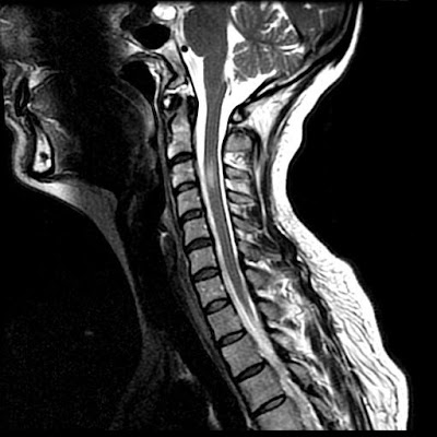
Figure 1: Sagittal T2-weighted image demonstrates an elliptical focus of high signal in the spinal cord from the mid C3 to the upper C4 vertebral body level. There is slight cord expansion.
Figure 2: Axial T2-weighted image at the C3 level demonstrates high signal in the right side of the spinal cord. There is slight extension of the abnormal signal across midline. Note the absence of the normal flow void in the right vertebral artery.
Figure 3: Aortic arch angiogram demonstrates no filling of the right vertebral artery consistent with occlusion.


Diagnosis: Sulcal artery syndrome following vertebral artery dissection


Most spinal cord infarctions involve the anterior two thirds of the spinal cord in an anterior spinal artery distribution. As both anterior horns are affected, patients typically present with bilateral extremity weakness and dissociated sensory deficits secondary to the involvement of cortical spinal and spinal thalamic tracts. Vibration and proprioception testing are normal as there is sparing of the posterior columns. In acute cases, MRI imaging demonstrate swelling of the cord with high T2-weighted signal intensity in the central aspect of the cord which creates the "owl's eye" appearance on the axial images due to involvement of the central gray matter.
A second, rarer cord ischemia syndrome is cord infarct due to occlusion of the posterior spinal artery. Patients present with deficits in vibration and proprioception testing as well as ataxia. Except for elevation in protein, CSF analysis is usually normal in all types of spinal cord infarction.

The rarest form of cord infarction involves only a unilateral sulcal artery. Unlike anterior spinal artery infarcts, where both anterior hemicords are involved, in unilateral sulcal artery infarctions only one-half of the anterior spinal cord is involved. Thus patients present with an acute Brown-Séquard-like syndrome without vibratory sensation or proprioception as the posterior columns are not involved. This type of lesion appears to involve only the upper cervical spinal cord. This may be due to the better vascular and possible collateral supply of the cervical cord than then thoracic spinal cord.

On axial imaging there is a unilateral central cord lesion with a linear extension to the contralateral central cord suggesting involvement of the crossing spinothalamic pathways. Contrary to the common belief that patients with spinal cord infarction have a poor prognosis, all patients with such sulcal artery infarction improve quickly and significantly with minimal or no deficits at the follow-up, most likely owing to the rapid development of collateral flow from other branches of the anterior or the posterior spinal artery. It is worth pointing out that dissection might not be the only mechanism to explain such a syndrome as similar sulcal artery syndromes have also been reported due to other etiology such as atherosclerosis or aortic emboli.

Our patient’s MRI findings are consistent with an infarction in the sulcal artery which is the penetrating branch of the anterior spinal artery. The sulcal arteries arise from the anterior spinal artery through the anterior median fissure. Successive sulcal arteries generally alternate in their distribution to the left or right side of the spinal cord but not both. The sulcal arteries supply the anterior two-thirds of the spinal cord at any cross-sectional level. The sulcal artery occlusion in this case could be due to an embolus traveling from vertebral artery dissection site via anterior radicular artery around the C3/4. A mechanism of hypoperfusion is unlikely for such an infarction in a terminal branch artery territory.

Review of the literature identified a few additional reported cases of sulcal artery territory infarction although other forms of spinal cord infarct following vertebral artery dissection are more common. All the patients with sulcal artery territory infarction had involvement of the upper cervical spinal cord. All had unilateral Brown-Séquard-like presentations with ipsilateral arm and leg weakness, contralateral sensory loss to temperature or hyperalgesia, and minimal or no loss of vibratory sensation and proprioception. All patients had significant improvement at short-term follow up with minimal or no neurological deficits. Spinal fluid examination was normal in two of the four patients tested. Three of the four patients had MRI and all showed a pencil like appearance on the sagittal plane. On axial image, two patients had a characteristic hemi-central cord lesion with anterior extension to the contralateral central counterpart.

lunedì 18 ottobre 2010

Basilar meningitis of unknown etiology


Findings

Figure 1,Figure 2, Figure 3, Figure 4: Axial and sagittal T1 weighted images postcontrast demonstrate thick and nodular predominantly basilar meningeal enhancement which on sagittal images encases the middle cerebral arteries. Cerebellar parenchymal enhancement (Figure 4) secondary to late subacute infarction and infectious/inflammatory exudates.
Figure 5: Axial T2 weighted image demonstrates slight increased size of lateral ventricles with more prominent frontal horn lateral ventricles and bilateral lateral ventricle atria in a 2 week follow-up study (initial study slightly motion degraded.)


Diagnosis: Basilar meningitis of unknown etiology (presumed MDR-Tuberculosis)


This young patient with immunocompromise and altered mental status presented initially with left posterior circulation infarctions of unclear etiology. In a young patient with posterior circulation infarctions, acute vertebral artery dissection/ injury in the setting of trauma or spontaneous etiology should be considered. However, when given a history of immunocompromise and HIV, other differential diagnostic considerations such as infectious (Tuberculous, fungal, or pyogenic), inflammatory (Neurosarcoidosis), vascular (infectious vasculitis from Neurosyphilis or HIV), or neoplastic (lymphoma, leukemia, leptomeningeal carcinomatosis) etiologies should all be taken into account. To date, laboratory and CSF values for this patient continue to be non-diagnostic as to the source of this patient's basilar meningitis/vasculitis. The top consideration after discussing with the neurology team is Multi-drug resistant tuberculosis (MDR-TB) due to unimpressive response to conventional treatment regimens and inconclusive microbiological testing.

Tuberculous CNS infections are mostly caused by M. tuberculosis; atypical organisms are rare except in immunosuppressed patients. 30% of patients are HIV positive (particularly IV drug users.) Due to hematogenous dissemination, lesions usually occur at the gray-white junctions of cerebral hemispheres, basal ganglia, or cerebellum (especially in children). Hematogenous dissemination is from a systemic source, most commonly the lung, but also possibly the GU system or GI tract.

Most common manifestations of CNS Tuberculosis are meningitis, seen predominantly in the basilar cisterns, and hydrocephalus. Acutely, cerebritis can be seen, which then can progress to ring-enhancing tuberculoma lesions. Tuberculosis can also result in vasculitis and cerebral infarctions. The thick and nodular basilar meningeal enhancement in association with hydrocephalus and left posterior circulation multifocal infarctions in our patient can all be seen in tuberculosis.

Leptomeningeal sarcoidosis must be distinguished clinically from carcinomatous, lymphomatous/leukemic, and infectious meningitis. Dramatic response can be seen in some cases with steroid therapy. Sarcoid has replaced syphilis as the great mimicker. Neurosyphilis can cause arteritis of intracranial and extracranial large and medium sized arteries

martedì 28 settembre 2010

Neurovascular compression syndrome of VII-VIII cranial nerves


Findings

High resolution T2 images at the level of the internal auditory meatus demonstrate the facial and vestibulocochlear cranial nerves entering the internal auditory meatus. The facial nerve lies anterior while the eighth cranial nerves courses posteriorly. With the left side used for reference, a tortuous basilar artery is seen posteriorly displacing the normal course of the right vestibulocochlear/facial nerve complex. The presumed point of symptomatic compression is encountered posteriorly as the nerve complex “bends” over posterior aspect of the internal auditory meatus. Although sometimes seen in asymptomatic individuals, when findings are viewed in the appropriate clinical setting, NVCS should be raised as a diagnostic possibility.


Diagnosis: Dolichoectasia of the basilar artery causing right sided sensorineural hearing loss


Hearing loss can be characterized as either conductive or sensorineural based on clinical exam and audiometry. Conductive hearing loss involves an abnormality of the external auditory canal to the oval window and is best evaluated with high resolution CT as it is able to display the external auditory canal and middle ear structures, particularly the ossicles. Sensorineural deafness implies an abnormality of the inner ear, vestibulocochlear nerve, or its central components which are best evaluated with MRI.

Neurovascular compression syndrome (NVCS) refers to a group of disorders in which an aberrant or tortuous vessel causes nerve compression with subsequent hyperexcitation and neuropathy. Vascular compression syndrome has been described as a causative etiology for cranial nerves III, V, VII, VIII, and IX. Controversy exists, however, because of the normal intimate apposition of nerves and vasculature around the brainstem and the frequency with which it is seen in asymptomatic patients.

Vestibulocochlear NVCS is symptomatic vascular compression of cranial nerve VIII. Clinical symptoms are often non-specific including tinnitus, vertigo, and sensineural hearing loss. A recent article in the American Journal of Neuroradiology failed to reliably determine neurovascular compression as a cause of tinnitus although some authors maintain it may still be considered when presenting with so called "typewriter" tinnitus. In decreasing order of frequency, vessels indicated in NVCS include the anterior inferior cerebellar artery, posterior inferior cerebellar artery, and vertebral artery.

The following MRI classification system for neurovascular compression has been proposed to aid in surgical planning.
- Type I: Point compression where a limited segment of the nerve is in contact with the vessel.
- Type II: Longitudinal compression in which the nerve and vessel traverse parallel to each other.
- Type III: A vascular loop encircling the neve.
- Type IV: The nerve contour is deformed and/or thinned.

Definitive treatment involves retromastoid craniectomy and microvascular decompression in which a small synthetic sponge is interposed between the offending vascular structure and nerve.