giovedì 22 aprile 2010

Pars flaccida cholesteatoma


Findings

There is an 8mm mass in Prussak’s space with erosion of the malleus and scutum.

Differential diagnosis:
- Pars flaccida cholesteatoma
- Cholesterol granuloma
- Paraganglioma
- Pars tensa cholesteatoma


Diagnosis: Pars flaccida cholesteatoma


Key points

A pars flaccida cholesteatoma often occurs when a patient has chronic middle ear inflammation and/or TM perforation. The cholesteatoma forms when there is an accumulation of stratified epithelial cells in Prussak's space. The cholesteatoma can be seen in all age groups, but tend to be more aggressive in children. Patients can present with aural discharge, conductive hearing loss, and otalgia. Early treatment with surgery can preserve hearing.


Radiologic overview of the diagnosis

A pars flaccida cholesteatoma appears as a mass in Prussak's space with erosion of the scutum and/or adjacent ossicle. Ossicle erosion is seen ~70% of the cases. There is no enhancement of the cholesteatoma itself, though surrounding granulation tissue may enhance.

High resolution temporal bone CT is the best modality to evaluate a suspected cholesteatoma. A pars tensa cholesteatoma is far less common and involves the sinus tympanum. A cholesterol granuloma appears blue on otoscopy and may have similar bony erosions as that of a cholesteatoma. A paraganglioma appears as a cherry red mass on otoscopy and usually does not erode bone.

In this case, there is an 8mm mass in Prussak's space with erosion of the malleus and scutum.

lunedì 19 aprile 2010

Leptomeningeal carcinomatosis from esophageal adenocarcinoma


Findings

Figure 1, Figure 2, Figure 3, and Figure 4: Postgadolinium T1 weighted images demonstrate linear contrast enhancement in the subarachnoid space, most notably interdigitating between the folia of the superior cerebellum and in the sulci of the parietal and occipital lobes. The T1 weighted precontrast images are unremarkable.
On FLAIR imaging in Figure 5, there is corresponding hyperintensity in the subarachnoid space.
Chest TC (not shown) demonstrates a high-attenuation central mass within the esophagus, which is expanding the lumen.


Diagnosis: Leptomeningeal carcinomatosis from esophageal adenocarcinoma


The leptomeninges consist of two layers; the pia mater and the arachnoid mater, which enclose the subarachnoid space and the CSF. The leptomeninges are a frequently missed site of metastastic involvement, especially for non-hematologic primary malignancies. There are many proposed routes of entry for tumor cells into the CSF including hematogenous spread via the arachnoid vessels or choroid plexus, direct extension from the skull, vertebrae, dura or retrograde perineural spread via the peripheral or cranial nerves. Once tumor cells reach the CSF, rapid dissemination can occur.

Up to 50% of patients with leptomeningeal metastases present with signs of increased intracranial pressure and/or hydrocephalus including headaches, back pain, nausea, vomiting and dizziness. These symptoms are most likely secondary to obstruction of CSF flow by tumor cells. Other clinical manifestations include seizures, focal cranial nerve deficits and meningeal signs such as nuchal rigidity and photophobia.

CSF cytology is the definitive test for diagnosis of leptomeningeal involvement. However, while highly specific, cytology is often falsely negative. The accuracy of a single lumbar puncture is approximately 50%, which increases to 90% with three LPs. Cytology remains negative in 10-20% of patients, presumably in situations in which the malignant cells are more adherent to the leptomeninges.

Contrast enhanced MR imaging is the diagnostic test of choice as an adjunct to CSF cytology when leptomeningeal metastases are suspected. Gadolinium enhanced MRI is more sensitive than a single lumbar puncture, but is less specific. The most common imaging findings include diffuse leptomeningeal contrast enhancement, multiple masses or nodules within the subarachnoid space and/or hydrocephalus. The diffuse leptomeningeal enhancement pattern has been referred to as sugar-coating or zuckerguss (German for icing or sugar-coating). Studies have shown that contrast-enhanced T1-weighted MR imaging is the most sensitive single sequence for detection of leptomeningeal metastases.

Prognosis for leptomeningeal metastases is poor. Without treatment, the average survival is 1-2 months. Treatment options include corticosteroids, intrathecal chemotherapy and radiation therapy. Despite aggressive therapy, the median survival in most randomized-controlled trials is 3-4 months.

martedì 6 aprile 2010

Acute infarction secondary to occlusion of artery of Percheron


Findings

Figure 1: Coronal DWI image demonstrates restricted diffusion within bilateral medial inferior thalami and superior midbrain.
Figure 2: Coronal DWI image, obtained 12 days later demonstrates relatively decreased diffusion hyperintensity.
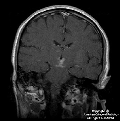
Figure 3: Coronal post-contrast coronal T1 weighted image demonstrates new enhancement involving bilateral inferior medial thalami and superior midbrain, secondary to breakdown of blood-brain barrier.
Figure 4: Diagram illustrating normal paramedian thalamic mesencephalic arterial supply, with many perforating vessels arising from bilateral P1 segments of the PCA. (Reprinted with permission from AJNR)
Figure 5: Diagram illustrating the variant, “artery of Percheron” a single perforating blood vessel arising from one P1 segment.


Diagnosis: Acute infarction secondary to occlusion of artery of Percheron


The thalami and the midbrain receive their blood supply from both the anterior and posterior circulations, and several variations in this supply are known to exist. The anterior circulation usually supplies the anteroinferior aspects of the thalami and midbrain, with thalamoperforator arteries arising from the posterior communicating arteries. The posterior circulation usually supplies the medial aspects of the thalami and midbrain via branches arising from P1 segments and the lateral and superior aspects with branches arising from P2 segments of the posterior cerebral arteries. Most of the perforating branches from the P1 segments have an ipisilateral distribution (78%); bilateral or even contralateral distributions may be observed in 22% of individuals.

Percheron studied the variations of this arterial supply and its distributions and described three different variations involving the paramedian thalamic-mesencephalic arterial supply: (1) small branches arising from both P1 segments, (2) an asymmetrical common trunk arising from a P1 segment (this variation is called the artery of Percheron), (3) or an arterial arcade emanating from an artery bridging the two P1 segments. In the second type, a common trunk arising from one of the P1 segments provides bilateral distribution. Occlusion of this trunk results in bilateral infarctions in the middle aspects of thalami and brain stem.

The thalami contain strategic nuclei and integrate several important cortical functions. Thus, infarcts at the mesencephalic-diencephalic junctions may result in complex clinical syndromes, with patients exhibiting a wide range of symptoms varying from motor deficits to behavioral and sensory alterations. The third nerve palsy demonstrated in this particular patient may have been due to involvement of the midbrain at the level of the Edinger-Westphal nucleus.

Performing conventional angiography may not be indicated, because lack of visualization of the artery does not exclude its presence (because it is occluded).

martedì 30 marzo 2010

Fahr disease


Findings

Figure 1 and Figure 2: Select axial non-contrast CT images demonstrate dense symmetric calcification in the basal ganglia (Figure 1) and dentate nuclei (Figure 2). Cortical atrophy is also seen.
Figure 3: Axial T1-weighted image demonstrates symmetric cerebellar hyperintense foci corresponding to calcification seen on CT.


Diagnosis: Fahr disease (Idiopathic familial basal ganglia calcification)


Fahr disease (Idiopathic familial basal ganglia calcification, bilateral striopallidodentate calcification, Familial cerebrovascular ferrocalcinosis) is a rare disorder characterized by idiopathic basal ganglia calcification with associated cognitive and neurobehavioral manifestations.

Calcification is found primarily in the globus pallidus, but the putamen, caudate, thalamus, cerebellum (especially dentate nucleus), corona radiata, and subcortical white matter can also be affected. There are no detectable abnormalities of calcium or phosphate metabolism. Patients develop progressive parkinsonism, dystonia, and neuropsychiatric disturbance.

There is a bimodal pattern of onset. Those affected in early adulthood may be asymptomatic in the first two decades, despite the presence of basal ganglia calcification. The disease presents with schizophreniform psychosis. A second peak of onset is seen in late middle age, when patients present with subcortical dementia or Parkinsonian symptoms, which are permanent and progressive. Paroxysmal dystonic choreoathetosis and seizures are common. Eventually patients develop symmetrical spastic paralysis progressing to a decerebrate state.

The disease process involves the deposition of calcium in the walls of the capillaries and larger arteries and veins. Other elements, including magnesium, zinc, aluminum, and iron have also been found deposited in the vessels. No definitive treatment is available.

In studying a three-generation family with an autosomal dominant form of the disease, Geschwind et al, established that the chromosomal locus, IBGC1, lies on chromosome 14, and found that this form of the disease demonstrates genetic anticipation. Autosomal recessive inheritance has also been documented. The disease demonstrates variable expressivity.

CT images demonstrate bilateral, symmetric calcification in the globus pallidus, cerebellum, and white matter. On T1-weighted MR images calcifications are hyperintense, while on T2-weighted and FLAIR images, calcification may be hypo- or hyperintense. T2 hyperintense regions in the white matter, which do not correspond to calcification can also be seen. This finding may reflect progressive inflammation.

Differential diagnosis for inherited and acquired basal ganglia calcification
Postinflammatory causes:
- Tuberculosis
- Toxoplasmosis
- Cystercercosis
- Congenital HIV

Endocrine causes:
- Hyperparathyroidism
- Hypoparathyroidism
- Pseudohypoparathyroidism
- Hypothyroidism

Congenital causes:
- Tuberous sclerosis
- Down syndrome
- MELAS
- Neurofibromatosis

Toxic causes:
- Exposure to carbon monoxide
- Chemotherapy
- Radiation therapy
- Lead intoxication

In addition, incidental basal ganglia calcification is seen frequently on CT imaging in patients over the age of 50.

venerdì 26 marzo 2010

Mechanical thrombectomy of the MCA


Findings

Figure 1: CT angiography of the head demonstrates a left MCA occlusion at the distal M1 area. There is visualization of distal vessels with filling by collaterals.

Figure 2, Figure 3, Figure 4, and Figure 5: CT perfusion images demonstrate increased transit time and poor flow in the left MCA territory. There is poor flow in the right posteroparietal region. There is generally preserved blood volume except for decreased volume in the left posterior watershed area. These findings are compatible with ischemia with some areas of infarct and fairly large area of preserved viability.

Diagnostic/therapeutic cerebral angiogram findings: Digital angiography confirms that the left M1 segment is occluded at its mid-portion (Figure 6). Subsequently, the Merci catheter is deployed in the region of the thrombus (Figure 7). Following Merci thrombectomy, there is recanalization of the left M1 segment of the middle cerebral artery (Figure 8).


Diagnosis: Mechanical thrombectomy of the MCA


Catheter-directed thrombectomy systems can be successfully applied towards salvaging areas of reversible ischemia detected on cerebral perfusion imaging.
The MERCI catheter thrombectomy technique involves insinuating the catheter’s coiled tip beyond the thrombus and then retracting the catheter, thereby extracting the clot material.

Until recent years, intravenous recombinant tissue plasminogen activator (TPA) administered within three hours following symptom onset was the only FDA approved treatment for acute stroke. However, mechanical thrombectomy has since emerged as an effective treatment option for acute cerebral ischemia. Indeed, satisfactory results have been achieved with mechanical thrombectomy in some patients that present beyond the time period for intravenous TPA administration has passed, up to about 8 hours. Specifically recanalization rates of nearly 70% have been reported using mechanical thrombectomy.

Various catheter-based devices and techniques have now been devised for mechanical thrombectomy in the cerebral arteries. Some of these include the use of a snare, the alligator retrieval system, the Phenox clot retriever, the Merci catheter, among others.

The Merci catheter thrombectomy technique consists of obtaining femoral artery access, guidewire insertion, and advancement of the catheter to the site of occlusion. Subsequently, the balloon is inflated and the coiled tip of the catheter is passed across the clot and engaged under angiography. The mesh helps trap thrombus material. Clot is retracted into the balloon guide catheter and out of the body. Finally, the balloon is deflated, thereby restoring blood flow.

The main complication of mechanical thrombectomy is intracranial hemorrhage, which occurs in an estimated 5 to 10% of patients.

martedì 23 marzo 2010

Focal cortical dysplasia, Taylor type


Findings

The MRI of the brain shows T2 and FLAIR hyper intense arrow-shaped left frontal white matter lesion pointing toward the frontal horn of the left lateral ventricle. There is no abnormal enhancement. There is mild hypo intensity on T1 sequences.

Differential diagnosis:
- Focal cortical dysplasia
- Gliosis
- Glial cell tumor
- Physiologic margination of white matter neurons


Diagnosis: Focal cortical dysplasia, Taylor type


Key points

Focal cortical dysplasia is considered to be in the spectrum of migrational disorders of the brain, and often manifests in the form of seizures, developmental delay and focal neurologic deficits. Focal cortical dysplasia is thought to represent the cause of between 5% and 25% of patients with focal epilepsy. This entity typically manifests in the first years of life. Antiepileptic medications are the first line of therapy, and no particular medications have been found to be more or less effective for focal cortical dysplasia. After two failed rounds of antiepileptic regimens, surgical interventions may be considered.


Radiology

MRI is the examination of choice for identifying focal cortical dysplasia. The typical appearance of the Taylor type of this disorder is that of T2/FLAIR hyper intensity within the sub cortical white matter, tapering toward the lateral ventricle. The most typical location of this finding is unilaterally within the frontal lobes. The lesions do not enhance.

TANTI AUGURI PROFESSO'

Happy Birthday to my great friend and teacher, Eytan Raz M.D. !!!