venerdì 24 dicembre 2010

Pseudotumor cerebri - Idiopathic Intracranial Hypertension (IIH)


Findings

T2W axial MRI (Figure 1) shows signs of increased ICP, but only increased fluid within the optic nerve sheaths, flattening of the posterior orbit, and a partially empty sella.
The 3D TOF MRV Towne and RPO projections (Figure 2 and Figure 3) show bilateral, right greater than left, focal transverse-sigmoid venous sinus junction narrowing’s. It is not a normal MRV given the pt’s history, with more explanation in the discussion. There is no aneurysm or collection of collateral blood vessels seen in these images.

The AP and lateral (Figure 4 and Figure 5) venous phase carotid arteriogram shows long segment stenosis at transverse-sigmoid venous sinus junction distal to the vein of Labbé. Pre procedure venography showed a venous pressure gradient across this lesion of 17 mmH2O with 37 mmH2O on transverse sinus side and 15 mmH2O on internal jugular vein side.

AP and lateral (Figure 6 and Figure 7) venous phase carotid arteriogram shows long segment stenosis at transverse-sigmoid venous sinus junction with a balloon crossing the gradient lesion.


Diagnosis: Pseudotumor cerebri - Idiopathic Intracranial Hypertension (IIH)


Pseudotumor cerebri is defined by typical clinical symptoms which occur in the setting of elevated “idiopathic” ICP and a normal composition of CSF. Classic clinical symptoms include diffuse recalcitrant headaches, vision changes (including vision loss), and hearing changes (e.g., tinnitus), and the disease is typically seen in obese women who are 20-50 years of age. Papilledema is the most common physical exam finding, but visual loss and sixth nerve palsy are also seen. Other symptoms include disabling headaches and blindness. LP opening pressure is greater than 25 cm H2O. Brain computed tomography (CT) and magnetic resonance imaging (MRI) are typically normal, however, the following suggestive non-pathognomonic findings are frequently present:

– Cerebral venous sinus stenoses
– Flattening of the bilateral posterior sclera
– Partially or fully empty sella; enlargement of the chiasmatic recess of the 3rd ventricle
– Distension of perioptic nerve subarachnoid space
– Intraocular protrusion of the optic nerve head
– Orbital optic nerve vertical tortuosity

Treatment for pseudotumor cerebri typically includes medical management with acetazolamide and pain control for headaches. Furosemide and corticosteroids have been used, as well. Surgical interventions to treat pseudotumor cerebri include lumboperitoneal shunt (LPS) and ventriculoperitoneal shunt (VPS), which often produce immediate results, however, eventual return of pseudotumor symptoms occur in approximately 50% within three years. Optic nerve sheath fenestration is also used to treat vision changes, with variable headache relief. Dominant transverse/sigmoid venous sinus angioplasty and stenting are relatively new methods for the treatment of pseudotumor cerebri for those who have significant dural sinus stenosis. Given that 80% of intracranial vascular compliance is provided from the venous vasculature, reduction of pressure in the sinuses reduces CSF pressure. Better results are achieved in patients with documented high pressure gradients, and greater efficacy is seen with regard to arrest of visual loss (>90%) than with headache relief (~50%). Long-term results are lacking. however.

In this case, cerebral angiography demonstrated bilateral high-grade transverse/sigmoid sinus stenoses distal to vein of Labbe insertions. Selective catheterization of the right transverse sinus revealed an estimated 80% narrowing to a luminal diameter of 1mm, and a pressure gradient across the stenosis of 13 mmHg (normal <5 mmHg). The contralateral sinus was smaller, but distally stenotic. A stent was placed across the right sided stenosis.
The patient was placed on antiplatelet medication to preserve stent patency immediately after the procedure. She had no headaches after the procedure and demonstrated objective visual improvement at her one- and six-week follow-up examinations.

mercoledì 22 dicembre 2010

Intracranial pseudotumor (Tolosa-Hunt syndrome)


Findings

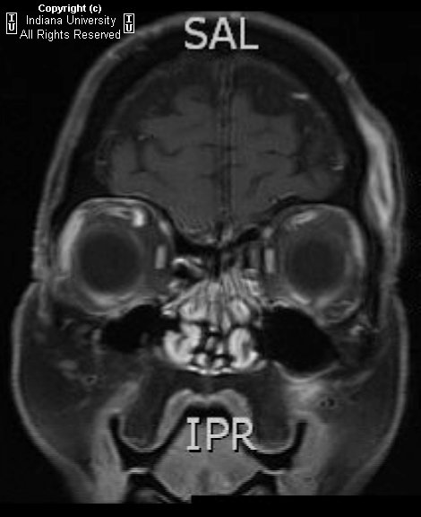
Increased CSF signal demonstrated by T2 hyper intensity within the right optic sheath. Nodular enhancement at the superior aspect of the right optic nerve at the orbital apex, which extends with prominent nodular enhancement posteriorly along the dural reflection of the right cavernous sinus. Asymmetric dilatation of the right superior ophthalmic vein.

Differential diagnosis:
- Meningitis
- Sarcoidosis
- En plaque meningioma
- Meningeal metastases
- Meningeal Non-Hodgkin's lymphoma
- Tolosa-Hunt syndrome


Diagnosis: Intracranial pseudotumor (Tolosa-Hunt syndrome)


Discussion

Intracranial pseudotumor (Tolosa-Hunt syndrome) is a part of a spectrum of "quasineoplastic" lesions that demonstrate orbital, intracranial, or pulmonary involvement, and include such other disorders as plasma cell granuloma and hypertrophic cranial pachymeningitis. It is a chronic granulomatous disease of unknown origin, which has been hypothesized to represent a low grade fibrosarcoma of inflammatory cells versus an autoimmune phenomenon. While Tolosa-Hunt syndrome is rare, orbital pseudotumor is the third most common ophthalmic disorder, and encompasses 5-8% of all orbital masses. Histologically, the lesions of Tolosa-Hunt demonstrate mixed lymphocytic and plasma cell infiltrate, with a variable degree of fibrosis. Lesions favor the cavernous sinus and basal meninges, although falcine and tentorial lesions have been described.

Patients with intracranial lesions are more frequently young adults, who present initially with chronic headaches or cranial nerve palsies. Patients with orbital involvement are more frequently middle-aged, and may present with painful proptosis and vision loss . Symptoms may be intractable, leading to severe disability. Untreated or unresponsive disease may progress to death. First-line treatment is invariably high-dose steroids, with radiotherapy or surgical resection reserved for patients with incomplete response to steroids .


Radiologic Overview of the diagnosis

The imaging hallmarks of Tolosa-Hunt syndrome are characterized by an enhancing, infiltrating meningeal mass, which favors the cavernous sinus or basal meninges, although falcine and tentorial involvement has been described . Focal meningeal thickening may range from just a few millimeters to a greater than 2 cm rind. With intracranial pseudotumor, orbital involvement is spared more than 90% of the time. Tolosa-Hunt remains a diagnosis of exclusion, once meningitis, en plaque meningioma, and meningeal metastases are ruled out.

The imaging modality of choice for imaging patients with Tolosa-Hunt remains MRI, although useful information may be gleaned from other modalities. On non-contrast enhanced CT (NECT), there are no specific findings to suggest the diagnosis; however, this modality may be of some value in differentiating the lesion from en plaque meningioma. On contrast enhanced CT (CECT), salient imaging findings include enhancing, thickened meninges or a curvilinear appearance of a single meningeal region. As aforementioned, MRI remains the primary modality for diagnosis of Tolosa-Hunt syndrome, and each sequence may provide critical information required to make the diagnosis. On T1WI, one may find focal thickening of the meninges that is isointense to gray matter. On T2WI, lesions are characterized as iso- to hypo intense regions of focal meningeal thickening, which may be more hypo intense as they become more fibrotic. FLAIR is of little help in making the diagnosis, but it is unlikely to demonstrate focal brain edema underlying the lesion . Contrast enhanced T1WI is the single most valuable sequence for evaluation of Tolosa-Hunt syndrome, and is characterized by diffusely enhancing region of meningeal thickening, which may range from a few millimeters to greater than 2 cm in some cases. Diffuse boney infiltrates may be appreciated on fat saturated contrast enhanced T1 sequence. While angiography is not considered a primary modality, severe disease may result in carotid artery narrowing, thus MRA may be a useful adjunct in the appropriate clinical setting.

The appropriate differential diagnosis of Tolosa-Hunt syndrome includes meningitis, sarcoidosis, en plaque meningioma, meningeal metastases, and meningeal Non-Hodgkin's lymphoma.

venerdì 17 dicembre 2010

Hypertensive intracranial hemorrhage


Findings

Axial CT of the head shows a large hyper dense focus with peripheral hypo density in the left frontal lobe, causing sulcal effacement at the frontal cortex but no significant midline shift. Surrounding rim of low density represents edema (image 1). Unenhanced MRI of the brain shows an the same mass-like focus in the left frontal lobe, which has an isointense center with a hyper intense rim (image 2). Post gadolinium-enhanced T1 image of the brain shows no internal enhancement of this lesion (image 3).


Differential diagnosis:
- Hypertensive intracranial hemorrhage
- Ruptured arteriovenous malformation (AVM)
- Hemorrhagic intracranial mass
- Posttraumatic cerebral contusion


Diagnosis: Hypertensive intracranial hemorrhage


Acute blood appears hyper dense of unenhanced head CT.
Without a history of trauma, intraparenchymal brain hemorrhage on head CT could represent a hemorrhagic mass, a ruptured AVM, or a hemorrhagic brain tumor (primary or metastatic).
MRI with and without contrast is the best diagnostic tool for determining if a mass lesion is present, and for evaluating the age of the intracranial hemorrhage.
Acute blood products on T1 appear hypo intense to isointense (image 2), whereas subacute and chronic hematoma are hyper intense.
Neoplasms should enhance on post contrast T1. The lack of enhancement in this patient on post contrast T1 excludes neoplasm (image 3).

giovedì 16 dicembre 2010

Sequestered disk


Findings

There is an non- enhancing ovoid mass slightly hyper intense to muscle on both T1 and T2 sequences, in the anterior epidural space at the L3 level, measuring approximatelyl 12 x 8 x 12 mm. This is not contiguous with any adjacent disks. No signal dropout on fat-saturated sequences. The mass causes severe stenosis of the left half of the spinal canal at the L3 level, compressing the left descending nerve roots. T1 and T2 hyper intensity at the endplates abutting L2-L3 disc space representing Modic Type II changes. There is intervertebral disk space height loss at L2-L3 with severe disk desiccation changes.


Differential diagnosis:
- Sequestered disk
- Extruded disk
- Failed back surgery
- Epidermoid
- Epidural abscess
- Epidural hematoma
- Lipoma


Diagnosis: Sequestered disk


A focal disk protrusion is an extension of intervertebral disc material (nucleus pulposus) beyond the vertebral margin (AP diameter < mediolateral diameter). An extruded disk is one in which the nucleus pulposus has herniated through a rent in the annulus fibrosis. The AP diameter > ML diameter, and the disk may migrate craniocaudally, but maintains attachment to the parent disk (frequently symptomatic).
When extruded disk material loses its attachment to the parent disk, it is referred to as a sequestered disk. Sequestered discs usually lodge in the anterior epidural space (AES), just anterior to the posterior longitudinal ligament, and migrate either cephalad or caudad (with equal frequency). Because there is a midline septum associated with the PLL in the AES, the fragment is usually just off midline (to the right or left). Rarely, the sequestered fragment may migrate beyond the PLL into the posterior epidural space, through the dural ( intrathecal location), or into the paraspinal muscles.
They usually resemble the parent disk on MR, with T1 hypo intense and T2 iso- / hypo intense. There may be surrounding T2 hyper intensity and a rim of enhancement from inflammatory changes.
This is a crucial diagnosis to make, as a sequestered disk is a contraindication to limited disk procedures (e.g. Percutaneous discectomy) and may result in failed back surgery.

martedì 14 dicembre 2010

Myxopapillary ependymoma


Findings

Figure 1: Sagittal T1-weighted images reveals an isointense lobulated intradural mass at the level of the conus medullaris.
Figure 2: Sagittal T2-weighted images shows a hyperintense lobulated intradural mass extending from T11 through L2 with numerous small flow voids.
Figure 3: Sagittal T1 post-contrast images demonstrates intense enhancement of the intradural mass centered around the conus.


Diagnosis: Myxopapillary ependymoma


Myxopapillary ependymoma is a slow-growing tumor arising from the ependymal cells of the filum terminale. These tumors compromise 13% of all spinal ependymomas, and they occur almost exclusively in the conus, filum terminale, and cauda equina although extradural occurence in the sacrum and presacral region has also been reported.

The lesions tend to span two to four vertebral segments, and appear as a well-circumscribed intradural masses. In most cases the tumor is intrinsic to the conus medullaris but this is often difficult to recognize on imaging as the bulk of the mass is extramedullary. Typical MR characteristics include T1 isointensity, T2 hyperintensity, and avid enhancement on post-contrast images. As these tumors are prone to hemorrhage, a hypointensity at the tumor margin is often seen indicative of hemosiderin. Calcification and cyst formation within the mass are not infrequent.

On radiography and CT, vertebral changes can be seen which include widened interpediculate distance, thinned pedicles, posterior vertebral scalloping, and intervertebral foraminal widening due to tumor extension.

They are more common in males (M:F=2:1) with a mean age of 35 at diagnosis. Clinically, they present with back pain, paraparesis, radiculopathy, and occasionally bowel and bladder dysfunction. Because these symptoms can mimic those of disc herniation, there is often a delay in diagnosis. Treatment consists of surgical resection, and the prognosis is excellent with complete resection. Leptomeningeal seeding metastasis in myxopapillary variety is not as frequent as it is in classic spinal cord ependymomas and associated with poorer prognosis when present. Radiotheraphy after surgery improves outcome.

venerdì 10 dicembre 2010

Capillary Telangiectasia


Findings

There is an ill-defined enhancing focus in the medial right temporal lobe on post gadolinium contrast T1-weighted imaging (Figure 4). There is no corresponding signal abnormality or mass on the precontrast T1-weighted, T2-weighted, or FLAIR images (Figure 1, Figure 2, and Figure 3, respectively). There is no mass effect. On susceptibility-weighted imaging (SWI) the lesion shows hypointensity (Figure 5).


Diagnosis: Capillary Telangiectasia


Brain capillary telangiectasias are benign vascular malformations which are often found incidentally.
They can be visualized by gadolinium contrast and gradient-echo susceptibility or susceptibility weighted imaging, but not through catheter angiography, and may often not be visible on conventional T1/T2, FLAIR, or diffusion-weighted imaging.
Often asymptomatic and usually no treatment is required.

Brain capillary telangiectasias (BCTs) are one of four major types of vascular malformations which occur in the brain (the other three are arteriovenous malformations, cavernous malformations (cavernous angiomas), and developmental venous anomalies (venous angiomas), and represent up to 20% of all intracranial vascular lesions. BCTs consist of multiple ectatic capillaries surrounded by normal brain parenchyma and are usually devoid of calcification, gliosis, extraluminal hemorrhage, and hemosiderin-laden macrophages. BCTs are most common in the midbrain, pons, medulla, and spinal cord, but they are found throughout the central nervous system. Multiple BCTs are possible, especially in certain syndromes (e.g.; ataxia telangiectasia, Osler-Weber-Rendu, or Sturge-Weber syndrome).

Often found incidentally, BCTs are usually benign, small in size, and rarely grow over time. They are rarely symptomatic and are not associated with any particular clinical feature but have been reported to be associated with headache, vertigo, and tinnitus.

BCTs are relatively well visualized through susceptibility weighted imaging where they demonstrate marked signal intensity loss due to deoxyhemoglobin present in slow flowing blood. They are also well visualized through gadolinium-enhanced T1-weighted imaging sequences where they are seen as small faint lesions. BCTs are difficult to visualize through conventional T1/T2, FLAIR, or diffusion-weighted imaging and are considered to be one of the “angiographically occult vascular malformations” due to their small size, tendency to occlude, and sluggish flow.

Wernicke’s Encephalopathy


Findings

On axial images, abnormal FLAIR signal is demonstrated at the pontomedullary junction adjoining the fourth ventricle, periaqueductal gray matter in the pons and midbrain (Figure 1), the superior aspect of the mamillary bodies (Figure 2), the tissue surrounding the third ventricle and the medial thalami (Figure 3).
On coronal slices, abnormal FLAIR signal again appears in the mamillary bodies (Figure 8), in the tissue surrounding the third ventricle (Figure 8 and Figure 10), medial thalami (Figure 10), and periaqueductal gray matter (Figure 11).


Diagnosis: Wernicke’s Encephalopathy


Wernicke’s encephalopathy is caused by thiamine deficiency, most often seen in chronic alcohol abuse. It has also been described in anorexia nervosa, prolonged starvation, hyperemesis gravidarum, patients on long-term hemodialysis, and patients with AIDS. Patients with this condition classically present with the triad of ataxia, acute mental confusion, and oculomotor dysfunction, although a minority (16-38%) of patients with the condition present with all three elements. If the symptoms also include amnesia and confabulation, then these manifestations are called Korsakoff syndrome. Wernicke’s encephalopathy is a significantly disabling and potentially lethal condition that can be prevented and reversed if treated early with thiamine supplementation.

On CT and MR imaging the brain demonstrates diffuse cerebral and cerebellar atrophy. Mamillary body enhancement or abnormal T2 signal may be the sole manifestation of Wernicke’s encephalopathy. Other typical MR findings include symmetric high T2 signal and variable enhancement within the periaqueductal gray matter of the midbrain, the tectal plate, the mamillothalamic tract, the thalami, and the tissue surrounding the third ventricle. The mamillary bodies may also show atrophy in patients with chronic Wernicke’s encephalopathy, though this finding can also be present in chronic alcoholic patients without Wernicke’s syndrome. Atypical changes may also be seen, almost always in non-alcoholic patients, and may include signal changes in cranial nerve nuclei, basal ganglia, cerebellum and dentate nuclei, the splenium, and frontal and parietal cortex. These atypical findings are very similar to the pattern seen in metronidazole-induced encephalopathy, and it is has been hypothesized that the two syndromes share a common metabolic pathway. The reason why these brain regions are more affected by thiamine deficiency is poorly understood, but it is speculated that they may be characterized by more intense thiamine metabolism.