

Findings
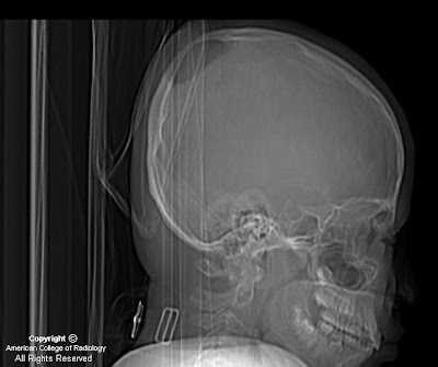
Scout view (lateral view) from a CT scan reveals a large, well-defined parietal calvarial defect.
Brain and bone windows demonstrate bilateral parietal large and symmetric rounded calvarial defects (Figures 2 and 3).
3D surface-shaded reconstructed CT shows to better advantage the 3D characteristics of the calvarial defects and also reveals sagittal and coronal suture synostosis.
Diagnosis: Giant parietal foramina
Giant parietal foramina (GPF) are also known as foramina parietalia permagna, fenestrae parietals symmetricae, enlarged parietal foramina, Catlin marks, and cranium bifidum. GPF is associated with cerebral venous and cortical anomalies, gentic syndromes, and gene mutations. GPS can be an isolated entity, or be inherited in an autosomal dominant fashion. Gene mutations have been linked to both GPS and craniosynostosis. The calvarial defects can be repaired by bone grafts and mesh plating systems in patients at risk for injury to protect the underlying brain from trauma.
There is essentially no differential diagnosis for giant parietal formaina if a good clinical history is obtained (no prior craniectomy, trauma, soft tissue mass associated with the defects, etc). This case illustrates the importance of recognizing the entity of giant parietal foramina. The radiologist should be aware of possible associated intracranial abnormalities such as cortical/cerebral venous anomalies that would be amenable to MRI, MRV, or CTV workup; 3D CT can be very helpful if there is any contemplation of surgical repair.
Nessun commento:
Posta un commento