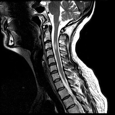
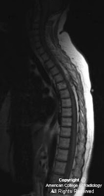
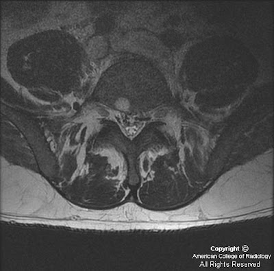
Visualizzazione post con etichetta Spine. Mostra tutti i post

lunedì 27 dicembre 2010

Lumbar disc extrusion with a wrapped disc


Findings

There is a left central disc extrusion at L5-S1 that causes mild to moderate left lateral recess narrowing and nerve root displacement without nerve root compression. At this level there is also contrast enhancement traversing the left laminectomy defect and encasing the disc extrusion, consistent with a wrapped disc. There is enhancement in the left lateral recess, suggesting post-operative fibrosis.

Differential diagnosis:
- Wrapped disc
- Peridural fibrosis
- Epidural abscess
- Epidural metastasis
- Nerve sheath tumor
- Disc pseudobulge
- Intervertebral disc protrusion
- Intervertebral disc extrusion
- Recurrent intervertebral disc herniation


Diagnosis: Lumbar disc extrusion with a wrapped disc


Key points: "Wrapped" disc


Disc herniation (protrusion, extrusion, or fragment) may be caused by trauma, repetitive or acute, and are a common source of pain and subsequent back surgery in the general population. In the acute phase, the herniated disc stimulates a fibrovascular response. A "wrapped disc" is the focal herniation (protrusion, extrusion, or fragment) that is encased in vascular scar tissue stimulated by this response and is evident by enhancement on contrast-enhanced T1-weighted images.

Asymptomatic or low back pain and/or radiculopathy are most common in the lumbar spine at L4-L5 and L5-S1. A wrapped disc is a post-surgical sequela, particularly following surgery for spinal stenosis in which the surgical procedure is more extensive, involving a laminectomy and a medial facetectomy.

Best imaging modality: MR (sequences: sagittal and axial T2WI and T1WI, as well as contrast-enhanced axial and sagittal T1WI)
Other imaging modalities: CT, myelography


Imaging findings

MR: Anterior extradural mass contiguous with the disc space extending into the spinal canal
*Contrast-enhanced T1WI: Peripheral enhancement surrounding the disc herniation or fragment with/without central canal, lateral recess, or foraminal stenosis and cord or nerve root impingement. (*most helpful MR sequence)
Non-enhanced T1WI: Isointense to parent disc
T2WI: Iso- to hyper intense to parent disc
General disc hypointensity and height loss at the level of the herniation, as well as postoperative changes (laminectomy defects, etc), degenerative facet disease, and osteophytes, are common associated findings.
CT:
Non-contrast CT: An anterior extradural soft tissue mass that may displace the nerve root / indent the thecal sac
Contrast-enhanced CT: Mild peripheral enhancement of the disc herniation/fragment
Myelography: An extradural mass that indents the thecal sac and nerve root sleeves
Imaging findings of other common differential diagnoses
Peridural fibrosis: Scar within epidural space after lumbar surgery that infiltrates epidural fat, causing homogeneous enhancement that diffusely surrounds the thecal sac and nerve root; increased in T2 signal relative to adjacent disc herniation
Epidural abscess: A distinct fluid collection in the epidural space with peripheral enhancement on post-contrast images, often associated with findings of diskitis
Epidural metastasis: Elongated (cranial-caudal orientation) enhancing mass with osseous involvement and may demonstrate paravertebral extension
Nerve sheath tumor: Avid enhancement surrounding the nerve root, some of which are in a "dumbbell" shape
Disc pseudobulge: Smooth generalized extension of the disc margin without a focal defect due to "uncovering" of disc related to spondylolisthesis
Intervertebral disc protrusion: Anterior extradural mass contiguous with disc space and triangular in shape with broader base than apex; no enhancement
Intervertebral disc extrusion: Anterior extradural mass contiguous with disc space by a "neck," in which this herniated disc material then widens in the epidural space
Recurrent intervertebral disc herniation: Extradural mass contiguous with intervertebral disc margin, demonstrating enhancement peripherally but without central enhancement
Treatment
Conservative: Anti-inflammatory and pain medications, avoid trauma
Surgical: Repeat surgery to remove herniated disc (protrusion, extrusion, fragment)

giovedì 16 dicembre 2010

Sequestered disk


Findings

There is an non- enhancing ovoid mass slightly hyper intense to muscle on both T1 and T2 sequences, in the anterior epidural space at the L3 level, measuring approximatelyl 12 x 8 x 12 mm. This is not contiguous with any adjacent disks. No signal dropout on fat-saturated sequences. The mass causes severe stenosis of the left half of the spinal canal at the L3 level, compressing the left descending nerve roots. T1 and T2 hyper intensity at the endplates abutting L2-L3 disc space representing Modic Type II changes. There is intervertebral disk space height loss at L2-L3 with severe disk desiccation changes.


Differential diagnosis:
- Sequestered disk
- Extruded disk
- Failed back surgery
- Epidermoid
- Epidural abscess
- Epidural hematoma
- Lipoma


Diagnosis: Sequestered disk


A focal disk protrusion is an extension of intervertebral disc material (nucleus pulposus) beyond the vertebral margin (AP diameter < mediolateral diameter). An extruded disk is one in which the nucleus pulposus has herniated through a rent in the annulus fibrosis. The AP diameter > ML diameter, and the disk may migrate craniocaudally, but maintains attachment to the parent disk (frequently symptomatic).
When extruded disk material loses its attachment to the parent disk, it is referred to as a sequestered disk. Sequestered discs usually lodge in the anterior epidural space (AES), just anterior to the posterior longitudinal ligament, and migrate either cephalad or caudad (with equal frequency). Because there is a midline septum associated with the PLL in the AES, the fragment is usually just off midline (to the right or left). Rarely, the sequestered fragment may migrate beyond the PLL into the posterior epidural space, through the dural ( intrathecal location), or into the paraspinal muscles.
They usually resemble the parent disk on MR, with T1 hypo intense and T2 iso- / hypo intense. There may be surrounding T2 hyper intensity and a rim of enhancement from inflammatory changes.
This is a crucial diagnosis to make, as a sequestered disk is a contraindication to limited disk procedures (e.g. Percutaneous discectomy) and may result in failed back surgery.

martedì 14 dicembre 2010

Myxopapillary ependymoma


Findings

Figure 1: Sagittal T1-weighted images reveals an isointense lobulated intradural mass at the level of the conus medullaris.
Figure 2: Sagittal T2-weighted images shows a hyperintense lobulated intradural mass extending from T11 through L2 with numerous small flow voids.
Figure 3: Sagittal T1 post-contrast images demonstrates intense enhancement of the intradural mass centered around the conus.


Diagnosis: Myxopapillary ependymoma


Myxopapillary ependymoma is a slow-growing tumor arising from the ependymal cells of the filum terminale. These tumors compromise 13% of all spinal ependymomas, and they occur almost exclusively in the conus, filum terminale, and cauda equina although extradural occurence in the sacrum and presacral region has also been reported.

The lesions tend to span two to four vertebral segments, and appear as a well-circumscribed intradural masses. In most cases the tumor is intrinsic to the conus medullaris but this is often difficult to recognize on imaging as the bulk of the mass is extramedullary. Typical MR characteristics include T1 isointensity, T2 hyperintensity, and avid enhancement on post-contrast images. As these tumors are prone to hemorrhage, a hypointensity at the tumor margin is often seen indicative of hemosiderin. Calcification and cyst formation within the mass are not infrequent.

On radiography and CT, vertebral changes can be seen which include widened interpediculate distance, thinned pedicles, posterior vertebral scalloping, and intervertebral foraminal widening due to tumor extension.

They are more common in males (M:F=2:1) with a mean age of 35 at diagnosis. Clinically, they present with back pain, paraparesis, radiculopathy, and occasionally bowel and bladder dysfunction. Because these symptoms can mimic those of disc herniation, there is often a delay in diagnosis. Treatment consists of surgical resection, and the prognosis is excellent with complete resection. Leptomeningeal seeding metastasis in myxopapillary variety is not as frequent as it is in classic spinal cord ependymomas and associated with poorer prognosis when present. Radiotheraphy after surgery improves outcome.

venerdì 3 dicembre 2010

Basilar invagination secondary to rheumatoid arthritis


Findings

Axial and sagittal CT images demonstrate severe basilar invagination (Figure 1). The tip of the odontoid process measures 2.3 cm above Chamberlain’s line (yellow line in Figure 2). McGregor's line (red line in Figure 2) is also shown. Incidentally noted are right-sided opacified mastoid air cells (Figure 1).
Once again, severe basilar invagination is evident. On the sagittal T2 image the foramen magnum is narrowed and obliteration of the CSF space is noted at the C2-C3 level (Figure 3). On the axial T2 weighted image increased T2 signal (Figure 4) is seen within the cord at the C2-C3 level indicating edema versus myelomalacia.


Diagnosis: Basilar invagination (impression) secondary to rheumatoid arthritis.


Basilar invagination refers to a condition in which the odontoid process protrudes upward into the intracranial space. Basilar invagination may be classified as primary (congenital) or secondary (acquired). Down syndrome, Klippel-Feil syndrome and Chiari malformations are congenital causes of basilar invagination. Acquired basilar invagination, also known as basilar impression, is associated with softening of the skull base and is often due to rheumatoid arthritis, Paget disease, osteomalacia, hyperparathyroidism and osteogenesis imperfecta. Basilar invagination is probably better described as a radiologic finding rather than a diagnosis. Once the finding is identified, a cause of basilar invagination should be diligently pursued.

Plain lateral radiographs with odontoid views, although not 100% sensitive, are often the initial study used to diagnose basilar invagination. MRI is the optimal study, which also assesses the cervicomedullary junction and cervical cord. Two craniovertebral junction lines are particularly useful in defining basilar invagination. Chamberlain’s line extends between the posterior pole of the hard palate and the posterior edge of the foramen magnum (opisthion). If the dens is >3.0 mm above this line basilar invagination is present. McGregor’s line, a modification of Chamberlain’s line was developed because the opisthion could not always be seen on plain radiographs. This line extends from the posterior pole of the hard palate to the undersurface of the occiput. If the dens extends >4.5 mm above this line basilar invagination is present.

Clinical manifestations of basilar invagination include posterior skull pain, headache, signs and symptoms of brainstem and upper cervical cord compression or disturbances of CSF circulation causing obstructive hydrocephalus. The brainstem may be compressed at the level of the foramen magnum possibly resulting in compromise of the autonomic centers resulting in labile blood pressures, arrhythmias, or sudden death. Neurosurgery is recommended in patients that are symptomatic with concomitant MRI findings indicating compression. Although asymptomatic patients are often followed conservatively, many authors favor surgery even if no symptoms of cord compression are evident in rheumatoid patients.

Although often appearing together, basilar invagination or impression should not be confused with platybasia; which literally means “flattening of the base of the skull”. Platybasia, which can be seen in Klippel-Feil anomalies, cleidocranial dysplasia and achondroplasia, is present when the basal angle formed by intersecting lines from the nasion to the tuberculum sellae and from the tuberculum along the clivus to the anterior aspect of the foramen magnum (basion) is greater than 143 degrees.

venerdì 12 novembre 2010

Idiopathic Thoracic Cord Herniation


Findings

These MR images demonstrate focal anterior displacement of the spinal in the mid thoracic spine. The cord (Images 1,2 and 3) appears to be either tethered anteriorly or compressed from the posterior aspect. The intradural space behind the cord is widened and has signal characteristics identical to CSF (Images 1,2 and 3).


Diagnosis: Idiopathic Thoracic Cord Herniation


Spinal cord herniation occurs when the cord herniates through a defect in the dura mater. These dural defects are typically located anteriorly or laterally, and occur most often in the mid-thoracic region. They may be idiopathic, post-traumatic or iatrogenic related to prior spinal surgery. Some have suggested that a herniated and calcified disk may cause thinning, erosion, or rupture of the dura, which may also be secondary to congenital weakening of the ventral dural fibers. The presence of free flow of cerebral spinal fluid dorsal to the herniated cord is key to differentiating a spinal cord herniation from an arachnoid cyst. Spinal cord herniation occurs most commonly in the middle-aged. Symptoms of myelopathy including chronic leg pain, gait disturbance, incontinence, and leg weakness are commonly seen and may slowly worsen over time if left untreated. The most common clinical feature reported is the Brown-Séquard syndrome consisting of hemiplegia and contralateral temperature sensation deficits and pain.

Typical imaging findings are focal anterior displacement of the spinal cord with expansion of the dorsal subarachnoid space. The preferred imaging modality in the setting of myelopathy is MRI, which is often sufficient for making the correct diagnosis. Myelography with CT may be required in ambiguous cases and to demonstrate the exact location of the dural defect. With cord herniation, myelography reveals uninterrupted flow of contrast and the absence of a filling defect posterior to the herniated cord segment. An arachnoid cyst will present during myelography as an early filling defect posterior to the displaced cord. Contrast may fill the cyst with time, so rapid acquisition of CT-myelograpgy after the initial myelographic images is essential. Phase contrast cine MR imaging may provide similar CSF flow information, in addition to restricted cord motion.

Treatment consists of surgically reducing the herniation by repositioning the protruding spinal cord back into the thecal sac followed by the repair of the defect in the dural mater in order to prevent recurring herniation. After surgery, symptoms typically improve and may completely resolve, even when longstanding. Patients whose symptoms are milder and non-progressive may be eligible for less invasive therapy or conservative management with monitoring.

giovedì 4 novembre 2010

Sulcal artery syndrome following vertebral artery dissection


Findings

Figure 1: Sagittal T2-weighted image demonstrates an elliptical focus of high signal in the spinal cord from the mid C3 to the upper C4 vertebral body level. There is slight cord expansion.
Figure 2: Axial T2-weighted image at the C3 level demonstrates high signal in the right side of the spinal cord. There is slight extension of the abnormal signal across midline. Note the absence of the normal flow void in the right vertebral artery.
Figure 3: Aortic arch angiogram demonstrates no filling of the right vertebral artery consistent with occlusion.


Diagnosis: Sulcal artery syndrome following vertebral artery dissection


Most spinal cord infarctions involve the anterior two thirds of the spinal cord in an anterior spinal artery distribution. As both anterior horns are affected, patients typically present with bilateral extremity weakness and dissociated sensory deficits secondary to the involvement of cortical spinal and spinal thalamic tracts. Vibration and proprioception testing are normal as there is sparing of the posterior columns. In acute cases, MRI imaging demonstrate swelling of the cord with high T2-weighted signal intensity in the central aspect of the cord which creates the "owl's eye" appearance on the axial images due to involvement of the central gray matter.

A second, rarer cord ischemia syndrome is cord infarct due to occlusion of the posterior spinal artery. Patients present with deficits in vibration and proprioception testing as well as ataxia. Except for elevation in protein, CSF analysis is usually normal in all types of spinal cord infarction.

The rarest form of cord infarction involves only a unilateral sulcal artery. Unlike anterior spinal artery infarcts, where both anterior hemicords are involved, in unilateral sulcal artery infarctions only one-half of the anterior spinal cord is involved. Thus patients present with an acute Brown-Séquard-like syndrome without vibratory sensation or proprioception as the posterior columns are not involved. This type of lesion appears to involve only the upper cervical spinal cord. This may be due to the better vascular and possible collateral supply of the cervical cord than then thoracic spinal cord.

On axial imaging there is a unilateral central cord lesion with a linear extension to the contralateral central cord suggesting involvement of the crossing spinothalamic pathways. Contrary to the common belief that patients with spinal cord infarction have a poor prognosis, all patients with such sulcal artery infarction improve quickly and significantly with minimal or no deficits at the follow-up, most likely owing to the rapid development of collateral flow from other branches of the anterior or the posterior spinal artery. It is worth pointing out that dissection might not be the only mechanism to explain such a syndrome as similar sulcal artery syndromes have also been reported due to other etiology such as atherosclerosis or aortic emboli.

Our patient’s MRI findings are consistent with an infarction in the sulcal artery which is the penetrating branch of the anterior spinal artery. The sulcal arteries arise from the anterior spinal artery through the anterior median fissure. Successive sulcal arteries generally alternate in their distribution to the left or right side of the spinal cord but not both. The sulcal arteries supply the anterior two-thirds of the spinal cord at any cross-sectional level. The sulcal artery occlusion in this case could be due to an embolus traveling from vertebral artery dissection site via anterior radicular artery around the C3/4. A mechanism of hypoperfusion is unlikely for such an infarction in a terminal branch artery territory.

Review of the literature identified a few additional reported cases of sulcal artery territory infarction although other forms of spinal cord infarct following vertebral artery dissection are more common. All the patients with sulcal artery territory infarction had involvement of the upper cervical spinal cord. All had unilateral Brown-Séquard-like presentations with ipsilateral arm and leg weakness, contralateral sensory loss to temperature or hyperalgesia, and minimal or no loss of vibratory sensation and proprioception. All patients had significant improvement at short-term follow up with minimal or no neurological deficits. Spinal fluid examination was normal in two of the four patients tested. Three of the four patients had MRI and all showed a pencil like appearance on the sagittal plane. On axial image, two patients had a characteristic hemi-central cord lesion with anterior extension to the contralateral central counterpart.

giovedì 23 settembre 2010

Filar cyst


Findings

Figure 1: Axial T2-weighted image demonstrates a cystic structure in the filum terminale.
Figure 2 and Figure 3: Coronal T2-weighted images demonstrate a cystic structure in the proximal filum terminale.


Diagnosis: Filar cyst


The human spinal cord develops through three distinct stages: neurulation, canalization, and retrogressive differentiation. The conus medullaris, filum terminale, and cauda equine are mainly developed and formed in the retrogressive differentiation stage as the caudal cell mass regresses.

Cystic structures within the distal spinal cord, conus medullaris, and filum terminale are commonly seen on routine lumbosacral spine sonography in the neonates. These patients often present with abnormal laboratory values and/or external body features that are suggestive of underlying spinal dysraphism or neural tube defects. The diagnostic consideration of a cystic lesion in this region includes: syrinx, neoplasm (ependymoma or astrocytoma), persistent ventriculus terminalis in the conus medullaris, and filar cyst in the filum terminale of the cord.

Filar cysts are a relatively common entity in the neonates usually detected on screening lumbosacral sonograms. However, it has not been extensively reported in the literature. It is considered a normal variant when found as an isolated finding. The exact etiology of a filar cyst has not been reported. Literature has suggested that filar cysts are developmentally similar to the septum pellucidum and ventriculus terminalis, which can regress with age. On ultrasound, it is usually describes as an anechoic, cystic structure completely contained within the filum terminale. MRI of the lumbosacral spine can be obtained in cases that are questionable on ultrasound. Filar cyst follows the typical characteristic of a simple cyst in all sequences of an MR study. Ventriculus terminalis is a normal developmental variant described as a nonenhancing dilation of the ependyma-lined central canal at the level of the conus medullaris. Persistent ventriculus terminalis deserves special attention such that it is often used interchangeably with filar cyst in the literature, in the setting of cystic lesions seen in the distal lumbar spinal cord proximal to the conus medullaris. A cystic lesion in the absence of a solid component makes a neoplastic process less likely. A syrinx isolated to the distal spinal cord is also less common as it usually has a superior extension.

Treatment options are primarily based on patient's symptomatology. In asymptomatic neonates/infants, no further imaging is needed.

mercoledì 18 agosto 2010

Spinal Cavernous Angiolipoma


Findings

Figure 1: Sagittal T1 image of the thoracic spine demonstrates a mixed signal soft tissue epidural mass posterior to the cord at the T4-T5 level causing posterior impression upon the thecal sac. There is also a T1-bright infiltrating lesion within the T5 vertebral body.
Figure 2 and Figure 3: Sagittal post-contrast T1 imaging shows an enhancing lesion with both bony and soft tissue components. This was localized to the right paraspinal soft tissues and there is compression of the cord. Bony involvement is seen in the vertebral bodies of T11 and L1 and in the posterior elements of multiple other thoracic levels.


Diagnosis: Spinal Cavernous Angiolipoma


Spinal angiolipomas are a rare entity that are histologically benign. These fatty tissue lesions have been quoted in the literature as representing 0.14-1.2% of all spinal axis tumors. Spinal angiolipomas are typically found in the thoracic vertebrae owing to the spine's regional variation in blood supply. The vast majority of epidural noninfiltrating angiolipomas are posterior or posterolateral in location. Patients are more commonly female and usually present in the fourth or fifth decades of life. Symptoms are slow and progressive over several months, with cord compression commonly seen at presentation. Other commonly seen symptoms include: back pain, sphincter dysfunction, progressive paraparesis, lower extremity paresthesias, and hyperreflexia. In rare cases, symptomatology may be acute due to factors such as spontaneous hemorrhage, venous thrombosis, or “steal” phenomenon.

Spinal angiolipomas are divided into two subtypes: the more common noninfiltrating type and the more aggressive (but still histologically benign) infiltrating type. An angiolipoma is termed infiltrating based on bony involvement.

Classically, spinal angiolipomas are T1 hypointense enhancing epidural lesions with or without bony involvement. The intraosseous component is usually heterogenous on T1 imaging. MR is frequently obtained due to patients symptoms of possible cord compression, and it is important to rule this entity out. Differential diagnosis would include hemangiomas, metastatic melanoma and lipid-rich metastases like liposarcoma and the clear cell variant of renal cell carcinoma. On CT, these are indistinguishable from hemangiomas. When viewed in bone windows, the typical “corduroy” appearance with linear streaks of high attenuation can be seen as well as the more stippled foci of increased density seen on axial imaging. To distinguish the two, a pathologic diagnosis is essential

The mainstay of treatment for patients with neurologic symptoms is cord decompression. When possible, surgical excision is preferred with an excellent prognosis for these patients. Heavy bleeding is typical, especially with the infiltrating type owing to the rich vascularity of these lesions. Typically, the neurosurgeon will have multiple blood products available at the time of surgery. For the case discussed above, the estimated blood loss at the time of surgery was 1800cc with 5 units of packed red blood cells administered.

venerdì 30 luglio 2010

Intramedullary spinal cord metastases


Findings

Figure 1: Sagittal T1 weighted image shows focal expansion of the cord substance in the region of the upper dorsal vertebrae (T3 and T4). The area is isointense to the cord.
Figure 2 and Figure 3: Sagittal T2 weighted and STIR images show a hyperintense lesion involving the dorsal spinal cord with edema, seen at its rostral and caudal ends. Incidentally seen is a hemangioma (Figure 1 and Figure 2) involving the T7 vertebral body.

Figure 4 and Figure 5: Post contrast axial and sagittal T1 weighted images show almost homogeneous enhancement in the lesion. Incidentally seen are pleural effusion (Figure 5) and pulmonary metastasis (Figure 5).


Diagnosis: Intramedullary spinal cord metastases (ISCM)


The most common neurological complications of breast cancer are brain metastases and spinal cord compression. In most instances spinal cord compression is caused by extra-dural soft tissue masses, however ISCM is a rare and a distinct diagnostic possibility.

ISCM is an unusual presentation of systemic malignancies. Close to 50% of all ISCMs arise from primary lung tumors, with small cell carcinoma being the most common. The remainder originate from primary cancers of the breast, colon, melanoma, lymphoma, and kidney. ISCMs are typically solitary and extend over a length of 2-3 vertebral segments.

The clinical manifestations of metastatic intramedullary spinal cord tumors are typically back pain, paresthesia, paraparesis, spasticity of the lower extremities, and autonomic dysfunction.

Magnetic resonance imaging is considered the gold standard for the diagnosis of tumors affecting the spinal cord. The typical ISCM seen on MRI is a small, isolated, oval-shaped lesion with or without slight deformation of the spinal cord profile. It is isointense on T1-weighted images with a nodular contrast enhancement and a pencil-shaped hyperintensity on T2-weighted sequences, most frequently extending proximal to the lesion. Cysts are rare, in contrast to primary intramedullary neoplasm.

External beam radiation with or without concomitant corticosteroids has been the most effective method of treating ISCM. In a small group of selected patients, surgical resection seems be a reasonable option, especially in cases presenting with previously undiagnosed or limited primary tumors and rapid neurologic deterioration. Also, when the primary tumor is well known to be radioresistant, as in the case of melanoma or renal cell carcinoma, surgical decompression or subtotal resection would be indicated.

Patients with ISCM have a very short life expectancy; their median survival is 3 to 4 months from the time of diagnosis. Those with breast cancer as the primary source of ISCM, tend to do better than other types of cancer; their median survival is 13 months.

lunedì 3 maggio 2010

IVC thrombosis with enlarged epidural veins causing a lumbar radiculopathy


Findings

Figure 1 and Figure 2: Sagittal T1 and T2 weighted images reveal the presence of signal in the IVC and iliac veins indicative of thrombus. Prominent epidural veins are appreciated here as well.
Figure 3 and Figure 4: Axial T2 weighted sequences reveal enlarged epidural veins as demonstrated by prominent flow voids in the epidural space. Note absent flow void in the IVC and iliac veins. Normal flow voids are seen in the associated arterial structures on these images.


Diagnosis: IVC thrombosis with enlarged epidural veins causing a lumbar radiculopathy


Lumbar radiculopathy and low back pain is a frequently encountered complaint in the general population. The most common etiology is structural spine disease, including disc herniation and spondylosis. However, nonstructural diseases, such as cytomegalovirus polyradiculopathy in immunocompromised individuals as well as other infections, neoplasms, and inflammatory conditions have also been shown to cause lumbar radiculopathy. Vascular causes are unusual as patients with vascular pathology tend to present with myelopathy.

Enlarged epidural veins are commonly a result of arteriovenous malformations, fistulas, and varicose veins. In the cervical spine, the epidural veins function as collateral pathways and become enlarged as they receive more blood flow when the jugular veins are compromised. Similarly, it can be inferred that in the case of an IVC obstruction or occlusion, the lumbar epidural veins become enlarged by the same mechanism; when IVC blood flow is compromised, blood is able to return to the heart through the azygos and hemiazygos veins via collateral pathways including the epidural venous system. While many epidural veins become enlarged as a result of this process, the vein implicated in causing the radiculopathy is the vein below the pedicle. This vein lies in close proximity to the existing nerve root and as the vein becomes larger, it will impinge directly upon the nerve root leading to the radiculopathy. Therefore, the best way to eliminate the radicular symptoms is by treating the underlying cause of the IVC thrombosis.

Risk factors for the development of IVC thrombosis and venous thromboembolism (VTE) include hypercoaguable states such as factor V Leiden mutation, prothrombin gene mutation, protein S deficiency, protein C deficiency, antithrombin deficiency, and dysfibrinogenemia. Acquired risk factors include a prior thrombotic event, recent major surgery, presence of a central venous catheter, trauma, immobilization, malignancy, pregnancy, oral contraceptives, heparin, myeloproliferative disorders, and antiphospholipid syndrome.

giovedì 25 febbraio 2010

Spinal meningioma


Findings

There is a contrast enhancing extramedullary, intradural lesion at T6.

Differential diagnosis:
- Meningioma
- Schwannoma
- Drop metastases
- Arachnoid cyst


Diagnosis: Spinal meningioma


Key points

Meningiomas are dural based, benign, slow growing tumors. They are usually solitary lesions and are 8 times more likely to occur in the brain than the spinal cord. When in the spinal cord, the thoracic spine is affected 80% of the time. On pathology, meningiomas are usually firm, round and well demarcated. Meningiomas have a peak incidence in the 5th and 6th decades of life. Presenting symptoms are usually due to mass effect the tumor has on adjacent neural tissue and can include pain, motor or sensory deficits.

Meningiomas are usually WHO grade I tumors (95%) and treated with surgical excision. In this case, the patient presented with back pain. She subsequently underwent resection of her tumor without recurrence.


Radiology

Meningiomas are well circumscribed, strongly enhancing lesions. On non-enhanced CT, meningiomas are difficult to recognize and usually isodense to mildly hyper dense when compared to surrounding brain parenchyma. With contrast, meningiomas enhance homogeneously. On T1 and T2 weighted images, meningiomas are Iso intense in relation to the spinal cord. Some meningiomas are well vascularized and may demonstrate flow voids on T2 images. Meningiomas have well defined borders and do not invade adjacent neural tissue.

In this case, there is a solitary, intradural, extramedullary, homogeneously enhancing lesion in the mid-thoracic spinal cord. In an older female patient, meningioma is the likely diagnosis. Other intradural, extramedullary lesions include schwannoma, drop metastases, epidermoid, arachnoid cyst, and paraganglioma.

Schwannomas are nerve sheath tumors and have imaging characteristics similar to meningiomas. They usually occur in younger patients and can have a dumbbell shape as the tumor encases the nerve root across the neural foramen. In drop metastases, multiple contrast enhancing lesions are seen in the cord. Arachnoid cysts can cause mass effect similar to meningiomas, but are fluid filled structures that demonstrate no contrast enhancement. Epidermoid cysts are lobulated structures that minimally enhance and rarely occur in the spine. Paragangliomas rarely occur in the spine and occur in the cauda equina when they do.

lunedì 22 febbraio 2010

Baastrup's disease


Findings

There is diffuse disk desiccation and multilevel narrowing of the lumbar spine disc spaces with multilevel disc bulges or protrusion. There is close approximation of the spinous processes of the lower lumbar spine with sclerosis (low signal) and flattening of the adjacent spinous processes. There are focal fluid collections seen as high signal on the T2 sequence between the opposing spinous processes in the expected locations of the interspinous ligaments. These correspond with adventitious bursae and surrounding inflammatory changes. There are cystic changes of the L4 spinous process at the pseudarthrosis.


Diagnosis: Baastrup's disease


Key points

The clinical syndrome of pain in the back when standing erect which is relieved by bending forward was described in 1929. In 1933, Christian Baastrup, a Danish radiologist described in detail the clinical and radiological features of the syndrome. It manifests clinically as localized midline lumbar tenderness and pain on spinal extension that can be relieved by spinal flexion, local anesthetic injection and excision of part of the involved spinous processes.

Baastrups's disease is characterized on plain films by close approximation of spinous processes ("kissing spines") with associated sclerosis, enlargement and squaring off/ flattening of the involved spines. MR may document the development of adventitious bursae between the spines, seen as high signal fluid between the processes. This condition usually arises from chronic postural hyperlordosis and regional loss of discal spacings. Hypertrophy of the tips of the spinous processes may occur in the elderly persons especially in those with an occupational history of long periods of back flexion.

Synonyms: Arthrosis interspinosa, diarthrosis interspinosa, kissing osteophytes, kissing spine, kissing spinous disease, osteoarthrosis processus spinosi vertebrarum lumbalum, osteoarthrosis interspinalis

giovedì 21 gennaio 2010

Klippel-Feil anomaly


Findings

There are multiple segmentation anomalies of the cervical spine, with fusion of C1-3.; left C6 hemivertebra.


Diagnosis: Klippel-Feil anomaly


Key points

Klippel-Feil anomaly refers to segmentation defects (congenital fusion) of the cervical spine.
Fusion of C2-3 and C5-6 most common
Frequently associated with Chiari malformation and syringohydromyelia.

Three categories:
- Group 1 = short, webbed neck, low hairline, complete lack of cervical segmentation
- Group 2 = isolated cervical segmentation defects
- Group 3 = segmentation defects affecting separate thoracic and/or lumbar level in addition of cervical involvement

Restricted motion at level of non-segmentation leads to accelerated disc degeneration.
50% of patients have at least partial dorsal splitting of the spinal cord. Defects in decussation of the corticospinal tracts can lead to "mirror movements" on physical exam.
Other CNS associations: occipital encephalocele, Dandy-Walker malformation, Duane syndrome, nasofrontal dermoid
Associated visceral anomalies: Sprengel deformity (20-30%, congenital elevation of the scapula, often with tethering omovertebral bone), cervical ribs, supranumary digits, tracheal and proximal bronchial stenosis, sickle sacrum, cleft palate, various renal anomalies.

martedì 22 dicembre 2009

Extramedullary hematopoiesis


Findings

There are at least three non-enhancing T1 mildly hypo intense, T2 hypo intense masses in the posterior epidural space of the mid thoracic spinal canal causing mass effect on the adjacent spinal cord. There is also abnormal T1 and T2 hypo intensity of the vertebral body marrow.

Differential diagnosis:
- Extramedullary hematopoiesis
- Epidural hematoma
- Epidural abscess
- Neurogenic tumor
- Lymphoma
- Metastases


Diagnosis: Extramedullary hematopoiesis


Key points

Extramedullary hematopoiesis (EH) is a compensatory response to deficient bone marrow blood cell production.
Either in response to continued RBC destruction (e.g. sickle cell disease, thalassemia, spherocytosis) or inability of normal RBC precursor to produce cells (e.g. iron deficiency, pernicious anemia, myelofibrosis, leukemia, lymphoma, diffuse osseous mets).
Liver and spleen most common sites followed by the spine. Other common sites include thymus, cardiac, adrenals, kidneys, lymph nodes, and GI tract.
Spinal involvement most common with thalassemia.
Epidural EH thought to arise from primitive rests in the dura mater or epidural space. Another theory attributes it to direct extension from vertebral bone marrow.
Typical imaging appearance is of a lobular multi-segmental mass in the mid thoracic posterior epidural space that is T1 isointense and T2 hypo intense to the cord with variable enhancement.
Cord and nerve root compression can occur.
Treatment: EH very radiosensitive with prompt response; surgery considered when severe symptoms or failed radiotherapy. Hydroxyurea and blood transfusions may also be used.