Visualizzazione post con etichetta Malformations. Mostra tutti i post

lunedì 14 marzo 2011

Band heterotopia

CASE 1 (7-month-old)


CASE 2 (Pre-adolescent)


Findings

Case 1: Axial and coronal T2-weighted images of the brain in a 7-month-old girl with seizures demonstrate a band of isointense signal within the subcortical white matter, characteristic of band heterotopia.
Case 2: Axial and coronal T1-weighted images demonstrate band heterotopia, better seen in this preadolescent girl due to completion of myelination.


Diagnosis: Band heterotopia


Band heterotopia is a rare neuronal migration anomaly which manifests as homogenous bands of gray matter are interposed between the lateral ventricles and cortical mantle with normal appearing white matter on either side. The overlying cortex may be normal, pachygyric, or display a simplified gyral pattern with short gyri and shallow sulci. At least six morphologically distinct subtypes have been described. Band heterotopias represent a subset of gray matter heterotopia which also includes subependymal and subcortical heterotopia subtypes.

Band heterotopia typically affects female patients as a result of an X-linked dominant inheritance pattern secondary to abnormal function of the doublecortin (DCX) gene (Xp22.3-p23) or less frequently the LIS1 (17p13.3) gene. Male patients can be affected due to sporadic mutations of these genes (41 reported cases in the literature by D’Agostino, et al in 2002). The rate of detectable mutations involving DCX or LIS1 in male patients (42%) is lower than the rate of 85% described in female patients. Dysmorphic features described in patients with band heterotopia include microcephaly (most common), wide nasal bridge, high arched palate, and short stature.

The clinical presentation of band heterotopia can range from normal to nearly normal intelligence and mild developmental delay to frank mental retardation. Seizures are often also present and may begin in the first decade, ranging from partial to generalized or multiple seizure types. The discovery of the underlying brain malformation is due to the onset of seizures in 65% of patients. Eventually 95% of patients with band heterotopias will develop epilepsy. Seizures associated with band heterotopia are often refractory to medical therapy, and surgical therapies such as callosotomy may be performed in these patients. In the series of 30 male patients published in 2002, 46% of patients were refractory to medical therapy and experienced up to 20-30 seizures daily despite trails of multiple therapeutic regimens. Affected male patients tend to have either mild or severe symptoms, whereas, female patients tend to have symptoms within the mild to moderate range of the spectrum from minimal cognitive impairment to severe mental retardation. Posterior involvement, in particular the partial posterior and intermediate posterior subtypes, occur more commonly in male patients. Frontal and diffuse subtypes are more often present in affected female patients.

venerdì 10 dicembre 2010

Capillary Telangiectasia


Findings

There is an ill-defined enhancing focus in the medial right temporal lobe on post gadolinium contrast T1-weighted imaging (Figure 4). There is no corresponding signal abnormality or mass on the precontrast T1-weighted, T2-weighted, or FLAIR images (Figure 1, Figure 2, and Figure 3, respectively). There is no mass effect. On susceptibility-weighted imaging (SWI) the lesion shows hypointensity (Figure 5).


Diagnosis: Capillary Telangiectasia


Brain capillary telangiectasias are benign vascular malformations which are often found incidentally.
They can be visualized by gadolinium contrast and gradient-echo susceptibility or susceptibility weighted imaging, but not through catheter angiography, and may often not be visible on conventional T1/T2, FLAIR, or diffusion-weighted imaging.
Often asymptomatic and usually no treatment is required.

Brain capillary telangiectasias (BCTs) are one of four major types of vascular malformations which occur in the brain (the other three are arteriovenous malformations, cavernous malformations (cavernous angiomas), and developmental venous anomalies (venous angiomas), and represent up to 20% of all intracranial vascular lesions. BCTs consist of multiple ectatic capillaries surrounded by normal brain parenchyma and are usually devoid of calcification, gliosis, extraluminal hemorrhage, and hemosiderin-laden macrophages. BCTs are most common in the midbrain, pons, medulla, and spinal cord, but they are found throughout the central nervous system. Multiple BCTs are possible, especially in certain syndromes (e.g.; ataxia telangiectasia, Osler-Weber-Rendu, or Sturge-Weber syndrome).

Often found incidentally, BCTs are usually benign, small in size, and rarely grow over time. They are rarely symptomatic and are not associated with any particular clinical feature but have been reported to be associated with headache, vertigo, and tinnitus.

BCTs are relatively well visualized through susceptibility weighted imaging where they demonstrate marked signal intensity loss due to deoxyhemoglobin present in slow flowing blood. They are also well visualized through gadolinium-enhanced T1-weighted imaging sequences where they are seen as small faint lesions. BCTs are difficult to visualize through conventional T1/T2, FLAIR, or diffusion-weighted imaging and are considered to be one of the “angiographically occult vascular malformations” due to their small size, tendency to occlude, and sluggish flow.

venerdì 5 novembre 2010

Pallister-Hall syndrome


60-year-old female with history of a childhood seizure disorder, polydactyly, esophageal narrowing and imperforate anus presents to the ER with acute onset headache.


Findings

Figure 1: T1 weighted coronal MRI demonstrates an isointense homogeneous suprasellar mass extending from the floor of the third ventricle.
Figure 3: T2 weighted coronal image, again demonstrating a solid homogeneous suprasellar mass with lack of any cystic components or surrounding edema.
Figure 3: T1 post-gadolinium coronal image shows a homogeneous mass that does not enhance


Diagnosis: Pallister-Hall syndrome


Pallister Hall syndrome is an extremely rare autosomal dominant disorder first described by Judith Hall and Phillip Pallister, both pediatricians and geneticists, in 1980. The underlying mutation causing this syndrome involves the GLI3 protein which participates in gene expression and early development. The manifestations of this disorder result from both the congenital anomalies associated with the genetic mutation and from the hypothalamic hamartomas. Described congenital anomalies include but are not limited to polydactyly, imperforate anus, bifid epiglottis, and renal abnormalities. Despite variability in presentations patients consistently present with polydactyly and frequently with imperforate anus, both of which were included in this patient's medical history.

Hypothalamic hamartomas typically involve a very specific region of the hypothalamus called the tuber cinereum, which consist of gray matter situated between the mammillary bodies and optic chiasm. This region of the brain secretes histamine in association with circadian rhythms. It is most easily identified on sagittal T1 sequences. MRI remains the best imaging modality for appreciating the position and characteristics of hamartomas. The classic image findings are a homogeneous mass in the region of the tuber cinereum which is isointense to gray matter and does not enhance after the administration of gadolinium. Following gadolinium administration, normal pituitary tissue should enhance and be easily identified separate from the tumor. Coronal sequences best demonstrate the mass extending from the floor of the third ventricle. On CT a mass which is isodense to gray matter may be seen in the suprasellar cistern, depending on the size of the lesion. The lesions typically do not calcify.

While specific treatment does not exist for Pallister-Hall syndrome certain measures should be taken in these patients. First, due to the autosomal dominant nature of the disease genetic counseling should be provided to these patients especially if considering conception. Considering the benign nature of the hypothalamic hamartomas, regardless of any syndrome association removal should be based on severity of symptoms. Patients who elect to not have the tumor removed should be evaluated regularly for visual disturbances, neurological changes, and hormonal derangement. Furthermore, consideration should be made for routinely scanning any patient with polydactyly for hypothalamic hamartomas, especially if the patient has other congenital anomalies.

lunedì 25 ottobre 2010

Infected 4th Branchial apparatus cyst


Findings

Figure 1: Neck CT, contrast enhanced, at level of pyriform sinus. The left pyriform sinus is effaced by an inflammatory mass.
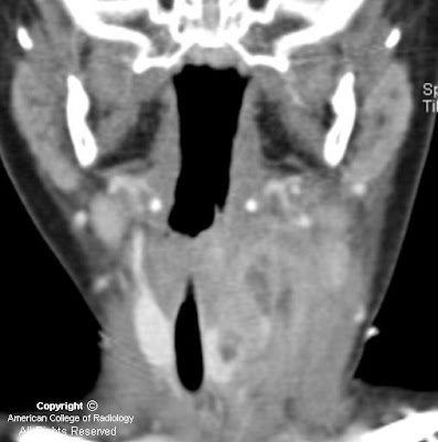
Figure 2: Neck CT, contrast enhanced, at level of subglottic trachea. The image demonstrates continuation of the large inflammatory mass with small areas of necrosis or abscesses. Note displacement of the trachea to the right and lateral displacement of the carotid sheath vessels. Reactive lymphadenopathy is present in the internal jugular chain.
Figure 3: Neck CT, contrast enhanced, at level of the thyroid gland. The image shows a mixed attenuation mass in the enlarged left lobe of the thyroid. This mass arises from extention of the extrinsic anterior and lateral inflammatory mass with phlegmon and abscesses from an infected 4th branchial apparatus cyst.
Figure 4: Neck CT coronal reformation, contrast enhanced. There is extensive phlegmon with multiloculated abscesses, extending from the left lower pharyngeal wall into the left lobe of the thyroid gland. Reactive lymphadenopathy in the left internal jugular lymph node chain is present.


Diagnosis: Infected 4th Branchial apparatus cyst


The main differential diagnostic considerations for a cystic neck mass in children include suppurative lymph nodes, abscess, thyroglossal duct cyst, lymphatic malformation, ranula, and branchial apparatus cyst. A branchial apparatus cyst (BAC) results from maldevelopment of an embryonic branchial apparatus (branchial cleft, arch, and pouch). Embryologically, 6 mesodermal branchial arches, separated by 5 external ectodermal branchial grooves (clefts) and 5 internal endodermal branchial pouches are present bilaterally. The majority of branchial apparatus anomalies are cysts that can arise from a remnant of a groove, arch, or pouch. A 2nd BAC is the most common and accounts for >90% all branchial cleft anomalies discovered in teens and adults. It represents 66%-75% of these anomalies discovered in children.

A 1st BAC is typically found as a cystic mass around the pinna or extending from external auditory canal (EAC) to the angle of the mandible. It can communicate with the external auditory canal. The 2nd BAC is typically found at or immediately caudal to the angle of the mandible, lateral to the carotid space and anteromedial to the sternocleidomastoid muscle. An associated fistulous track may extend from the cyst between the external & internal carotid arteries to the palatine tonsil. The cyst can extend to the carotid bifurcation, producing a beaked configuration, which has been called the "notch sign" and which is considered pathognomonic for a 2nd BAC. The 3rd BAC is typically found in the posterior cervical space behind the carotid sheath in the upper neck and along the anterior border of sternocleidomastoid muscle in the lower neck.

A 4th BAC is rare and seen more often in female infants. It can occur anywhere from the apex of pyriform sinus to the ipsilateral thyroid lobe. Involvement with the thyroid can be understood by noting that the thyroid gland arises from the 4th branchial arch. The most typical imaging finding of a non-infected 4th BAC is a unilocular thin-walled cyst found adjacent to or within the superior lateral aspect of the left thyroid lobe. Ninety-four percent of BACs involve the left side of the neck. These cysts normally show minimal or no peripheral contrast enhancement and no calcification. When infected, a thickened cyst wall is seen and often enhances with intravenous contrast media. Infected cysts often develop higher attenuation than noninfected cysts on CT images. Associated thyroiditis/thyroid abscess is not uncommon. An esophagram may demonstrate fistulous communication between the pyriform sinus and a 4th BAC, providing a pathway for spread of infection. Surgical resection of the cyst and its associated sinus or fistulous tract is necessary for complete cure. Medically treated or incompletely resected cysts/tracts are prone to recur.

mercoledì 13 ottobre 2010

Intraorbital Lymphatic Malformation


Findings

Figure 1: There is a multilobulated mass lesion in the retroorbital region with a fluid- fluid level.
Figure 3: There is an intraconal multilobulated mass with a fluid-fluid level and mild right globe proptosis.


Diagnosis: Intraorbital Lymphatic Malformation


Vascular lesions account for 5-20% of all orbital masses, and the two most common orbital vascular lesions are venous malformations (formerly known as cavernous hemangiomas) and lymphatic malformations (LM) ( formerly known as lymphangiomas). LMs are relatively uncommon in the pediatric population and account for only 4% of all childhood orbital masses. LMs are benign and most frequently found in the head and neck. Intraorbital LMs can arise in any orbital space, but are most commonly intraconal with frequent extraconal and preseptal expansion. Histologically, a vascular malformation can contain venous and lymphatic components, hence the name lymphaticovenous malformation. Of note, 70% of orbital lymphaticovenous malformations are associated with ipsilateral, noncontiguous, intracranial vascular abnormalities.

Intraorbital venous-lymphatic malformations are present at birth, but tend not to be discovered clinically until early childhood when they enlarge as a result of either intralesional hemorrhage or lymphoid hyperplasia and result in acute proptosis. Approximately one-half of all patients with orbital LMs also complain of limited ocular mobility. Additionally, conjunctival, facial, or oral vesicles may also be observed.

Radiologic imaging of intraorbital LMs demonstrates unencapsulated, irregular, lobulated, and multicompartmental masses. These lesions can have cystic as well as more solid components. The cystic elements of these masses commonly exhibit fluid-fluid levels as a result of intralesional hemorrhage. Additionally, LMs are frequently both pre- and postseptal and intra- and extraconal. They often display orbital expansion with irregular margins that traverse tissue planes. Ultrasound images of LMs demonstrate heterogeneous, ill-defined lesions with anechoic cystic portions and extraconal extension. On CT, these masses exhibit ill-defined borders, irregular attenuations, and variable enhancement with peripheral rim enhancement in cystic regions. Additionally, calcified phleboliths can be seen on CT in venous portions of these lesions. MR imaging is the preferred imaging modality to evaluate the location, vascular components and evolving blood products of venous-lymphatic malformations. LMs demonstrate iso- to slightly high signal intensities on T1-weighted images and very high signal intensities on T2-weighted images. MR imaging also allows for the simultaneous evaluation of the brain in an effort to detect any associated intracranial vascular anomalies.

The differential diagnosis for pediatric orbital tumors can be divided into osseous and non-osseous lesions. Pediatric osseous lesions of the orbit include dermoid inclusion cysts, which are most common, fibrous dysplasia, juvenile ossifying fibroma, osteosarcoma, Langerhans cell histocytosis, granulocytic sarcoma and neuroblastoma bone metastases. Non-osseous lesions of the pediatric orbit include most commonly rhabdomyosarcoma, but additionally infantile fibromatosis, infantile hemangioma, and LM.

Orbital lymphaticovenous malformations are histologically benign, but can demonstrate aggressive behavior, such as vision loss, as they expand. The treatment of such lesions is focused on ameliorating pain, alleviating optic nerve compression, maintaining ocular alignment and improving cosmetic appearance. Surgical resection is the preferred treatment for these lesions, and while complete removal can often be achieved with well-demarcated extraconal lesions, more diffuse intraconal lesions tend to be treated with subtotal resection.

martedì 24 agosto 2010

Florid Cemento Osseous Dysplasia


Findings

Figure 1, Figure 2, Figure 3, and Figure 4: Mandibular and maxillary cystic lesions with central calcification, bony expansion and selective involvement of the periapical regions are noted.


Diagnosis: Florid Cemento Osseous Dysplasia


Florid cemento-osseous dysplasia is a slow growing benign lesion which is extremely aggressive and infiltrative. It involves the periapical regions of both the maxilla and mandible with a diffuse distribution of mixed lucent-opaque osseous changes. The lesions are benign fibro-osseous lesions, which histologically, represent normal bone replaced by highly cellular fibrous connective tissue and cementum. Cemento-osseous dysplasia predominantly affects females greater than males (10-14:1) with a predilection for African American patients. Treatment of these lesions should be further radiographic follow-up without surgery unless the lesions become symptomatic, necrotic, or super-infected. Odontoma would also be in the differential as it is a hamartomatous malformation composed of odontogenic tissues. These lesions are usually discovered in the 2nd decade of life and can cause impaction, malpositioning or resorption of adjacent teeth. Ameloblastoma is the most common odontogenic tumor arising from follicular epithelium, dental lamina or enamel. It is a benign lesion but extremely aggressive and infiltrative. It most commonly occurs as an expansile lesion in the posterior mandible in the region of the third molar within the 3rd to 5th decades of life. The unicystic variant occurs in adolescents and has a soap bubble like appearance and involves the ramus and posterior body of the mandible. Large tumors may erode the bony cortex and infiltrate adjacent soft tissues. More malignant processes such as osteosarcoma exhibit a more pronounced periosteal reaction, bone destruction and the presence of a soft tissue mass. Multifocal osteosarcoma is rare. Also, chronic diffuse osteomyelitis is less likely, as it is usually unilateral, associated with pain and fever, exhibits poorly defined borders and would not be exclusively confined to tooth bearing areas.

mercoledì 23 giugno 2010

Trigonocephaly


Findings

CT 3D surface reconstructions show near-complete fusion of the metopic suture with trigonocephaly and "beaking" of the frontal bone along the midline. The sagittal, lambdoid, and coronal sutures are normal.


Diagnosis: Trigonocephaly


Craniosynostoses are calvarial dysmorphsims that result from premature closure of one or more sutures. Trigonocephaly, which involves the metopic suture, comprises less than 10% of all forms of craniosynostoses. It manifests prominent frontal keel, narrow forehead, and hypotelorism, which produces the appearance of a triangular skull. 3D CT reconstructions of the skull are well-suited for imaging the cranial sutures and osseous deformities. Trigonocephaly usually only poses an aesthetic concern and is amenable to reconstructive surgery. This craniosynostosis is infrequently associated with intracranial pathology or genetic disease such as Opitz "C" syndrome.

lunedì 17 maggio 2010

Focal cortical dysplasia


Findings:

Figure 1 and Figure 2: Axial FLAIR and T2W MR images demonstrate blurring of the grey-white junction with high signal intensity in the subcortical white matter of the right frontal lobe. Adjacent cortical ribbon thickening is also present. These findings are characteristic of focal cortical dysplasia.


Diagnosis: Focal cortical dysplasia


Focal cortical dysplasia (FCD) is a congenital disease in which the neurons arrange abnormally in focal areas of the cerebral cortex. It is a common cause of intractable and drug resistant epilepsy, and while seen mostly in the pediatric population, it is not uncommon to see the disease manifest itself in adulthood. Radiologic diagnosis is important for pre-surgical planning and can have prognostic implications.

FCD can be further sub-typed based on histopathology. In Type I FCD there is architectural distortion of the cerebral cortex alone without any abnormal cells. In Type II FCD, however, pathology will demonstrate both architectural distortion and dysmorphic neurons. This includes the characteristic elliptically shaped neuron with a displaced nucleus and lack of axons, known as the “balloon cell”. While the pathologic diagnosis is made retrospectively, clinically those with Type I FCD generally are responsive to medical therapy, while those with Type II FCD are resistant.

Regardless of the pathologic type of FCD, patients that demonstrate preoperative imaging findings tend to have a better outcome after surgery than those who do not demonstrate any findings. The characteristic MR abnormalities in FCD include blurring of the grey-white junction, abnormal high T2 signal in the subcortical white matter, and adjacent focal cortical thickening. Often these findings are very subtle and may not be detected on MR imaging alone. In such cases MR/FDG-PET fusion has been utilized for more sensitive detection. Surgery is the mainstay of treatment for these patients.


Extra: Balloon cells are large elliptical shaped cells with displaced nuclei and are characteristic of focal cortical dysplasia.

venerdì 7 maggio 2010

Aplasia of right submandibular gland with compensatory parotid and sublingual gland hypertrophy


Findings

There is aplasia of the right submandibular gland, and severe atrophy of the left submandibular gland. There is hypertrophy of the sublingual glands bilaterally, with herniation of the right sublingual gland through the mylohyoid boutonniere. There is also hypertrophy of the parotid glands bilaterally, without a discrete mass and without ductal dilatation or sialolithiasis.

Differential diagnosis:
- Parotiditis
- Parotid gland tumor
- Parotid hyperplasia
- Lymphadenopathy
- Amyloidoma


Diagnosis: Aplasia of right submandibular gland with compensatory parotid and sublingual gland hypertrophy


Key points

Congenital absence of a major salivary gland is extremely rare. It is reported to be associated with other congenital craniofacial anomalies and absence of all four major salivary glands is the most common pattern.
Most patients are asymptomatic. Hypertrophy of other salivary glands may result in an asymptomatic neck mass.
Symptoms such as dysphagia, xerostomia, dental caries, and local pain have been reported. Symptomatic patients generally have aplasia of multiple glands.
Agenesis of the major salivary glands may be associated with other first and second branchial arch defects.
The cause is not known , but is thought to be a result of a fetal development disturbance in the 4th-8th weeks of gestation
Enlargement of the other salivary glands is felt to be compensatory in etiology.

martedì 23 marzo 2010

Focal cortical dysplasia, Taylor type


Findings

The MRI of the brain shows T2 and FLAIR hyper intense arrow-shaped left frontal white matter lesion pointing toward the frontal horn of the left lateral ventricle. There is no abnormal enhancement. There is mild hypo intensity on T1 sequences.

Differential diagnosis:
- Focal cortical dysplasia
- Gliosis
- Glial cell tumor
- Physiologic margination of white matter neurons


Diagnosis: Focal cortical dysplasia, Taylor type


Key points

Focal cortical dysplasia is considered to be in the spectrum of migrational disorders of the brain, and often manifests in the form of seizures, developmental delay and focal neurologic deficits. Focal cortical dysplasia is thought to represent the cause of between 5% and 25% of patients with focal epilepsy. This entity typically manifests in the first years of life. Antiepileptic medications are the first line of therapy, and no particular medications have been found to be more or less effective for focal cortical dysplasia. After two failed rounds of antiepileptic regimens, surgical interventions may be considered.


Radiology

MRI is the examination of choice for identifying focal cortical dysplasia. The typical appearance of the Taylor type of this disorder is that of T2/FLAIR hyper intensity within the sub cortical white matter, tapering toward the lateral ventricle. The most typical location of this finding is unilaterally within the frontal lobes. The lesions do not enhance.

martedì 9 marzo 2010

Hydrancephaly


Findings

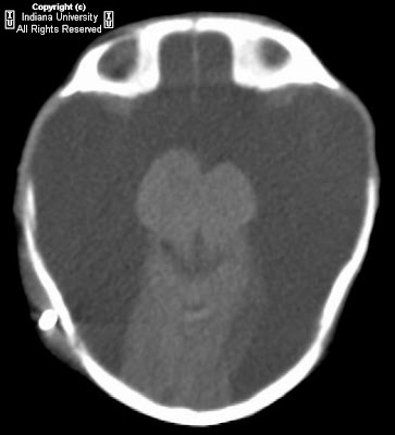
There is near complete absence of the cerebrum, with small amount of residual occipital lobe. Thalami, cerebellum, and brainstem are present.

Differential diagnosis:
- Hydranencephaly
- Hydrocephalus
- Bilateral schizencephaly
- Alobar holoprosencephaly


Diagnosis: Hydrancephaly


Key points

Hydranencephaly - In utero destruction of cerebral parenchyma with intact falx and preservation of posterior fossa structures.
Cerebrum replaced with CSF.
Caused by in utero occlusion of bilateral supraclinoid internal carotid arteries. Etiology unclear (hereditary thrombophilic states, infection, maternal irradiation/toxin exposure, twin-twin transfusion, intrauterine anoxia).
Rarely unilateral
Occurs approximately <1:10000 births, greatest incidence in teenage mothers.
Clinically, present with macrocephaly, developmental delay, irritability, hyperreflexia, seizures.
Often seen with prenatal ultrasound (anechoic cranial vault).
On CT and MR (best characterized with MR) – CSF attenuation/signal intensity replacing the supratentorial brain parenchyma with sparing of thalami, brain stem, cerebellum, and choroid plexus.
Falx is intact (distinguishes from holoprosencephaly).
No thin rim of cerebral parenchyma (distinguishes from severe hydrocephalus).
Prognosis is poor – usually death in infancy.
Treatment is supportive care, shunting to decrease head growth.

giovedì 4 febbraio 2010

Septo-optic dysplasia


Findings

Absence of the septum pellucidum. Hypoplastic pituitary. Nonvisualization of the pituitary stalk. Hypoplasia of the optic nerves and optic chiasm.

Differential diagnosis:
- Septo-optic dysplasia
- Kallman syndrome
- Holoprosencephaly


Diagnosis: Septo-optic dysplasia


Key points

Also known as de Morsier's syndrome
Manifested by hypoplasia (underdevelopment) of the optic nerve, hypopituitarism and absence of the septum pellucidum
In a severe case, this results in pituitary hormone deficiencies, blindness, and mental retardation. However, there are milder degrees of each of the three problems, and some people only have one or two of the three.
Majority have other brain abnormalities as well, schizencephaly is most common
Ocular anomalies (coloboma, anophthalmia, microphthalmia)

Imaging findings:
- Absent septum pellucidum
- Flat roof of frontal horn, inferior aspect of frontal horn point down
- Small optic chiasm
- Thin pituitary stalk
- Posterior pituitary ectopia
- Callosal-forniceal continuation or fused midline fornices
- Thin corpus callosum
- Heterotopias, schizencephaly

giovedì 21 gennaio 2010

Klippel-Feil anomaly


Findings

There are multiple segmentation anomalies of the cervical spine, with fusion of C1-3.; left C6 hemivertebra.


Diagnosis: Klippel-Feil anomaly


Key points

Klippel-Feil anomaly refers to segmentation defects (congenital fusion) of the cervical spine.
Fusion of C2-3 and C5-6 most common
Frequently associated with Chiari malformation and syringohydromyelia.

Three categories:
- Group 1 = short, webbed neck, low hairline, complete lack of cervical segmentation
- Group 2 = isolated cervical segmentation defects
- Group 3 = segmentation defects affecting separate thoracic and/or lumbar level in addition of cervical involvement

Restricted motion at level of non-segmentation leads to accelerated disc degeneration.
50% of patients have at least partial dorsal splitting of the spinal cord. Defects in decussation of the corticospinal tracts can lead to "mirror movements" on physical exam.
Other CNS associations: occipital encephalocele, Dandy-Walker malformation, Duane syndrome, nasofrontal dermoid
Associated visceral anomalies: Sprengel deformity (20-30%, congenital elevation of the scapula, often with tethering omovertebral bone), cervical ribs, supranumary digits, tracheal and proximal bronchial stenosis, sickle sacrum, cleft palate, various renal anomalies.

martedì 12 gennaio 2010

Optic disc coloboma


Findings

A posterior protrusion of vitreous is seen in the left globe, at the insertion of the optic nerve head. There is no hemorrhage, retrobulbar colobomatous cyst, or optic nerve atrophy. The right globe is normal.

Differential diagnosis:
- Coloboma (chorioretinal or optic disc)
- Peripapillary staphyloma
- Morning glory disc anomaly (MGDA)
- Buphthalmos (ox eye)


Diagnosis: Optic disc coloboma


Key points

Coloboma (Greek koloboun, "to mutilate") is the result of incomplete closure of the choroidal fissure.
Funnel or cone-shaped protrusion from the posterior aspect of the globe, often involving the optic nerve head insertion.
Two general types: Optic disc coloboma (ODC = excavated/everted region involves the posterior globe at the optic disc insertion only). Chorioretinal colobomas are more broad-based, extending beyond the margins of the optic disc.
May be associated with large retrobulbar colobomatous cysts (apparently encapsulated outpouchings of vitreous) which may be larger than the actual globe, may cause significant proptosis, or even globe atrophy / microphthalmia.
Majority of colobomas are sporadic and unilateral.
Associated with midline craniocerebrofacial clefting, choanal atresia, basal (particularly sphenoidal) encephalocele, corpus callosal agenesis, olfactory hypoplasia, cardiac anomalies, retardation, genital hypoplasia, and ear anomalies.

Differential diagnosis:
- Peripapillary staphyloma – more diffuse / broad based than coloboma, must feature posterior uveoscleral thinning and lack enhancement.
- Morning glory disc anomaly – fundoscopic description of ODC. Funnel or square shaped posterior defect at the optic nerve head.
- Buphthalmos ("ox eye") – 2/2 congenital glaucoma. Assoc. with Marfan's, NF-1, Sturge-Weber. Entire globe enlarged.