Visualizzazione post con etichetta Infectious. Mostra tutti i post

martedì 8 febbraio 2011

Subdural empyema


Findings

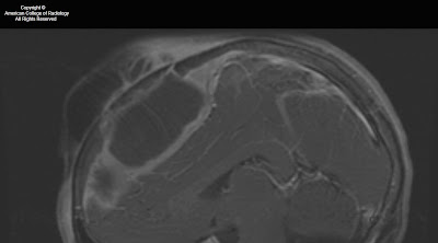
A large left middle cranial fossa subdural empyema is demonstrated, with a relatively thin rim of enhancement. Internally, there is a large quantity of debris. There is mass effect, with a modest midline shift and effacement of the left lateral ventricle. Inflammatory changes are demonstrated in the left temporal bone which is likely the source of the abscess. There is diffusion restriction, not marked, consistent with abscess. There is extensive dural enhancement, along with considerable surrounding edema.

Differential diagnosis:
- Subdural empyema
- Chronic subdural hematoma
- Subdural effusion
- Subdural hygroma
- Dural metastasis


Diagnosis: Large left middle cranial fossa subdural empyema; left mastoiditis


Key points

Loculated collection of pus in subdural space
Best diagnostic clue: Extra-axial collection with contrast enhancing rim
Supratentorial typical
Infratentorial (up to 10%), often associated with mastoiditis
Crescentic typical; may be lens shaped on coronal images
CT demonstrates extra-axial collection, iso-to hyper dense to CSF on noncontrasted CT; shows strong peripheral enhancement with contrast
Best imaging tool: MR with DWI to demonstrate presence, nature, extent and complications
T1W image shows:
Extra-axial collection hyper intense to CSF
Crescentic extra-axial collection
T2WI demonstrates a lesion that is Iso-to hyper intense to CSF,
FLAIR shows a crescentic fluid collection which is hyper intense to CSF, underlying brain may be hyper intense
DWI shows restricted diffusion (increased signal intensity); Differentiates subdural empyema from subdural effusions
T1WI post contrast shows:
Prominent enhancement at margin related to granulomatous tissue and inflammation
Encapsulating membranes enhance strongly, may be loculated with internal fibrous strands
May see enhancement of adjacent brain parenchyma
MRV may show venous thrombosis seen as a lack of flow
CT may miss small collections
Complications include cerebritis and brain abscess, cortical vein and dural sinus thrombosis, and cerebral edema
Subdural empyema is much more common than epidural empyema
In older children, adults: Related to paranasal sinus disease (>2/3), in infants and young children it can be a complication of bacterial meningitis
Most common signs/symptoms include fever, headaches, meningismus, sinusitis, cerebritis
Sinus or ear infection in > 75% of cases
Confused with meningitis which may lead to delayed diagnosis
Can occur at any age
Rare, yet high mortality rate.
If subdural or epidural abscess is discovered, look also for sinusitis, otomastoiditis, dural sinus thrombosis and brain abscess
Progresses rapidly, neurosurgical emergency
Surgical drainage via wide craniotomy is gold standard

venerdì 29 ottobre 2010

Pott's Puffy Tumor


Findings

Figure 1: The image shows a large collection anterior to the frontal bone and a large epidural abscess with peripheral enhancement.
Figure 2: Erosion of the frontal bone. Sinus tract of the bone into the large anterior collection.
Figure 3: Large collection anterior to the frontal bone.
Figure 4: Large epidural abscess.
Figure 5: Large epidural abscess with peripheral enhancement.
Figure 6: Large collection anterior to the frontal bone. Large epidural abscess. Normal bone marrow signal. Abnormal bone marrow signal in the frontal bone.
Figure 7: Large collection anterior to the frontal bone with peripheral enhancement. Large epidural abscess with peripheral enhancement.
Figure 8: Abnormal bone marrow signal in the frontal bone. There is a lack of enhancement of the frontal bone. The abnormal dark bone marrow signal on T1 images and lack of enhancement on T1 post gad fat sat is consistent with dead necrotic bone from severe osteomyelitis.


Diagnosis: Pott's Puffy Tumor


Pott’s puffy tumor is a subperiosteal abscess of the frontal bone that appears as a localized swelling of the forehead associated with frontal osteomyelitis. Pott’s puffy tumor is a complication of frontal sinusitis or trauma, which is predominatly seen in the adolescent age group. However, there are a few case reports in adults. Pott’s puffy tumor is a rare complication of frontal sinusitis in the post antibiotic era but can be seen in patients with undiagnosed or partially treated sinusitis. Patients will typically present with frontal scalp swelling, headache, fever, nasal drainage, and frontal sinus tenderness. Ocassionally, Pott’s puffy tumor can mimic findings of preseptal or orbital cellulitis. In severe cases, there will be neurologic decompensation. Varying degrees of hemiparesis, obtundation, papillary dilatation or aphasia have been described in case reports.

Imaging is necessary to exclude intracranial complications such as epidural abscess. Pott’s puffy tumor can also be associated with dural sinus thrombosis, meningitis, subdural empyema, epidural abscess, brain abscess, and rarely seizure. Intracranial infection is caused by posterior extension from the frontal sinus while preseptal and orbital cellulitis is caused by downward spread from the frontal sinus to the orbit. Younger children who do not have pneumatized frontal sinuses, are more likely to have ethmoid sinusitis. Orbital cellulitis is a more common complication in patients with ethmoid sinusitis.

Patients must be treated with a combination of surgery and long-term antibiotic therapy.

lunedì 25 ottobre 2010

Infected 4th Branchial apparatus cyst


Findings

Figure 1: Neck CT, contrast enhanced, at level of pyriform sinus. The left pyriform sinus is effaced by an inflammatory mass.
Figure 2: Neck CT, contrast enhanced, at level of subglottic trachea. The image demonstrates continuation of the large inflammatory mass with small areas of necrosis or abscesses. Note displacement of the trachea to the right and lateral displacement of the carotid sheath vessels. Reactive lymphadenopathy is present in the internal jugular chain.
Figure 3: Neck CT, contrast enhanced, at level of the thyroid gland. The image shows a mixed attenuation mass in the enlarged left lobe of the thyroid. This mass arises from extention of the extrinsic anterior and lateral inflammatory mass with phlegmon and abscesses from an infected 4th branchial apparatus cyst.
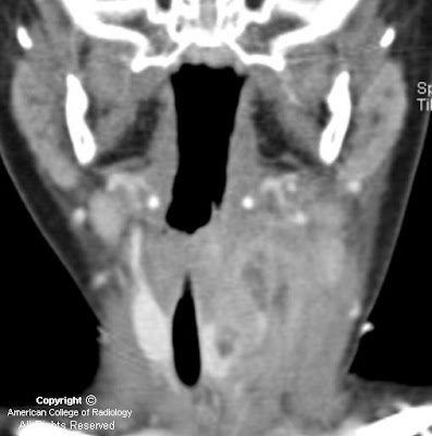
Figure 4: Neck CT coronal reformation, contrast enhanced. There is extensive phlegmon with multiloculated abscesses, extending from the left lower pharyngeal wall into the left lobe of the thyroid gland. Reactive lymphadenopathy in the left internal jugular lymph node chain is present.


Diagnosis: Infected 4th Branchial apparatus cyst


The main differential diagnostic considerations for a cystic neck mass in children include suppurative lymph nodes, abscess, thyroglossal duct cyst, lymphatic malformation, ranula, and branchial apparatus cyst. A branchial apparatus cyst (BAC) results from maldevelopment of an embryonic branchial apparatus (branchial cleft, arch, and pouch). Embryologically, 6 mesodermal branchial arches, separated by 5 external ectodermal branchial grooves (clefts) and 5 internal endodermal branchial pouches are present bilaterally. The majority of branchial apparatus anomalies are cysts that can arise from a remnant of a groove, arch, or pouch. A 2nd BAC is the most common and accounts for >90% all branchial cleft anomalies discovered in teens and adults. It represents 66%-75% of these anomalies discovered in children.

A 1st BAC is typically found as a cystic mass around the pinna or extending from external auditory canal (EAC) to the angle of the mandible. It can communicate with the external auditory canal. The 2nd BAC is typically found at or immediately caudal to the angle of the mandible, lateral to the carotid space and anteromedial to the sternocleidomastoid muscle. An associated fistulous track may extend from the cyst between the external & internal carotid arteries to the palatine tonsil. The cyst can extend to the carotid bifurcation, producing a beaked configuration, which has been called the "notch sign" and which is considered pathognomonic for a 2nd BAC. The 3rd BAC is typically found in the posterior cervical space behind the carotid sheath in the upper neck and along the anterior border of sternocleidomastoid muscle in the lower neck.

A 4th BAC is rare and seen more often in female infants. It can occur anywhere from the apex of pyriform sinus to the ipsilateral thyroid lobe. Involvement with the thyroid can be understood by noting that the thyroid gland arises from the 4th branchial arch. The most typical imaging finding of a non-infected 4th BAC is a unilocular thin-walled cyst found adjacent to or within the superior lateral aspect of the left thyroid lobe. Ninety-four percent of BACs involve the left side of the neck. These cysts normally show minimal or no peripheral contrast enhancement and no calcification. When infected, a thickened cyst wall is seen and often enhances with intravenous contrast media. Infected cysts often develop higher attenuation than noninfected cysts on CT images. Associated thyroiditis/thyroid abscess is not uncommon. An esophagram may demonstrate fistulous communication between the pyriform sinus and a 4th BAC, providing a pathway for spread of infection. Surgical resection of the cyst and its associated sinus or fistulous tract is necessary for complete cure. Medically treated or incompletely resected cysts/tracts are prone to recur.

lunedì 18 ottobre 2010

Basilar meningitis of unknown etiology


Findings

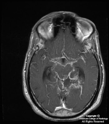
Figure 1,Figure 2, Figure 3, Figure 4: Axial and sagittal T1 weighted images postcontrast demonstrate thick and nodular predominantly basilar meningeal enhancement which on sagittal images encases the middle cerebral arteries. Cerebellar parenchymal enhancement (Figure 4) secondary to late subacute infarction and infectious/inflammatory exudates.
Figure 5: Axial T2 weighted image demonstrates slight increased size of lateral ventricles with more prominent frontal horn lateral ventricles and bilateral lateral ventricle atria in a 2 week follow-up study (initial study slightly motion degraded.)


Diagnosis: Basilar meningitis of unknown etiology (presumed MDR-Tuberculosis)


This young patient with immunocompromise and altered mental status presented initially with left posterior circulation infarctions of unclear etiology. In a young patient with posterior circulation infarctions, acute vertebral artery dissection/ injury in the setting of trauma or spontaneous etiology should be considered. However, when given a history of immunocompromise and HIV, other differential diagnostic considerations such as infectious (Tuberculous, fungal, or pyogenic), inflammatory (Neurosarcoidosis), vascular (infectious vasculitis from Neurosyphilis or HIV), or neoplastic (lymphoma, leukemia, leptomeningeal carcinomatosis) etiologies should all be taken into account. To date, laboratory and CSF values for this patient continue to be non-diagnostic as to the source of this patient's basilar meningitis/vasculitis. The top consideration after discussing with the neurology team is Multi-drug resistant tuberculosis (MDR-TB) due to unimpressive response to conventional treatment regimens and inconclusive microbiological testing.

Tuberculous CNS infections are mostly caused by M. tuberculosis; atypical organisms are rare except in immunosuppressed patients. 30% of patients are HIV positive (particularly IV drug users.) Due to hematogenous dissemination, lesions usually occur at the gray-white junctions of cerebral hemispheres, basal ganglia, or cerebellum (especially in children). Hematogenous dissemination is from a systemic source, most commonly the lung, but also possibly the GU system or GI tract.

Most common manifestations of CNS Tuberculosis are meningitis, seen predominantly in the basilar cisterns, and hydrocephalus. Acutely, cerebritis can be seen, which then can progress to ring-enhancing tuberculoma lesions. Tuberculosis can also result in vasculitis and cerebral infarctions. The thick and nodular basilar meningeal enhancement in association with hydrocephalus and left posterior circulation multifocal infarctions in our patient can all be seen in tuberculosis.

Leptomeningeal sarcoidosis must be distinguished clinically from carcinomatous, lymphomatous/leukemic, and infectious meningitis. Dramatic response can be seen in some cases with steroid therapy. Sarcoid has replaced syphilis as the great mimicker. Neurosyphilis can cause arteritis of intracranial and extracranial large and medium sized arteries

lunedì 10 maggio 2010

Parapharyngeal abscess


Findings

The right parapharyngeal space contains a hypo attenuating collection near simple fluid in attenuation. Collection extends from just below the level of the hyoid to just below the right mandibular condyle, is anterior to the carotid space inferiorly, anteromedial to the carotid space superiorly. No significant rim enhancement. No inflammatory fat stranding is present. Right submandibular gland is along the anterolateral margin of this collection. Multiple prominent lymph nodes were visible.

Differential diagnosis:
- Abscess
- Lymphangioma
- Second branchial cleft cyst
- Ranula


Diagnosis: Parapharyngeal abscess

mercoledì 17 marzo 2010

Subacute Sclerosing Panencephalitis (SSPE)


History: 18-year-old male with 3 months of personality change, gradual decrease in responsiveness, rigidity and abnormal EEG findings (the patient is HIV negative and does not report a recent viral or respiratory infection or vaccination).


Findings

Figure 1: T2-weighted axial image demonstrates multiple areas of hyperintense signal in the bilateral subcortical and deep white matter.
Figure 2: T2-weighted FLAIR axial image demonstrates areas of hyperintense signal in the posterior limb of the right internal capsule (blue arrow) and right aspect of the splenium of the corpus callosum (yellow arrow).
Figure 3: T1-weighted FLAIR post contrast image demonstrates no areas of abnormal enhancement.
Figure 4: There is no restricted diffusion.


Diagnosis: Subacute Sclerosing Panencephalitis (SSPE)


SSPE is a chronic progressive encephalitis that can very rarely occur in people 2-10 years following infection with the measles virus. Early stages are characterized by personality, behavioral and intellectual changes. There is then further neuropsychiatric deterioration progressing to ataxia, chorea, dystonic rigidity, seizures, myoclonus, optic atrophy and cortical blindness. The terminal stages include unresponsiveness, autonomic dysfunction, coma and death within 1 to 3 years of the onset of symptoms.

The white matter is predominantly affected with patchy demyelination and initial sparing of the subcortical U fibers. In the first stage of the disease, T2WI/FLAIR typically demonstrates asymmetrical gray and adjacent subcortical white matter hyperintensity in the parieto-occipital regions. Enhancement and mass effect may occur early in the disease. The basal ganglia (particularly the putamen), cerebellum, pons and frontal white matter may also be involved. In the second stage of the disease, cortical/subcortical lesions regress and T2WI/FLAIR periventricular hyperintensity develop. In the terminal stage, periventricular white matter disease progresses and profound parenchymal loss occurs.

Diagnosis is made by characteristic CSF and EEG findings. In SSPE, CSF will typically have normal cellular components, glucose and total protein, but markedly elevated values of gammaglobulin and anti-measles antibodies. Typically, serum anti-measles antibody titers are also grossly elevated. A characteristic “burst-suppression” pattern on EEG is nearly always seen.

Treatment is the immunomodulator interferon and antivirals ribavirin and inosine pranobex which, if given early and throughout the patient’s life, may limit progression of disease.

martedì 16 marzo 2010

Progressive multifocal leukencephalopathy (PML)


History: Woman with lung transplant and weakness.
Additional clinical information: The patient has CSF positive for JC virus and EBV virus.


Findings

Earlier CT showed an ill-defined process of the left cerebellar hemisphere, thought to represent ischemia, but was shown to progress over serial scans, making ischemic process unlikely. The MR shows T2 prolongation within the left cerebellar white matter extending across the middle cerebellar peduncle and into the left anterior pons. There is also a chronic right frontal lobe infarct.

Differential diagnosis:
- Progressive multifocal leukencephalopathy
- Encephalomalacia
- CMV encephalitis
- Lymphoma
- Toxoplasmosis
- Abscess


Diagnosis: Progressive multifocal leukencephalopathy (PML)


Key points

JC virus infection of oligodendrocytes causes demyelinating lesions
Often occurs as reactivation of latent virus in an immunosuppressed patient
Most prevalent in AIDS, leukemia, and organ transplant patients
3rd most common cause of encephalopathy in AIDS patients after toxoplasma encephalitis and HIV encephalitis
Responds to immune-strengthening treatment, such as HAART in AIDS; 8% spontaneous resolution


Clinical presentation

Insidious onset of focal symptoms, behavioral, speech, cognitive, motor, and visual impairment, over weeks
More rapid progression than AIDS dementia complex
Conjugate gaze abnormalities are common and are the initial presentation in more than 30% of patients


Radiology

Contrast-enhanced CT
- Multifocal, nonenhancing white matter hypodensities without mass effect or edema.
- Rapid change in size or number of lesions

MRI
- Hypointense T1-weighted appearance with cortical sparing; typically nonenhancing; occasionally, mild peripheral enhancement
- T2-weighted images reveals hyperintense subcortical lesions

mercoledì 3 febbraio 2010

Hemorrhagic meningoencephalitis


Findings

Figure 1: Coronal US image of the brain shows areas of cystic necrosis and hemorrhagic abscess.
Figure 2: Coronal US image of the brain shows ventriculomegaly.
Figure 3: Coronal US brain image shows periventricular cystic changes, parenchymal hemorrhage and abscess.
Figure 4: Coronal US brain image shows periventricular cystic change and a calcified hemorrhagic abscess.
Figure 5: Axial nonenhanced CT scan image of the brain shows multiple calcified hemorrhagic abscesses.
Figure 6: Axial CT scan image of the brain shows diffuse white matter destruction. The cortex is spared initially in the disease process.


Diagnosis: Bacillus cereus hemorrhagic meningoencephalitis


Group B beta-hemolytic streptococci and E coli are the most common causes of meningoencephalitis in the neonatal period. Serratia marcescens, or Citrobacter, may be considered as a causal micro-organism in cases of hemorrhagic meningoencephalitis. It is important to note that Bacillus cereus, though rare, can also cause hemorrhagic meningoencephalitis which is most often fatal. B cereus is a gram-positive, aerobic, spore-forming rod which is ubiquitous in the environment. The presence of this organism in blood or CSF is often regarded as a contaminant of specimens received by the microbiology laboratory and is therefore often disregarded by clinicians and microbiologists.

Acquisition of an infection by Bacillus cereus is thought to be nosocomial and is linked to the use of central or peripheral catheters, contaminated dressings, hospital linens, and ventilator equipment. Premature infants in ICU units are most vulnerable to this infection. Vasculitis, vasospasm, hydrocephalus, and diminished cerebrovascular autoregulation phenomena which are often seen in premature infants, are some of the risk factors for the infection.

Bacillus cereus is known to swarm out of the veins and cause extensive tissue damage and liquefactive necrosis by producing toxins, which include a necrotising enterotoxin, phospholipases, proteases, and haemolysins. In the brain, the destruction initially involves the subcortical and periventricular white matter extensively, sparing the cortex. The cortex can become involved in later stages and when this occurs, it preferentially involves the occipital lobes. Involvement of the basal ganglia is rare and can occur as a part of extensive brain destruction.

Brain sonograms obtained in the first 2-3 days can be normal but days later show asymmetrical white matter hyperechogenicity due to widespread destruction. This is later followed by development of multiple cysts, often with rims of hyperechogenicity around them. Multiple areas of parenchymal or intraventricular hemorrhage may also be demonstrated on ultrasound. Sometimes, brain sonography shows intraventricular septa and thickened ependyma due to ventriculitis. CT features include diffuse hypodensity of the white matter with multiple areas of hemorrhage and abscesses. The abscesses may undergo cavitatory necrosis and calcification. On MRI, multiple, confluent white matter hyperintensities are seen on T1W images and hypointensities on T2W images, both consistent with hemorrhagic destruction. DWI show restricted diffusion in the brain parenchyma due to cytotoxic edema and may also show lesions that were not detected on conventional MR images. The rate at which the findings develop sequentially in cases of suspected Bacillus cereus meningoencephalitis can be rapid and hence it’s worthwhile to consider serial sonography.


Differential diagnosis

PVL: PVL is an ongoing process which in many cases starts with symmetrical flaring in the periventricular white matter within the first days of life sometimes evolving into cystic destruction of the white matter. The condition is bilateral and almost always symmetrical, preferentially damaging posterior frontal and parietal regions. This can be confused with B cereus meningoencephalitis in the premature neonate. B cereus meningoencephalitis shows a dramatic change on serial sonograms performed whereas in PVL, the rate of evolution of the cystic changes is much slower. The presence of multiple abscesses in meningoencephalitis and the detection of the causative organism in the CSF may help differentiate the two conditions.

Asphyxia: shows transition from abnormal hyperechogenicity in major arterial areas to slow (in the course of a few weeks) destruction. This is in contrast to very rapid destruction of an initially normal brain in cases of bacterial encephalitis.

Herpes simplex viral encephalitis: The cortex and deep gray nuclei may also be affected, because of infarction caused by vasculitis and obstruction of small vessels, as well as neuronal apoptosis.

Deep cerebral vein thrombosis: Deep cerebral vein thrombosis often causes destruction of the white matter, the deep gray matter, basal ganglia, and thalamus. Moreover, thrombus is often visualized on US or CT involving the sagital sinus or the deep cerebral veins. The areas of destruction in B cereus are not compatible with arterial or venous infarction and do not correspond to arterial/ venous territories that would be involved in arterial or venous infarction.

venerdì 22 gennaio 2010

Neurocysticercosis (Calcified stage)


Findings

CT with contrast images showed a 7 mm calcified enhancing lesion within the posterior aspect of the left frontal lobe with a small amount of surrounding oedema. MRI images confirm the presence of a calcified ring enhancing lesion within the left posterior frontal lobe.

Differential diagnosis:
- Neurocysticercosis
- Tuberculoma


Diagnosis: Neurocysticercosis


Discussion

Cysticercosis is caused by larvae of the pork tape worm Taenia solium. Infestation occurs via the fecal-oral route. It is the most common parasitic infection involving the central nervous system in developing countries with 90% of patients present with seizures. Parenchymal cysticercosis is the most common type with lesions most commonly being peripherally distributed near the grey-white matter junction. The parasite goes through different stage of involution, each of which has different imaging features on CT and MRI.

These include the following:
- Vesical stage: CT shows hypodense non enhancing lesions. On MRI cysts follow CSF signal; T2 hyper intense scolex may be seen. No edema. Usually no enhancement.
- Colloidal stage: CT shows hypodense/isodense lesion with peripheral enhancement and perilesional edema. On MRI Cysts are hyperintense to CSF; surrounding edema, cyst wall enhances.
- Granular stage: CT shows nodular enhancing lesions. On MRI the cyst wall thickens and retracts, there is a decrease in edema, and there is nodular or ring enhancement.
- Calcified stage: When the parasite dies, nodular parenchymal calcifications are seen. These findings are best seen on CT.

giovedì 12 novembre 2009

Submandibular sialadenitis


Findings

The left submandibular gland is hypervascular, inflamed and markedly enlarged (Figure 1 and Figure 2). There is dilatation of the submandibular duct leading to a calculus within the distal aspect of the duct (Figure 3 and Figure 4). There are no drainable fluid collections. There is injection and stranding of the overlying dermis (Figure 5). The right submandibular gland is unremarkable (Figure 2).


Diagnosis: Submandibular sialadenitis


Acute sialadenitis may be secondary to a bacterial/viral infection or an obstructing lesion such as a calculus or tumor at the floor of the mouth.
Associated conditions include HIV, sarcoidosis, Sjogren syndrome, dehydration, diabetes mellitus and immunocompromised/postoperative patients.
Imaging is often helpful to delineate the location of the calculus and the presence/absence of subsequent complications (abscess formation, osteomyelitis, etc).

Inflammation of the submandibular gland accounts for approximately 10-15% of cases of sialadenitis involving any of the major salivary glands. Risk factors for submandibular sialadenitis include immunocompromised/postoperative patients, debilitation, elderly patients, dehydration, diabetes mellitus, hypothyroidism, hypercalcemia, radiation/chemotherapy, eating disorders (bulimia, anorexia nervosa), and other concomitant medical problems (malignancy, head and neck infections). Associated conditions also include HIV, sarcoidosis, Sjogren syndrome, tuberculosis, mumps, and cat scratch disease. It is rare in pediatric patients.

Most commonly, acute sialadenitis is a result of a bacterial infection (common organisms include Staphylococcus aureus, Streptococcus, Haemophilus influenzae, and Pseudomonas). Less commonly, the infection may be related to a virus such as mumps, coxsackie virus, herpes and influenza. On the other hand, chronic sialadenitis is usually a result of salivary stasis, ductal stenosis, calculi or other obstructive lesions such as a tumor at the floor of the mouth. Sialolithiasis is most common in the submandibular gland, accounting for approximately 80% of cases. The majority of calculi are radio-opaque, vary in size, and can be single or multiple. Often, the calculus obstructs a duct, resulting in secondary inflammation of the affected salivary gland which then becomes suppurative.

Although plain film radiography can depict the majority of calculi, CT is often first-line imaging. The affected submandibular gland is enlarged, hypervascular and there may be associated cellulitis/myositis. Calculi are easily identified and described as being either distal (towards the ductal opening) or proximal (towards the submandibular hilum). Chronic sialdenitis manifests as a small, fatty gland.

Complications of sialadenitis include abscess formation, bacteremia/septicemia, osteomyelitis, cranial nerve involvement (facial nerve paralysis), and respiratory complications. Management of acute sialadenitis includes both medical (conservative) and surgical options. Antibiotics, analgesics, sialogogues, warm compresses, glandular massage and intravenous fluids are the mainstay of medical management. Surgical options include duct cannulation with subsequent removal of the calculus and complete gland excision.