Visualizzazione post con etichetta Trauma. Mostra tutti i post

mercoledì 1 settembre 2010

Bilateral ectopia lentis


Findings

Figure 1: Axial noncontrast CT of the head at the level of the orbits demonstrates posterior dislocation of both lenses, which now rest dependently in the vitreous. The etiology in this particular patient was repetitive trauma from serial falls.


Diagnosis: Bilateral ectopia lentis


The crystalline lens of the eye is designed to refract the light entering the iris and project/focus it onto the retina. The lens itself contains no vasculature, nerves, or connective tissue. It sits behind the iris and the front of the lens is in contact with the aqueous fluid of the anterior chamber while the posterior surface of the lens is in contact with the vitreous. The lens is held in place by zonular fibers, otherwise known as suspensory ligaments. These fibers connect to the cilliary body around the circumference of the lens.

Subluxation (partial dislocation) or luxation (complete dislocation) of the crystalline lens, otherwise known as ectopia lentis, is caused by dysfunction or disruption of these zonular fibers. Trauma is the most common cause of this disorder. The absence of a traumatic history should prompt consideration of hereditary causes of zonular fiber dysfunction; predisposing conditions include Marfan syndrome, homocystinurea, tertiary syphilis, and Weil-Marchesani syndrome.

Patients will complain of monocular diplopia, markedly decreased visual acuity in the affected eye(s), and/or poor near vision.

Treatment is determined by lens position, with anterior chamber dislocation often being a surgical emergency. As the aqueous humor of the eye flows in the anterior chamber, around the iris from the cilliary body to the canal of Schlemm, this route can become acutely obstructed with anterior dislocation leading to acute glaucoma. The cornea and iris are also at risk for damage. Posterior dislocation may be treated conservatively depending on lens position, but may also lead to uveitis or glaucoma in some cases.

mercoledì 30 giugno 2010

Diving Ranula


Findings

Figure 1 and Figure 2 Axial contrast-enhanced CT demonstrates a thin-walled, well-defined hypodense lesion in the right sublingual space (Figure 1) which herniates posteriorly into the submandibular space (Figure 1). This lesion also tracks into the sublingual space lateral to the right genioglossus muscle (Figure 2). It is located anterior and lateral to the right internal and external carotid arteries and effaces the submandibular gland.


Diagnosis: Diving Ranula


A diving ranula is an extravasation pseudocyst of a simple ranula in the sublingual space rupturing out into the submandibular and or parapharyngeal space. It originates from trauma or inflammation of the sublingual gland or minor salivary glands in the sublingual space. With obstruction of the gland duct, the duct dilates and eventually ruptures, allowing its secretions to leak into the surrounding soft tissue.

CT is the imaging study of choice. On CT, the lesion demonstrates water content and thin walls with subtle or no wall enhancement. If the lesion is infected, it may show thick, enhancing walls. The characteristic shape is a collapsed cystic portion in the sublingual space - “tail sign,” with its head extending into the submandibular space. The sublingual spaces are located on the floor of the mouth on either side of the midline genioglossus muscles. They are separated from the submandibular space by the mylohyoid muscle that extends from the medial inferior aspect of the mandible to the hyoid bone. Thus, the sublingual space is superomedial to the mylohyoid muscle and the submandibular space is inferolateral to the muscle. Ultrasound and MR can also be used to characterize these lesions. Ultrasound demonstrates a well-defined hypoechoic mass in the sublingual and submandibular space. MR images show the lesion with signal intensity of water, though if infected, signal intensity can vary according to the protein content or presence of hemorrhage with some wall enhancement.

The differential diagnosis would include epidermoid or dermoid cyst, lymphangioma or cystic hygroma, abscess, submandibular gland cyst (mucocele), and second branchial cleft cyst. These lesions differ in appearance on CT.

The treatment of a diving ranula is removal of the ipsilateral sublingual gland via the cervical or intraoral approach. It is not necessary to excise the pseudocyst since it puts the surrounding structures at risk for damage. Biopsy can be done to confirm the diagnosis.

mercoledì 27 gennaio 2010

Injured petrous internal carotid with proximal occlusion and clot from stab wound


Findings

Figure 1: Transaxial CT scan of the brain. Knife entering the superolateral aspect of the left nasal cavity.
Figure 2, Figure 3, and Figure 4: Transaxial CT scan of the brain. Knife traversing the midline.
Figure 5: Transaxial CT scan of the brain. Knife traverses the carotid canal with tip at the level of the internal auditory canal.
Figure 6: Transaxial CT scan of the brain. Postoperative pneumocephalus and posttraumatic infarction in the distribution of the right middle cerebral artery. Knife has been removed.
Figure 7: Angiogram of the right internal carotid artery in an oblique projection. Knife tip in close proximity to the right internal carotid artery with little flow seen intracranially. Spasm noted at the catheter tip in the internal carotid artery.
Figure 8: Angiogram of the right internal carotid artery in an AP projection. The knife traverses the midline with the knife tip in the right carotid canal.


Diagnosis: Injured petrous internal carotid with proximal occlusion and clot from stab wound


Gunshot wounds to the head have become the leading or second leading cause of head injury in many cities in the United States; penetrating head injuries can also be the result of stab wounds, and motor vehicle or occupational accidents. Knives are the most frequent weapon used in stab wounds to the cranium although bizarre craniocerebral-perforating injuries have been reported that were caused by nails, metal poles, ice picks, keys, pencils, crochet needles, chopsticks, and power drills. A mortality rate of 17% has been reported for cranial stab wounds, mostly related to vascular injury and massive intracerebral hematomas. The orbital surfaces and squamous portions of the temporal bones are the most frequently affected since these bones are thin and easily traversed. The morbidity from stab wounds to the squamous portion of the temporal bone is more severe due to the short distance to the deep brain stem and vascular structures.

CT scans are vital in the work up of patients with penetrating head injuries since they do show the extent of intracranial injury and fracture and rule out the presence of radiopaque foreign bodies. CT angiography is limited in the presence of metallic foreign bodies because of artifact; in these cases, conventional angiography is important in ruling out vascular injury and preoperative planning. Potential signs of vascular injury include transection, pseudoaneurysm and arterial-venous fistula. Occlusion is a sign, albeit a nonspecific sign, of an underlying vascular injury. In the case of a vascular injury, injection of the contralateral artery should be done to demonstrate crossfilling via collateral circulation in the circle of Willis prior to intervention. Cerebral vasospasm is another noteworthy finding since it can portend a poor outcome.

Patients with penetrating head injuries are often taken to surgery to remove hematomas which may be causing mass effect, to remove dead brain tissue to prevent further swelling and necrosis, and to control active bleeding. Metallic foreign bodies are often removed to prevent infection, the potential for fibroglial scarring with the ensuing development of epilepsy and intracranial migration. Knives should only be removed in the OR under direct visualization. Clot was noted in the proximal ICA below the presumed level of injury; coils were placed proximal to the clot to prevent migration of the clot. In the case of an obvious direct transection, coils may also be used to prevent exsanguinations when the knife is removed. Occlusion balloons can also be used.

mercoledì 13 gennaio 2010

Diffuse axonal injury (DAI)


The patient was in a motor vehicle accident.


Findings

Non-contrast head CT demonstrates few punctuate hyper attenuating foci, including one near the gray-white junction in the right frontal lobe. MRI brain demonstrates multiple punctuate foci of T2/FLAIR prolongation, with corresponding restricted diffusion, within the subcortical frontal, temporal, and parietal lobes bilaterally. Most occur near the gray-white junction. The corpus callosum, deep gray matter structures, and brainstem are spared.

Differential diagnosis:
- Diffuse axonal injury.
- Multifocal nonhemorrhagic lesions:
Aging/microvascular ischemic disease
Demyelinating disease (MS, ADEM, etc.)
Marchiafava-Bignami syndrome
- Multifocal hemorrhagic lesions:
Cerebral amyloid angiopathy
Chronic hypertension
Cavernous malformation
Hemorrhagic tumors


Diagnosis: Diffuse axonal injury


Key points

Clinical Presentation
- Usually occurs in setting of high-speed MVC
- Transient loss of consciousness and retrograde amnesia common in minor traumatic brain injury
- LOC at moment of impact seen in moderate/severe TBI

Pathophysiology:
- Does not require direct impact to skull
- Occurs due to shear forces from differential acceleration/deceleration and rotational forces
- Cortex moves at different speed relative to underlying deep brain structures, creating axonal stretching.
- Non-traumatically disrupted axons are also damaged by traumatic depolarization, ion fluxes, and ultimately toxic edema.

50-80% of initial CT studies appear normal.
Hyper dense petechial hemorrhage, especially near gray-white junctions, seen in approximately20% of cases.
MRI is most-sensitive routine study for detection, especially when GRE sequences are utilized.

Typical MRI Findings:
- T1WI: Usually normal. May see hyper intense hemorrhage between 3-14 days.
- T2WI: Hyper intense foci seen at expected locations (gray-white junctions, corpus callosum, brainstem, uncommonly deep gray matter, internal/external capsules, corona radiata). Hemorrhagic lesions appear hypo intense on T2WI.
- FLAIR: Both hyper intense and hypo intense lesions at expected locations.
- DWI: Hyper intense foci of restricted diffusion.
- T2 GRE: Most sensitive sequence. Hypo intense foci due to susceptibility from blood products.

Increasing severity of traumatic force correlates with deeper brain involvement.

Staging based on Adams and Gennarelli system:
- Stage 1: Involves gray-white junctions of frontal and temporal lobes (mild)
- Stage 2: Involves corpus callosum and lobar white matter (moderate)
- Stage 3: Involves midbrain and pons (severe)

mercoledì 28 ottobre 2009

Carotid cavernous fistula


Diagnosis: Carotid cavernous fistula


Discussion

Carotid Cavernous fistula (CCF) is a direct communication between the intracavernous portion of the carotid artery and the venous cavernous sinus. Usually is ipsilateral.
Most often results from significant trauma, penetrating or nonpenetrating.
Spontaneous rupture happens in the elderly.

Spontaneous rupture of CCF also associated with:
- Osteogenesis imperfecta
- Ehlers-Danlos syndrome
- Psuedoxanthoma elastica

Clinical presentation: acute onset pulsating exophthalmos, orbital bruit, dilated conjunctival vessels, and glaucoma. No pain whereas psuedotumor is almost always associated with pain.


Radiology

Unilateral diffuse enlargement of extra ocular muscles.
Proptosis.
Dilation of Superior ophthalmic vein (SOV) and venous structures within the carotid cavernous sinus due to backpressure. Our case was unusual in that the venous engorgement was on the opposite side of the CCF.
Irregularity of SOV may represent thrombus (not seen in this case).
Bowed convexity to the Cavernous sinus usually unilateral (our case demonstrated right cavernous sinus bowing due to the venous engorgement from the opposite carotid-very atypical)
US: reversal of flow in SOV.
Carotid Angio: filling of ophthalmic veins due to decompression of arterial flow with retrograde filling of orbital veins.
Repair: important to determine the extent of contralateral flow in the cavernous sinus without the CCF if carotid must be sacrificed. Glue embolization of the SOV and coiling are the primary treatments.
Diagnosis first by CT orbit findings then proceed to conventional angiogram for diagnosis and treatment.
Treatment: glue embolization and coiling. Approach varies but may go through the SOV from the internal jugular or direct approach by interventional neuroradiologist or neurosurgeon through the SOV as in our case.


Pearls

With carotid cavernous fistula look for enlarged superior opthalmic vein.(SOV) in addition to unilateral rectus enlargement and proptosis.
Need cerebral angiogram to definitively diagnose-may not see enlarged carotid cavernous sinus on CT.
SOV lives between the superior rectus and the optic nerve.
Treatment of CCF is with glue embolization and coiling

giovedì 2 luglio 2009

Subperiosteal cephalohematoma


Findings

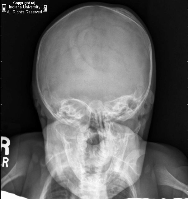
Lenticular left parietal calvarial mass-like lesion with peripheral sclerosis/calcification and central lucency. The lesion does not cross suture lines.

Differential diagnosis:
- Calcified cephalohematoma
- Caput succedaneum
- Subgaleal hematoma
- Subperiosteal osteoid osteoma
- Leptomeningeal cyst


Diagnosis: Subperiosteal cephalohematoma.


Cephalhematomas are subperiosteal hematomas that frequently occur after using instruments (forceps) during delivery. They are not associated with skull fractures and are limited by suture lines. They typically occur in the parietal region and may not appear until 2-3 days after birth. Treatment is usually not necessary. Complications include infection. Caput succedaneum is a subcutaneous hemorrhage that occurs after vaginal delivery, does cross suture lines and requires no intervention. A subgaleal hemorrhage covers a much larger area than a cephalohematoma and is potentially life threatening. It usually occurs from rupture of emissary veins in the subaponeurotic space, often secondary to vacuum suctioning.


Radiologic overview

Often seen as soft tissue subperiosteal elevation on conventional radiography. Frequency of location: parietal > occipital > frontal. The outer border may calcify. The skull at the hematoma site my remain thickened for years. It does not cross suture lines as it is bounded by the periosteum.

giovedì 18 giugno 2009

Os Odontoideum


Findings

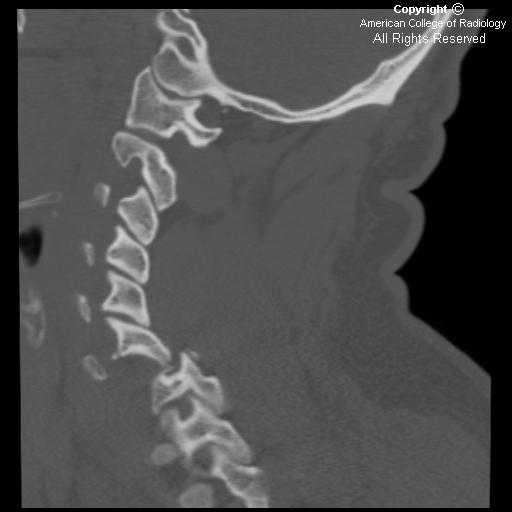
The coronal CT neck image in Figure 1 shows a small, round os odontoideum separated from the body of C2 by a wide radiolucent gap. It has smooth and uniform cortical margins.
The sagittal CT neck image in Figure 2 shows signs of C1 anterior arch hypertrophy. There is no paravertebral swelling. The posterior atlanto-dens interval (PADI) also known as the space available for cord (SAC) is 12mm (less than 13mm has a poor prognosis).


Diagnosis: Os Odontoideum


Os odontoideum describes a rare condition in which the dens is separated from the axis body. Two types of os odontoideum have been identified. An orthotropic os odontoideum is located in the normal position of the odontoid process, whereas a dystrophic os odontoideum is either attached to the anterior arch of C1 or the clivus.

The etiology of os odontoideum is still debated. The leading hypothesis suggests that trauma in early childhood causes injury to the soft tissues between C1 and C2. This is often seen when babies fall from their cribs or toddlers fall from stairs. Radiographic images taken after this injury usually do not show any significant pathology besides retropharyngeal swelling.

Injury results in avascular necrosis of the odontoid process over a period of months or years. Therefore, the cephalad ossification centers do not fuse with the C2 body. With growth, the alar ligaments that attach the tip of the dens to the occiput pull the ossicle upwards giving it its characteristic appearance.

Os odontoideum may also occur in patients with congenital anomalies including Down's syndrome, multiple epiphyseal dysplasia, Klippel-Feil malformation and Morquio syndrome. These patients are born with deficient odontoid processes.

Symptoms are variable with vague pain in the neck and shoulders being the most common. Less commonly reported symptoms include headaches, torticollis, weakness and paresthesias. The most serious complications of an os odontoideum occur when C1-C2 instability causes spinal cord compression or vertebral artery compromise. This may result in brain stem symptoms, quadraparesis, transient bouts of unconsciousness and sudden death.

The best way to evaluate an os odontoideum is by obtaining open-mouth anterior-posterior and flexion-extension lateral plain films. A criteria often used to make the diagnosis is the Posterior AtlantoDens Interval (PADI) also known as the Space Available for Cord (SAC). This is the distance between the posterior border of the dens and the anterior border of the posterior ring of the atlas. A PADI of less than 13mm is associated with neurologic decline. C1-C2 translation of more than 5mm on flexion-extension radiographs also has a poor prognosis.

The most common treatment for a symptomatic os odontoideum is C1-C2 posterior arthrodesis. This is particularly indicated in patients who exhibit signs of cord compression. Medical management is indicated for patients with only mechanical symptoms.

venerdì 5 giugno 2009

Bilateral jumped facets with spinal cord transection


Findings

Axial CT scan of the cervical spine demonstrates the naked facet sign, with uncovered articulating processes. A fracture is noted anteriorly on the left.
Sagittal image shows marked subluxation of C6 on C7 with marked narrowing of the spinal canal.
Sagittal images of the right facets. The facets are jumped.
Sagittal T2 and STIR show marked anterior subluxation of C6 on C7, transection of the spinal cord, edema and hemorrhage in the prevertebral soft tissues and spinal cord edema extending from C4 to T1.


Diagnosis: Bilateral jumped facets with spinal cord transection


Bilateral facet joint dislocation is a severe unstable cervical flexion injury. Flexion- rotation injuries can cause dislocation of the vertebral bodies with jumped/locked facets which can be unilateral or bilateral. If the dislocation is not complete, the facets may appear “perched” upon one another. Unilateral facet dislocation more characteristically has a rotatory component and carries a better prognosis.

With complete dislocation, the facets are said to be jumped and/or locked. The integrity of all the spinal ligaments including the posterior longitudinal ligament, the posterior portions of the annulus fibrosus, and the capsular, interspinous and supraspinous ligaments is disrupted. Destabilization of the ligamentous support of the cervical spine allows for the charactersitic >50% anterolisthesis observed. Unfortunately, severe narrowing of the spinal canal and intervertebral foramen occurs with subsequent severe neurologic deficits (in approximately 75% of cases), as seen in this case with spinal cord transection.

The naked facet sign, also referred to as the, “Hamburger Sign” refers to the CT appearance of uncovered articulating processes. Normally, on axial CT, the vertebral facet joint looks like a hamburger with the superior articular process of the vertebra below forming the, “bun” on top of the “meat patty,” and the inferior articular process of the vertebra above forms the bun beneath the patty. When the joint is dislocated, the superior articular facet (top bun of the Hamburger) now lies posteriorly. This is the Hamburger Sign, which can be unilateral or bilateral.

Associated osseous injuries include avulsion of the spinous process above the locked level and small triangular fractures of the anterosuperior corner of the inferior involved vertebral body.

giovedì 4 giugno 2009

Anterior subluxation of C4 on C5, with other imaging demonstrating unilateral locked left facet and perched right facet at C4-C5.


Findings

Figure 1: Lateral Cervical spine plain film demonstrates anterior subluxation of C4 on C5 and widening of spinous processes.
Figure 2: Sagittal MRI image shows anterolisthesis of C4 on C5 with central disc herniation. The spinal cord demonstrates normal signal intensity. Increased T2 signal intensity is present posteriorly, consistent with ligamentous disruption with edema and hematoma of posterior soft tissues.


Diagnosis: Anterior subluxation of C4 on C5, with other imaging demonstrating unilateral locked left facet and perched right facet at C4-C5.


Hyperflexion injuries can occur during trauma and are described as a combination of distraction and flexion that disrupts the ligamentous structures between two adjacent vertebrae, but the anterior longitudinal ligament remains intact. The height of the anterior interspace is narrowed and the posterior height is widened. There is also a possibility of disruption of the facet joints. Hyperflexion can result in injury to the posterior and middle columns whereas hyperextension may lead to injury to the anterior and/or middle columns.

A unilateral locked facet is secondary to excessive flexion and rotation and is considered a stable injury. The inferior facet of the superior vertebra moves up and anterior to the inferior vertebra thereby causing them to sit in a “locked” position. In unilateral locked facets there is typically anterolisthesis of the superior vertebral body of <50% of the width of the vertebral body. If there is translation of more than 50%, then there is most likely bilateral locked facets, which is an unstable injury.

Cervical spine CT with sagittal reconstructions or lateral cervical plain films can be used for measuring cervical translation. Lines are drawn along the involved posterior vertebral bodies and the transverse distance is measured between the lines at the level of the inferior endplate of the upper vertebra.

MRI may be used in patients with acute cervical spine trauma to evaluate neurologic or ligamentous integrity. It is indicated in patients with negative radiographs and negative CT who have neurologic symptoms, and also in patients with fracture or unstable injury. It has recently been noted that MR imaging has high sensitivity for most ligamentous injuries, in particular posterior longitudinal ligament, interspinous ligaments, disc annulus and facet capsular ligaments. Sensitivity is somewhat lower for anterior longitudinal ligament and ligamentum flavum injury after comparing with intraoperative findings. It should also be noted that, while sensitive, MR findings were generally not found to be highly specific with respect to intraoperative findings and can result in overestimation of ligamentous injury.

venerdì 6 marzo 2009

Sphenoid wing dysplasia


Findings

Figure 1: CT head without contrast demonstrates partial protrusion of the left temporal lobe and associated dura and CSF through the bony defect producing distortion of the optic nerve and proptosis. Also of note is a contusion in the left temporal lobe and left preseptal soft tissue swelling.
Figure 2, Figure 3, Figure 4, Figure 5, and Figure 6: CT head without contrast demonstrates absence of the left sphenoid wing. There is also a left occipital and right petrous bone fracture.
Figure 7 and Figure 8: Anterior and posterior volume rendered 3-D CT images demonstrate a gaping bony defect in the posterior aspect of the left orbit. The normal anatomical landmarks of the optic nerve canal, superior and inferior orbital fissures are not identified, and the anterior clinoid is not present. The margins of the bony defect are smooth and regular suggesting that this is developmental in nature.


Diagnosis: Sphenoid wing dysplasia


Sphenoid wing dysplasia can occur as an isolated finding or in patients who have Neurofibromatosis type 1. Approximately 50% of cases are associated with neurofibromatosis type 1. Sphenoid dysplasia has been reported to occur in 1 to 7% of patients with neurofibromatosis. Sphenoid wing lesions are usually asymptomatic, but patients can present with pulsating exophthalmos or enopthalmos. Others can present with vision impairment, extra-ocular muscle impairment or inflammation of the conjunctiva.

The radiological features of sphenoid wing dysplasia are a defect in a part or all of the greater wing, elevation of the lesser wing and distortion of the sella. There may be elevation of the superior orbital fissure, enlargement of the temporal fossa in all directions and lifting of the sphenoid ridge out of the orbit.

The most likely cause of sphenoid dysplasia is defective ossification of the sphenoid bone. In patients with NF-1, sphenoid wing dysplasia was initially thought to be congenital and non-progressive. It is now believed to be progressive in some patients with NF-1. Sphenoid wing dysplasia is one of six clinical criteria for the diagnosis of NF-1.

venerdì 13 febbraio 2009

Uncal herniation with posterior cerebral artery occlusion and resulting infarct


Findings

Image 1: Acute intraparenchymal hemorrhage with midline shift.
Image 2: Effacement of the basal cistern, quadrigeminal plate with enlargement of the contralateral lateral ventricle.
Image 3: Followup study 2 weeks later demonstrates hypo density in the left posterior cerebral artery distribution.

Differential diagnosis:
- Uncal herniation with posterior cerebral artery occlusion
- New thrombotic infarct in the posterior cerebral artery distribution
- Edema from infarct not seen with initial imaging


Diagnosis: Uncal herniation with posterior cerebral artery occlusion and resulting infarct


Discussion

With increased intracranial pressure, the brain can herniate transtentorially, cutting off circulation to the posterior cerebral artery and causing infarct of the ipsilateral occipital lobe. Radiologic evidence of descending transtentorial herniation includes obstruction of the CSF draining system and contralateral enlargement of the lateral ventricle, the uncus extending into the suprasellar cistern, and ipsilateral prepontine cistern widening. The clinical evidence for herniation itself includes an ipsilateral dilated pupil and contralateral hemiparesis, though these may be difficult to discern in the setting of already increased intracranial pressure due to the underlying etiology. The sequelae of PCA infarct include acute vision loss, confusion, new onset posterior cranium headache, paresthesias, limb weakness, dizziness, nausea, memory loss and language dysfunction.


Radiologic overview of the diagnosis

There will be evidence for increased intracranial pressure such as a space-occupying lesion, hemorrhage or edema; and, more specifically, evidence for transtentorial herniation including mass effect with uncal extension into the suprasellar cistern and contralateral lateral ventricle expansion. Over a period of time, there will eventually develop ipsilateral or bilateral signs of infarct on MR or CT in the distribution of the posterior cerebral artery within the occipital lobe.

martedì 20 gennaio 2009

Orbital blow out fracture


Findings

Figure 1: The x-ray demonstrates a fracture of the right orbital floor and an air-fluid level in the right maxillary sinus suspicious for hematoma.
Figure 2: Coronal CT image demonstrates a fracture of the left orbital floor along with herniation of the orbital fat. Air-fluid level in the left maxillary sinus containing high attenuation is consistent with hematoma.


Diagnosis: Orbital blow out fracture


An orbital blow-out fracture is a fracture of the orbital floor caused by blunt trauma to the orbit. Blow-out fractures are usually caused by round or oval object with a diameter slightly larger than the orbital diameter such as a baseball, snowball, tennis ball or fist. The mechanism of fracture is controversial. The two most accepted theories are: 1) The "retropulsion" theory which proposes the fracture is a result of a sudden increase in intraorbital pressure when the globe is pushed posteriorly. 2) The "buckling" theory states that the fracture is secondary to the force causing the orbital rim to buckle and transmitting the force into the orbital bones.

One may expect medial wall fractures to be more common than orbital floor fractures as the medial wall is thinner; however, the reverse is true. Proponents of the "retropulsion" theory attribute this to the honeycomb structure of the ethmoid air cell septae, which support the lamina papyracea, thus allowing it to withstand the sudden rise in intraorbital hydraulic pressure better than the orbital floor. Proponents of the “buckling” theory propose that the orbital floor is particularly vulnerable as the infraorbital canal further weakens the floor’s already delicate bony structure.

Routine facial views obtained should include the Caldwell and Waters view. The Caldwell projection is used to evaluate the lateral orbital wall and ethmoid bone, while the Waters view is useful for visualizing the inferior orbital wall and maxillary sinuses. The teardrop sign is secondary to opacification of the upper maxillary sinus from herniated orbital fat.

CT has become the modality of choice to evaluate orbital fractures. Herniation of orbital fat, the inferior rectus muscle, and the inferior oblique muscle may occur and herniated extraocular muscles entrapped between the fragmented bone segments may lead to diplopia. Edema and hemorrhage from the trauma can also cause diplopia; however this should resolve in a few days in the absence of entrapment. Immediate surgical intervention may be required in cases of entrapped extraocular muscles or acute enopthalmos. Chronic enopthalmos may also develop in fractures with extensive orbital fat herniation.

giovedì 18 dicembre 2008

Retinal hemorrhage in non accident trauma


Findings

Figure 1: T2*GRE axial image through the globe demonstrates nodular low signal in both retinas representing bloom artifacts from blood products suggesting retinal hemorrhage.
Figure 2: T2*GRE axial image shows low signal in the posterior parietal subdural space suggesting subdural hemorrhage.
Figure 3: Axial T1 image shows left frontal subdural fluid collection similar to CSF signal suggesting subdural hygroma.


Diagnosis: Retinal hemorrhage in non accident trauma


Child physical abuse is an unfortunately common occurrence that may manifest as any pattern of injury. Some patterns of injury (metaphyseal fractures, posterior rib fractures, subdural hemorrhages, retinal hemorrhages) are more suggestive and specific than others. Child abuse is often misdiagnosed and under-recognized by physicians and caregivers.

Non-accidental head injury (NAHI) or shaken baby syndrome (SBS), (some authors prefer the term shaken-impact syndrome), occurs in 12% of the physically abused children and is a major cause of neurological disability and death during infancy. Radiological imaging plays a crucial role in evaluating cranial injury, both for guiding medical management and the forensic aspects of abusive trauma. The neurologic injuries are more devastating than other injuries. NAHI is the leading cause of morbidity and mortality in abused children. MRI is the test of choice over CT for detecting different ages of intracranial hemorrhages, shearing injuries, and retinal hemorrhages.

Subdural hemorrhage is the most common intracranial lesion in shaken baby syndrome. Other findings are cerebral edema, subarachnoid hemorrhage, intraparenchymal hemorrhage, intraventricular hemorrhage, diffuse axonal injury, shearing injury, ischemia and brain herniation. Retinal hemorrhages may be found in 50%-100% of shaken infants, the prevalence of retinal hemorrhages in victims who die approaches 100%. Retinal hemorrhage is a characteristic and diagnostic feature of SBS. The severity of retinal hemorrhage is strongly correlated with intracranial injury in SBS. It is therefore very important to recognize retinal hemorrhage on neuroimaging.