


Findings
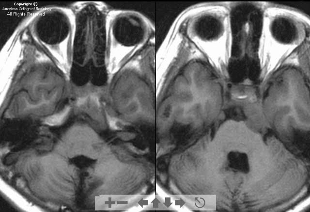
Axial T1 precontrast images demonstrate masses on the bilateral tentorium and in the internal auditory canals (Figure 1).
Axial T1 postcontrast images better demonstrate the masses (Figure 2).
Sagittal T1 and T2 images of the thoracic spinal cord demonstrate syringhydromyelia (Figure 3 and Figure 4).
An enhancing spinal cord mass is shown to be the etiology of the syrinx on the fat saturated sagittal T1 postcontrast images (Figure 5).
Diagnosis: Neurofibromatosis type 2 (NF-2)
Patients with NF-2 may develop meningiomas, schwannomas, and spinal ependymomas (or combinations of these). The genetic defect is localized to chromosome 22, which results in an abnormal tumor suppressor gene, causing these patients to have higher rates of certain tumors. Although common in adults, meningiomas are relatively rare in children and should raise a red flag for possible NF-2.
Some examples of criteria for the diagnosis of NF-2 include bilateral acoustic schwannomas, acoustic schwannoma and meningioma or schwannoma elsewhere, and first degree relative with NF-2 and varying combinations of two or more of the following: schwannoma, meningioma, and juvenile posterior subcapsular lenticular opacities.
Nessun commento:
Posta un commento