giovedì 18 maggio 2006

Sturge-Weber syndrome (SWS)


Findings

Plain films (Figure 1 and Figure 2) show “tram track” calcification in the right frontal region. The gyral or subcortical calcification is better seen in the CT image (Figure 3).
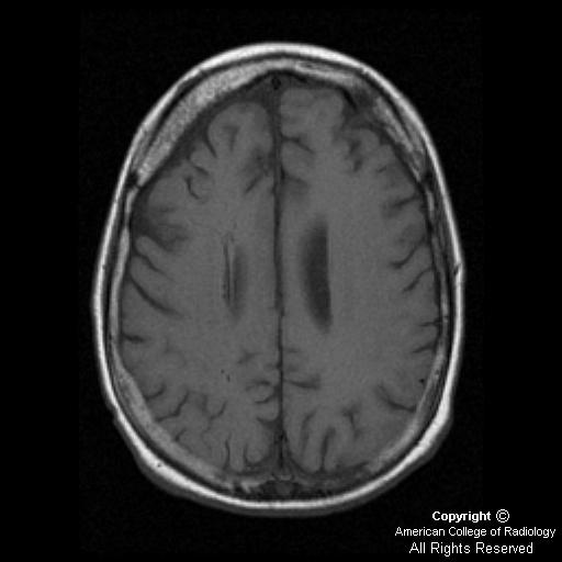
Axial T1 image (Figure 4) demonstrates volume loss in the right frontal area with thickened diploic space in this region. Sagittal SE image (Figure 5) shows multiple round signal voids, which are dilated deep medullary veins.
Postcontrast images (Figure 7 and Figure 8) show leptomeningeal enhancement, suggestive of increased venous collaterals and prominence of the right transmantle (transcerebral) veins (Figure 6). Also note the enlarged ipsilateral right choroid plexus (Figure 7).
GRE (Figure 7) shows “blooming” in the right frontal area corresponding to the area of calcification seen on CT.
MRV (Figure 9) findings demonstrate a hypoplastic right transverse sinus as well as a very small right-sided jugular vein.


Diagnosis: Sturge-Weber syndrome (SWS)


SWS, also known as encephalotrigeminal angiomatosis or meningiofacial angiomatosis, is a rare neurocutaneous syndrome that includes a facial port wine stain and associated leptomeningeal angiomatosis. The pial angiomatosis may be bilateral in 20% of cases.

It is generally considered nonhereditary. Embryologically, the abnormality most likely develops at gestational weeks four to eight. It is hypothesized that impaired venous outflow, caused by loss of normal connections between cortical veins and dural and calvarial circulation, results in persistent, primitive vascular plexus. This results in poor venous drainage from the cerebral cortex and progressive cortical damage.

Seizures often begin in the first year of life. It is the presenting feature in 80% of patients and may be generalized or partial. Hemiparesis may occur in up to 30% of cases resulting from hemiatrophy of brain. Developmental delay is common.

The port wine stain (nevus flammeus) usually occurs in the distribution of first and second division of trigeminal nerve. The choroid of eye may be involved with patients developing glaucoma.

The impaired venous drainage results in progressive ischemia of underlying brain and, eventually, progressive cortical atrophy and calcification. The cortical calcifications are rarely identified at birth. The most common area involved is the parieto-occipital region. Skull radiographs may show the typical “tram track” calcifications in apposing gyri. CT may also show enlargement of adjacent diploic space, hyperpneumatizaton of ipsilateral sinuses, and mastoid air cells.

MRI may show signs of ischemia and gliosis in early stages, but parenchymal atrophy over time. Gadolinium-enhanced MRI is highly sensitive to meningeal enhancement. Calcifications are better detected with gradient echo techniques. Enlargement of the ipsilateral choroid plexus may be secondary to hyperplasia or angiomatous involvement.

MR venogram may show enlarged deep collateral (medullary or subependymal) veins with lack of superficial cortical veins.

PET may show increased metabolism in early stages, which can be helpful in surgical planning.

martedì 9 maggio 2006

Pachygyria


Findings

CT (Figure 1) and MR (Figure 2) imaging demonstrate the classic patterns of pachygyria, including:
- Smooth and markedly thickened cortex (Figure 1 and Figure 2)
- Broad, flat gyri with shallow sulci (focal or widespread)
- Vertically-oriented and shallow Sylvian fissures
- “Figure-of-eight” appearance of the brain on axial imaging


Diagnosis: Pachygyria


The findings in this case are consistent with pachygyria, which falls under the classification of an embryologic neuronal migration disorder. With pachygyria (synonymous with incomplete lissencephaly), there are focal areas of broad, flat gyri interrupted by a smooth and markedly thickened cortex. The etiology in some cases is linked to genetic abnormality, including mutations in chromosome 17 or Xq22, while others demonstrate no genetic abnormalities. Alternative etiologies for the development of this disorder include intrauterine insult (ischemic, metabolic, or viral such as CMV) during the 12th to 24th week of embryologic development. This results in damage to the germinal matrix and radial glial cells. Radial glial cells act as mechanical guides by which neurons are transported to the outer cortex during development. Damage to these cells results in subsequent malfunction of neuronal cell migration to the outer cortex.


Clinical findings

Onset and severity of symptoms varies, depending on the amount of cortical involvement. However, muscle spasms, hyperreflexia, refractory epilepsy, developmental delay, and mental retardation are common in children afflicted with the disorder.


Radiographic findings

Although CT is adequate in making the diagnosis, as in this case, MRI is more effective in evaluating and differentiating between neuronal migration disorders.

CT and MR imaging demonstrate the classic patterns of pachygyria, including:
- Smooth and markedly thickened cortex
- Broad, flat gyri with shallow sulci (focal or widespread)
- Vertically oriented and shallow Sylvian fissures
- “Figure-of-eight” appearance of the brain on axial imaging

venerdì 5 maggio 2006

Subarachnoid hemorrhage (SAH) due to left PICA aneurysm


Findings

High attenuation within basilar cisterns and subarachnoid spaces, extending to the Sylvian fissures consistent with hemorrhage. SAH was not seen to extend over the convexities (Figure 1, Figure 2 and Figure 3).
Vertebral artery injection digital subtraction angiography demonstrates an aneurysm of the Left PICA origin.
Figure 6 shows classic “teat” at site of rupture.


Differential diagnosis for subarachnoid hemorrhage (CT findings are diagnostic):
- Trauma
- Aneurysm rupture
- AVM
- Angioma
- Neoplasm
- Cortical thrombosis
- Dissection from intraparenchymal hematoma

Differential diagnosis for cerebral aneurysm (angiographic findings are diagnostic)
DD for saccular aneurysms:
- Developmental/degenerative
- Traumatic pseudoaneurysm
- Mycotic
- Oncotic
- Flow related
- Vasculopathy related
- Drug related)

DD for fusiform aneurysms: Atherosclerosis

DD for dissecting aneurysms:
- Trauma
- Vasculopathy


Diagnosis: Subarachnoid hemorrhage (SAH) due to left PICA aneurysm


SAH is most commonly due to trauma. In the absence of trauma history or correlative findings, presence of SAH necessitates investigation for a cause. Nearly two thirds of these cases will be due to ruptured aneurysm. Clinically, patients experience severe headache. Patients often describe a “sentinel” headache, or prodrome indicating earlier bleeding. Less often, focal or global neurologic findings are present.

SAH is graded based on clinical presentation:
Grade 1) being mild headache
Grade 2) severe headache
Grade 3) mild mental status changes
Grade 4) obvious altered mental status or neurologic change
Grade 5) comatose or posturing

Long-term outcome directly correlates with grade at initial presentation. Of all patients with SAH, roughly 40% die within 24 hours and an additional 10% to 25% within six months. Of the survivors, half will have major long-term neurologic deficits.

Radiographically, high attenuation within subarachnoid spaces (sulci, ventricles, basal cisterns) on a noncontrast CT is the principal finding. Multidetector CT is up to 98% sensitive in the first 24 hours. Degree of SAH is also graded and has prognostic value, as does location of hemorrhage.

Complications of SAH include hydrocephalus, rebleeding, and vasospasm. Hydrocephalus is due to obstructing clot within the ventricular system and may be early or delayed. Early evidence is enlargement of the temporal horns. Rebleeding most often occurs within the first 24 hours, but can occur up to 2 weeks after initial insult. This is found in 20% to 30% of untreated patients and carries up to an 85% mortality. Vasospasm is the most feared complication, resulting in the greatest degree of morbidity and mortality, and affects nearly 40% of all patients. Peak time frame, according to the literature, is four to 12 days, although anecdotal experience from our institution suggests this is often seen earlier. Vasospasm can lead to ischemic events and progression of neurologic deficits.

As discussed, primary nontraumatic SAH is most commonly due to aneurysm rupture. Saccular aneurysms are true aneurysms and are the most common. These are thought to be congenital areas of weakening in arterial walls, which develop over many years into an aneurysm. Fusiform and dissecting are the other two major categories, and are much less common. Aneurysms have a fairly predictable distribution: 35% anterior communicating artery, 30% posterior communicating artery, 20% MCA bifurcation, and 15% vertebrobasilar system. Location of SAH is often a clue to aneurysm location. While no aneurysm is too small to exclude rupture, 4 to 7 mm appears to be the critical size. Aneurysms larger than 10 mm are at much higher risk. A patent aneurysm appears as an outpouching of contrast, while a thrombosed aneurysm may have a normal appearance. Lobulation or irregularity of aneurysm dome (“teat”) is an indication of possible rupture site, especially in the presence of multiple lesions.

Treatment of aneurysms has significantly progressed with intraarterial coil embolization. In the past, the only option was open surgical intervention. Timing of treatment is divided into two phases: early (within two to three days) and late (after ten to 14 days), avoiding the peak incidence of vasospasm. No significant outcome difference has been proven, however, proponents of early intervention for low-grade aneurysms cite the ability for aggressive treatment to lower risk of vasospasm once the aneurysm is controlled. Treatment for asymptomatic, unruptured aneurysms is even more controversial, due to postprocedural complications. Typically, lesions less than 5 mm are followed, while larger lesions are often treated.

venerdì 28 aprile 2006

Labyrinthitis ossificans


Findings

There is osseous obliteration of the visualized cochlea (Figure 1), vestibule, and lateral semi-circular canal (Figure 2).


Diagnosis: Labyrinthitis ossificans


The most common causative factor is inflammation of the inner ear, secondary to bacterial meningitis and subsequent purulent labyrinthitis. Other, less common causes include labyrinthectomy, temporal bone trauma, autoimmune inner ear disease, sequela of mumps or measles, and otitis media with associated suppurative labyrinthitis.

Ossification in labyrinthitis ossificans commonly occurs primarily at the basal turn of the cochlea, which is the insertion site of the electrodes of cochlear implant devices, and subsequently may interfere with full insertion and successful results. MRI is used to assess for early fibrosis prior to the formation of calcification.

mercoledì 26 aprile 2006

Superficial siderosis


Findings

Figure 1 and Figure 2: T2 and gradient echo hypointensity is demonstrated along the pial surfaces of the brain, and subarachnoid 7th and 8th cranial nerves. The hypointensity seen on the gradient echo images is more prominent than that on the T2/FLAIR sequences, consistent with blooming due to hemosiderin deposition.
Figures 3, Figure 4, and Figure 5: T2 and gradient echo hypointensity is demonstrated along the pial surfaces of the brain, spinal cord, and the subarachnoid courses of the visualized nerve roots. As in Figure 1 and Figure 2, the hypointensity seen on the gradient echo images is more prominent than that on the T2/FLAIR sequences, consistent with blooming due to hemosiderin deposition.


Diagnosis: Superficial siderosis


Superficial siderosis (SS) of the central nervous system is the sequela of chronic recurrent subarachnoid hemorrhage. Hemosiderin, a blood product of chronic hemorrhage, is deposited along the leptomeningeal surfaces of the brain and spinal cord, as well as the subarachnoid surfaces of the cranial and spinal nerves. The hemosiderin deposition is not benign. The leptomeninges become thickened and the affected nervous tissue can undergo neuronal loss, reactive gliosis, and demyelination. Classic locations for hemosiderin deposition in SS are the peripheral folia of the cerebellum. Hemosiderin deposited here is usually associated with Bergmann gliosis and death of Purkinje cells. This leads to cerebellar atrophy, especially involving the vermis.

The cranial nerves are often affected with preferential involvement of cranial nerves VIII and I. These nerves seem to be more frequently affected due to their relatively long courses through the subarachnoid space. As the hemosiderin can be neurotoxic, patients with SS can present with sensorineural hearing loss and anosmia. Symptomatic involvement of cranial nerves V and VII also occur with some frequency. SS is associated with hydrocephalus, as the subarachnoid granulations become impaired from the chronic subarachnoid hemorrhage.

The MR findings of SS are characteristic. Pial T2 and gradient echo (GRE) hypointensity is demonstrated along the leptomeningeal surfaces of the brain and spinal cord. The pons and cerebellum are two of the most frequently affected regions. T2 and GRE imaging also show hypointensity along the subarachnoid portions of the cranial and spinal nerves. The hypointensity demonstrated on the T2 “blooms” on GRE imaging due to hemosiderin MRI susceptibility.

The etiology of SS includes trauma, vascular anomalies, and neoplasm. Any process producing subarachnoid hemorrhage can result in SS. Between 80% and 90% of nontraumatic subarachnoid hemorrhage is the result of intracranial aneurysm rupture. Other vascular malformations that have significant frequencies of hemorrhage are AVMs and cavernous angiomas. Diagnosis of SS mandates an evaluation for the source of the hemorrhage and should include both intracranial MRI/MRA, as well as total spine MRI. Traditional cerebral - spinal angiography may be necessary to identify subtle vascular abnormalities. The etiology of SS is not identified in 25% to 46% of patients.

venerdì 21 aprile 2006

Multiple hemorrhagic intracranial metastases from primary lung carcinoma


Findings

Noncontrast enhanced CT of the brain reveals multiple high-density brain lesions with surrounding vasogenic edema, suggestive of hemorrhagic metastases (Figure 1 and Figure 2). Post-contrast images demonstrate enhancement of several of the lesions (Figure 3 and Figure 4).
Medialstinal and lung windows from the CT of the chest (not shown)demonstrated the primary lung carcinoma.


Diagnosis: Multiple hemorrhagic intracranial metastases from primary lung carcinoma


Metastatic spread to the brain is usually arterial, although less commonly spread via the Batson venous (pelvic, GI tumors) may occur. Most metastatic lesions are well-demarcated and tend to cluster at the gray-white matter junction.

Most of the brain metastases originate either from a primary lung tumor or from metastatic lesions in the lungs, since most large aggregates of tumor cells are filtered by the lung capillary network. Single tumor cells, however, can pass through the lung capillary network, and larger tumor emboli can pass from the venous to arterial circulation through a patent foramen ovale or a right to left shift.

The most likely malignancies to produce hemorrhagic metastases are melanoma, renal cell carcinoma, choriocarcinoma, thyroid, lung, breast, and germ cell tumors. Due to their increased prevalence, however, bronchogenic lesions are the most common source of hemorrhagic lesions.

Up to 20% of patients with metastases to the brain present with seizures.

Mnemonic MR CT BB
M: Melanoma
R: Renal cell carcinoma

C: Choriocarcinoma
T: Thyroid, terratoma

B: Bronchogenic carcinoma
B: Breast carcinoma

giovedì 13 aprile 2006

Vertebral plana due to Langerhan's cell histiocystosis


Findings

Figure 1: Reconstructed sagittal CT scan through the thoracic spine demonstrates a flattened vertebral body. The disk spaces are maintained. The remaining vertebral bodies are unremarkable.
Figure 2: Sagittal T2-weighted MRI demonstrates a focal kyphosis at the level of the flattened vertebral body, with a ventral epidural soft tissue component resulting in thoracic spinal cord compression.
Figure 3: Sagittal T1 post-contrast MRI with fat saturation again demonstrates a focal kyphosis at the level of the flattened vertebral body. The ventral epidural soft tissue component is better demonstrated than on the T2 series

Differential diagnosis for vertebral plana = “MELTS”
- M: Metastasis, myeloma
- E: Ewing’s sarcoma
- L: Langerhan’s cell histiocystosis, leukemia, lymphoma
- T: Trauma, tuberculous osteomyelitis
- S: Steroids


Diagnosis: Vertebral plana due to Langerhan's cell histiocystosis


LCH predominantly affects children and is comprised of three conditions: eosinophilic granuloma, Hand-Schuller-Christian disease, and Letterer-Siwe disease. All three conditions are characterized by the presence of abnormal histiocytes called Langerhan’s cells. Eosinophilic granuloma is the mildest form and is the most common, comprising approximately 70% of the disease. It consists of single or multiple bony lytic lesions, and most commonly involves the skull, mandible, spine, ribs, and long bones. Hand-Schuller-Christian disease (20% of LCH) is a multisystem disease comprised of diabetes insipidus, exopthalmos, and one or multiple bony lesions. It is fatal in approximately 30% of patients. Letterer-Siwe disease (10% of LCH) is the most severe form and it affects children less than three years old with liver, spleen, skin, and bony involvement. It is usually fatal within two years of diagnosis.

Eosinophilic granuloma is a benign condition that has a variable prognosis. Specifically, the prognosis for vertebral plana is favorable. The lesions may regress spontaneously, taking up to two years. The treatment is often conservative, but surgery, radiation, chemotherapy, and steroids may be used if conservative treatment fails.