martedì 11 aprile 2006

Pineal cyst


Findings

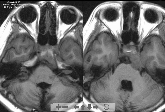
A cystic lesion (2.0 x 1.8 x 1.2 cm) with thin and well-defined enhancing walls (Figure 1 and Figure 2) places mild mass-effect on the tectum. No hydrocephalus is present. The cystic lesion (Figure 1, Figure 2, Figure 3 and Figure 4) is homogeneously isointense to CSF on T1-weighted imaging, uniformly hyperintense on FLAIR sequences, and isointense to CSF on T2-weighted imaging. No diffusion restriction is seen on diffusion-weighted imaging. These findings are most compatible with a pineal cyst. Less likely, this may represent a pinecytoma.

Differential diagnosis:
- Pineal cyst
- Normal pineal
- Pineocytoma
- Epidermoid cyst
- Arachnoid cyst
- Germ cell neoplasm


Diagnosis: Pineal cyst


Pineal cysts are usually asymptomatic and are often discovered incidentally. They have been discovered in up to 40% of patients on autopsy studies. Patients with large pineal cysts may experience headaches secondary to obstructive hydrocephalus. Rarely, pineal cysts can produce Parinaud syndrome (paralysis of upward gaze, lid retraction, and abnormal pupillary movements). If the cyst is hemorrhagic, acute hydrocephalus can develop. Young females (aged 21-30) are affected more than males (female to male ratio is 3:1). The cysts commonly enlarge with puberty and then regress with age.

On CT, the pineal cyst contents are usually isodense relative to CSF. The wall may be calcified. The cyst wall (nodular or thin) may enhance with contrast. Pineal cysts have a variable appearance with MR. Cystic contents are most often hyperintense relative to CSF, but may be isointense on T1-weighted imaging. On T2-weighted sequences, the cystic contents are either isointense or hypointense to CSF. On FLAIR sequences, the cystic contents are hyperintense. No restriction is seen with diffusion-weighted imaging. The cyst walls may enhance (60%). On delayed images, the cystic contents may enhance.

The key management issue regarding a pineal cyst, is the fact that they can be indistinguishable from a pinecytoma. Therefore, a histological diagnosis may be needed to make the distinction. If biopsy or surgery is not performed, long-term follow-up imaging should be obtained. Some suggest that clinical follow-up without imaging is an acceptable alternative. In one follow-up series, the cysts did not change in size in 75% of patients.

martedì 4 aprile 2006

Branchial cleft cyst


Findings

Contrast-enhanced CT examination of the neck demonstrates an ovoid, well-circumscribed low attenuation lesion in the right neck.
The images show a branchial cleft cyst, which is well-circumscribed with a thin enhancing rim. The branchial cleft cyst is anteromedial to the sternocleidomastoid muscle (Figure 1 and Figure 2) and lateral to right internal and external carotid arteries (Figure 1 and Figure 2).

Differential for a cystic neck mass in the adult:
- BCC
- Thyroglossal duct cyst
- Lymphangioma
- Thymic cyst
- Abscess
- Suppurative lymph node
- External laryngocoele
- Necrotic squamous cell carcinoma


Diagnosis: Branchial cleft cyst


During normal embryologic development, the branchial arches arise at four weeks. The second branchial arch overgrows the third and fourth arches during development.
The branchial arches involute by week seven. If an arch fails to involute completely, it may form an epithelial-lined cyst. These congenital cysts may be identified in the early years with a second peak incidence in the second and third decades. BCCs are typically a painless compressible mass. They may occasionally become infected and present with fever, induration, and tenderness.

The second BCC is the most common, accounting for 90% of BCCs. A second BCC is typically found at or slightly below the angle of the mandible. The second BCC is centered lateral to the carotid sheath and anteromedial to the sternocleidomastoid muscle. Medial beaking of the cyst can be seen if it extends between the external and internal carotid arteries. The less common first BCC is seen in a periauricular or periparotid location. It can communicate with the external auditory canal and can be associated with otorrhea and parotid gland cysts. A third BCC is seen in the posterior cervical space.

venerdì 31 marzo 2006

Metastatic squamous cell carcinoma


History: The patient is a 63-year-old white man with a painless right neck mass and a history of resection of multiple cutaneous squamous cell carcinomas of the facial region.


Findings

Figure 1: Demonstrates an intraparotid soft tissue mass with an irregular hypodense area centrally consistent with necrosis.
Figure 2: Demonstrates a second intraparotid soft tissue mass that is homogeneous.
In light of the patient's history, this constellation of findings is most consistent with intraparotid nodal metastases.


Diagnosis: Metastatic squamous cell carcinoma


Malignant neoplasms of the parotid glands generally present with a palpable, discrete, painless mass. More specific indications of malignancy include facial nerve paralysis or dysfunction, and cervical adenopathy; however, these symptoms are present in only 24% and 6% of cases, respectively.

Evaluation of parotid malignancies must include localization to the superficial or deep aspect, determination of degree of involvement of the facial nerve, and whether there has been spread of tumor through the stylomastoid foramen, to determine the extent of surgical intervention required. If the skull base has been breeched, the cartilaginous external auditory canal may require dissection and possible resection. MRI is the most effective imaging modality to fully evaluate malignant parotid tumors, and provides the best demonstration of perineural, vascular, or dural invasion.

mercoledì 29 marzo 2006

Posterior reversible encephalopathy syndrome (PRES)


Findings

Diffuse hyperintensity is present in the cortical and subcortical parietal, occipital, and frontal white matter (all figures). No diffusion restriction is present on the DWI (images not shown). The ADC signal is increased (images not shown). Hypodensity is seen in the corresponding areas on the CT of the brain. These findings are most compatible with PRES.

Differential diagnosis:
- Posterior reversible encephalopathy syndrome (PRES)
- Acute PCA infarction
- Hypotensive stroke
- Severe hypoglycemia
- Progressive multifocal leukoencephalopathy
- Gliomatosis cerebri


Diagnosis: Posterior reversible encephalopathy syndrome (PRES)


PRES has been associated with several conditions, such as acute glomerulonephritis, thrombotic thrombocytopenic purpurpa, and hemolytic uremic syndrome, drug toxicity, cryoglobinemia, and systemic lupus erythematosis. Drug toxicity from Cisplatin, interferon, erythropoietin, tacrolimus, and cyclosporin has been documented as a cause of PRES. Most cases are thought to be the result of inability of the posterior circulation to autoregulate blood flow in the setting of acute hypertension. This in turn leads to endothelial and blood brain barrier injury with resultant vasogenic edema. Patients typically have signs or symptoms of headache, mental status changes, acute hypertension, and seizures. When imaging findings are compatible with PRES, one should also search for potential complications, including hemorrhage and stroke. Follow-up imaging should be performed when the hypertension is reversed. Most patients respond well with hypertensive management and treatment of the underlying etiology.

Cranial computed tomographic images frequently show low attenuating cortical or subcortical lesions, primarily in the posterior parietal and occipital lobes. Hypodense foci may be seen in the frontal and temporal lobes. Additional symmetric lesions may occur in the basal ganglia and brainstem with less frequency. MR will show hypointense lesions on T1-weighted imaging with corresponding hyperintensity on the T2-weighted imaging - FLAIR images. ADC images will have increased diffusion coefficients secondary to vasogenic edema. Diffusion-weighted imaging is usually normal. If diffusion restriction is present with decreased ADC values, cytotoxic edema may be present. With contrast, patchy enhancement may be seen. Diffusion tensor imaging shows increased brain water diffusion.

mercoledì 22 marzo 2006

Agenesis of the corpus callosum


Findings

Noncontrast CT of the head demonstrating absence of the corpus callosum with widely spaced parallel lateral ventricles, and dilatation of the trigones and occipital horns of the lateral ventricles (Figure 3).


Diagnosis: Agenesis of the corpus callosum


Agenesis of the corpus callosum is a congenital abnormality affecting approximately 0.7% of all births. Patients may present with refractile seizures, macrocephaly, mental retardation, hypothalamic dysfunction, or may be asymptomatic and of normal intelligence. The development of the corpus callosum begins at approximately the eighth week of gestation. Formation is initiated at the anterior genu and proceeds through the posterior splenium. The rostrum is the final portion to form at about week 20. Callosal abnormalities can vary from hypogenesis to complete agenesis. When hypogenetic, the structures that are formed the latest are the areas that are affected, including the posterior body, splenium, and rostrum.

There are several characteristic radiographic findings with agenesis of the corpus callosum. The corpus callosum is a relatively firm structure due to the compact nature of the axons within it, and thus in the normal brain, gives the ventricles their shape. In its absence, only loose white matter surrounds the ventricles and they expand. Anteriorly, the firm caudate heads maintain the shape of the frontal horns. Therefore, in callosal agenesis, only the posterior portions of the lateral ventricles enlarge, manifested by dilatation of the trigones and occipital horns. This is referred to as colpocephaly. In addition, the bodies of the lateral ventricles appear straight, widely spaced, and parallel. The lateral ventricles may also appear crescent-shaped. The white matter fiber tracks that would normally cross at the corpus callosum are redirected and run parallel to the interhemispheric fissure along the medial aspect of the lateral ventricles. The tracks leave an impression along the medial borders of the lateral ventricles, giving them a crescent shape. These white matter tracks are referred to as the longitudinal callosal bundles of Probst. With callosal agenesis, the third ventricle is often dilated and described as “high-riding,” as its location is more superior, lying between the lateral ventricles. Also, when the corpus callosum fails to form, the cingulate gyri, which lie just superiorly, do not invert and there is subsequent absence of the cingulate sulcus.

Agenesis of the corpus callosum may be associated with other midline abnormalities, including an interhemispheric cyst or pericallosal lipoma. There are many syndromes in association with agenesis of the corpus callosum. Aicardi syndrome is an x-linked dominant disorder that includes agenesis of the corpus callosum, mental retardation, seizures that are often resistant to anticonvulsants, and abnormalities of the retina. Corpus callosal agenesis is also associated with Chiari malformations, Dandy-Walker syndrome, and Trisomies 13, 15, and 18. In addition, it is the most common brain abnormality seen in fetal alcohol syndrome.

martedì 7 marzo 2006

Neurofibromatosis type 2 (NF-2)


Findings

Axial T1 precontrast images demonstrate masses on the bilateral tentorium and in the internal auditory canals (Figure 1).
Axial T1 postcontrast images better demonstrate the masses (Figure 2).
Sagittal T1 and T2 images of the thoracic spinal cord demonstrate syringhydromyelia (Figure 3 and Figure 4).
An enhancing spinal cord mass is shown to be the etiology of the syrinx on the fat saturated sagittal T1 postcontrast images (Figure 5).


Diagnosis: Neurofibromatosis type 2 (NF-2)


Patients with NF-2 may develop meningiomas, schwannomas, and spinal ependymomas (or combinations of these). The genetic defect is localized to chromosome 22, which results in an abnormal tumor suppressor gene, causing these patients to have higher rates of certain tumors. Although common in adults, meningiomas are relatively rare in children and should raise a red flag for possible NF-2.

Some examples of criteria for the diagnosis of NF-2 include bilateral acoustic schwannomas, acoustic schwannoma and meningioma or schwannoma elsewhere, and first degree relative with NF-2 and varying combinations of two or more of the following: schwannoma, meningioma, and juvenile posterior subcapsular lenticular opacities.

lunedì 6 marzo 2006

Acute adult epiglottitis


Findings

Lateral x-ray view of the neck soft tissues. There is thickening of the epiglottis with narrowing of the airway.
CT: Diffuse edema of the epiglottis and aryepiglottic folds is noted (Figure 2 and Figure 3). Edematous changes are noted in the midline in the base the tongue, perhaps related to endotracheal tube placement or perhaps part of the inflammatory process involving the epiglottis (Figure 2). A tracheostomy tube is noted in place. Bilateral soft tissue emphysema is noted (Figure 2 and Figure 3).

Differential diagnosis:
- Epiglottitis
- Tonsillitis
- Abscess
- Angioedema
- Foreign body
- Anaphylaxis
- Pharyngitis
- Trauma


Diagnosis: Acute adult epiglottitis


Epiglottitis is a rapid onset of inflammation in the supraglottic region of the oropharynx with inflammation of the epiglottis, aryepiglottic folds, and arytenoids. Epiglottitis is an uncommon disease. Incidence in adults is about 1 case per 100,000 per year. Adult epiglottitis is most frequently a disease of men, occurring during the fifth decade of life. Occurrence has decreased since introduction of the Haemophilus influenzae B vaccine. Epiglottitis is generally more common in nations that do not immunize against H influenzae type B. Risk of death is high due to sudden airway obstruction and difficulty intubating patients with extensive edema. Adult mortality rate ranges between 6%-7%. Mortality rate in pediatric cases is less than 1%. Male-to-female ratio is approximately 3:1. In the pediatric population, epiglottitis is most common in ages 3-7, although any age may be affected.

Symptoms encountered include: sore throat, odynophagia/dysphagia, usually no prodromal upper respiratory infection, fever, drooling, cervical adenopathy, stridor, tripod position (sitting up on hands with the tongue out and the head forward), hypoxia, and respiratory distress.

The causative agents are type B Haemophilus influenza, Staphylococcus, Streptococcus, and viruses. Swelling and edema cause airway compromise that often necessitates tracheal intubation to protect the airway. Diagnosis of epiglottitis is based upon clinical exam and history, radiography, and direct visualization of the epiglottis. Treatment includes intubation, or immediate formal tracheostomy or cricothyrotomy may be performed in the operating room, if the case is less severe. Those without signs of airway compromise may be managed without immediate airway intervention by close monitoring in ICU.