giovedì 27 ottobre 2005

Arteriovenous malformation


Findings

MR findings include serpiginous high and low signal (depending on flow rates) within feeding and draining vessels (Figure 1 andFigure 3). High T2 signal can be seen in the adjacent brain parenchyma secondary to gliosis, edema, or ischemia, as is seen in this case (Figure 3).
MRA reveals AVM with feeding artery branching from the left PCA. Nidus and tangled vessels are visualized (Figure 4).


Diagnosis: Arteriovenous malformation


Cerebrovascular malformations are divided into 4 major categories including arteriovenous malformation, venous angioma, capillary telangiectasia, and cavernous angioma. AVMs can be visualized with angiography, computed tomography, or magnetic resonance imaging. AVMs are the most common symptomatic vascular malformation and occur in about 0.1% of the general population. The clinical presentation is variable, including headache, seizure, and focal neurological deficits. They are occasionally discovered as an incidental finding.

AVMs are a direct communication between arterial and venous circulations and consist of a nidus of tangled dilated vessels. There are 3 subtypes that depend on the blood supply: these are pial, dural, and mixed pial-dural types. Aneurysm is associated with the feeding arteries in approximately 10% of cases. Arteriovenous shunting is usually rapid so that most vessels appear as flow voids. An AVM replaces normal brain tissue without causing mass effect, unless complicated by hemorrhage and edema. Adjacent parenchymal atrophy is common secondary to vascular steal and ischemia.

3-D TOF MRA can be diagnostically useful in demonstrating feeding arteries, the nidus, and draining veins. Occasional pitfalls in MRA evaluation include signal void in tortuous vessels, nonvisualization of draining veins resulting from spin saturation, and difficulty with differentiation of blood flow from blood clot. Conventional angiography may be necessary for treatment planning.

mercoledì 26 ottobre 2005

Thyroglossal duct cyst


Findings

Figure 1 demonstrates a cystic mass embedded in the strap muscles of the neck. Tract of thyroid tissue between cystic mass and thyroid gland is seen (Figure 2and Figure 3).


Diagnosis: Thyroglossal duct cyst


Thyroglossal duct cyst is the most common congenital neck mass and accounts for 70% of congenital neck abnormalities. Most patients present in the second decade with an enlarging, painless mass. Thyroglossal duct cysts are located in the midline (75%) or slightly off midline (25%), however, they are always within 2 cm of midline. Most cysts are located either at (15%) or below (65%) the level of the hyoid bone. On CT, the cysts usually appear as a smooth, well-circumscribed mass with a thin wall and homogeneous attenuation, measuring fluid in attenuation. Elevated attenuation levels may reflect increased protein content and can correlate with history of prior infection. Peripheral rim enhancement is usually observed on contrast-enhanced scans.

Thyroid gland development begins in the third week of fetal life as a median outgrowth from the primitive pharynx at the level of the foramen cecum, which lies at the junction of the anterior two thirds and posterior one third of the tongue. The primitive thyroid descends in the neck and passes anterior to the hyoid bone and laryngeal cartilages. The gland reaches its final position in the inferior part of the neck by the seventh week after descending anterior to the thyrohyoid membrane and strap muscles. During the migration, the analage of the thyroid gland is connected to the tongue by a narrow tubular structure, the thyroglossal duct. This structure usually involutes by the eighth to tenth week of gestation. If any portion of the duct persists, secretions from the epithelial lining may give rise to cystic lesions.

The differential diagnosis includes obstructed laryngoceles and branchial cleft cysts. Laryngoceles are dilated laryngeal saccules. They may appear as a well-defined smooth mass in the lateral aspect of the superior paralaryngeal space. They may occur deep to the strap muscles, however, they arise within the larynx and can be visualized to connect back to the larynx. Failure of complete obliteration of an embryonic branchial cleft in the eighth to ninth week of fetal development results in a branchial cleft cyst, 95% of which derive from the second branchial cleft. Branchial cleft cysts usually manifest in the young adult as a mass near the mandibular angle (submandibular space), however, they can occur anywhere along the residual cleft tract extending from the suprclavicular region to the tonsillar fossa. Typically, the cyst is a round or oval mass that displaces the sternocleidomastoid muscle posteriorly or posterolaterally, the carotid artery and internal jugular vein medially or posteromedially, and the submandibular gland anteriorly.

mercoledì 19 ottobre 2005

Colloid cyst


Findings

There is a hyperdense cyst at the roof of the third ventricle at the foramen of Monro (Figure 1, Figure 2, Figure 3 andFigure 4). There is no enhancement of the cyst. The ventricles are nondilated.


Diagnosis: Colloid cyst


Colloid cysts are rare cystic lesions that are classically located at the anterior roof of the third ventricle at the foramen of Monro. They can be pedunculated, which can make the cyst mobile and lead to sudden ventricular obstruction at the foramen of Monro. Patients are typically in their third to fourth decade of life and present with intermittent or persistent headaches due to the increased intracranial pressure from obstruction of CSF outflow. Colloid cysts are treated microsurgically, endoscopically, or with biventricular shunts in nonsurgical candidates. These are surgical emergencies in cases of acute hydrocephalus.

Colloid cysts are typically hyperdense on noncontrast CT and typically do not demonstrate any enhancement. Case reports have documented rare cyst wall enhancement. Their MR characteristics are variable due to the variations in protein, mucin, and water content of the cyst fluid. Calcifications and hemorrhage are rare.

venerdì 30 settembre 2005

Periventricular leukomalacia


Findings

Figure 1 (26-day-old neonate). An arrow points to cysts within the brain parenchyma anterior to the right frontal horn. An even larger area of cysts is seen anterior and lateral to the left frontal horn. The cysts are consistent with a diagnosis of cystic periventricular leukomalacia. The ventricles are normal in size. There is no evidence of intraventricular hemorrhage.
Figure 2 (4-day-old neonate). The brain appears normal. Choroid plexus is seen in each normal-sized lateral ventricle. The anterior watershed area cysts seen 22 days later (Figure 1) are not seen. Ventricles do not parallel each other, but rather extend toward each other suggesting that lines drawn through them would meet if extended anteriorly. This fact goes against the diagnosis of agenesis of the corpus callosum.
Figure 3(26-day-old neonate). Cysts are noted within the brain parenchyma in the right peritrigonal area. No ventriculomegaly is seen.
Figure 4 (4-day-old neonate). Some bright echogenicity is seen anteriorly in an area just lateral to the frontal horn of the right lateral ventricle. A larger area of bright echogenicity is seen in the right ventricle's peritrigonal region. Whether this is normal echogenicity that may be caused by the anisotropic effect as the sound beam crosses normal white matter fibers in the region at 90 degrees or this is very subtle echogenic PVL, could not be determined at the time of the US examination.


Diagnosis: Periventricular leukomalacia


Perhaps the most significant pathologic injury that can occur to the premature brain during neonatal life is periventricular leukomalacia (PVL). It is an ischemic abnormality thought responsible for the later development of spastic diplegia and intellectual deficits including learning disorders. It is a major cause of cerebral palsy in children. It is due to necrosis of cerebral white matter at arterial borders (watershed areas). Optic radiations at the ventricular trigone and cerebral white matter just lateral to the frontal horns are the most commonly affected areas noted on US. It was only first described on CT in 1978 and US in 1983. PVL has been well-documented in association with obvious perinatal problems, but has also been noted in newborns without them.

PVL may be difficult to diagnose during its acute phase when affected areas are echogenic, but may appear similar to the normal echogenic periventricular white matter seen surrounding the lateral ventricles, particularly on coronal views. Asymmetry, when comparing right to left sides may be the only hint something is amiss. The echogenic peritrigonal area seen in normal neonates is thought to be due to the anisotropic effect of the US beam passing from its entry at the anterior fontanelle to the white matter fibers in the peritrigonal area at a 90 degree angle.

Although the echogenic phase of PVL may be difficult to separate on US imaging from areas of normal brain, the diagnosis becomes quite easy within 2-3 weeks, when the cystic phase of PVL develops. The cysts are far more readily seen and are quite distinguishable from normal solid brain parenchyma, as noted in our test images. The best areas to note this on US are on either side of the brain, anterior and lateral to the frontal horns (an anterior watershed area) and posterior to the lateral ventricles in the peritrigonal area (a posterior watershed area).

These cystic areas eventually are filled in by gliosis. When this occurs, US will no longer show the abnormality. This suggests that timing is of the essence in making the diagnosis. Some pediatric radiologists recommend head US exams to be performed on at risk premature infants who are asymptomatic at least 2-3 times during their neonatal course. An exam in the first week (day 4-7) is to be performed to analyze for IVH, one at 2-3 weeks for posthemorrhagic hydrocephalus or cystic PVL, and one at 12 weeks if head growth chart readings are of concern and there is, therefore, further concern to diagnose late hydrocephalus.

lunedì 26 settembre 2005

Neurosarcoidosis


Findings

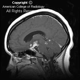
There is a suprasellar mass lesion identified, which is soft-tissue signal on the T2-weighed sequence (Figure 1)
The mass enhances (Figure 2 and 3) along with nodular enhancement in the Sylvian fissures bilaterally (Figure 2), the prepontine region (Figure 3), and the superior vermian region (Figure 3). The pituitary stalk demonstrates dense thick enhancement (Figure 3).


Diagnosis: Neurosarcoidosis


Sarcoidosis is inflammatory disease of unknown etiology, which affects multiple organ systems including the central nervous system. Females are affected more than males and this disease has a 10:1, black to white predominance. Central nervous system involvement occurs in 15% of patients, while symptomatic neurologic involvement is seen in approximately 5%. Dysfunction of cranial nerves and diabetes insipidus are common neurologic presentations. The MR appearance in these patients includes thickening and enhancement of the meningeal surfaces, solitary masses or multiple small, scattered meningeal or parenchymal lesions. When MR imaging shows thickening and enhancement of the pituitary stalk and associated leptomeningeal disease as in this case, sarcoidosis is a very strong possibility, along with meningitis, particularly fungal. Relative T2 hypointensity can be seen commonly, although this can also be seen in cases of fungal diseases, lymphoma, and meningioma.

The diagnosis can be established by performing a Kveim-Siltzbach skin test or by identifying other organ involvement with the intent to biopsy.

lunedì 19 settembre 2005

Spinal intradural extramedullary neurenteric cyst


Findings

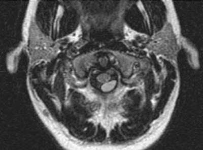
Axial FLAIR MRI sequence demonstrates a well-defined intradural extramedullary lesion posterior to the upper cervical spinal cord (Figure 1).
Axial T1 and T2 images show that the lesion is similar, but not identical, to cerebral spinal fluid signal (Figure 2 and Figure 3)
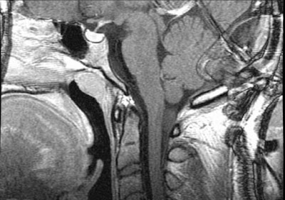
Axial and sagittal T1 post-contrast images demonstrate no associated post-contrast enhancement (Figure 4 and Figure 5).


Diagnosis: Spinal intradural extramedullary neurenteric cyst


Neurenteric cysts of the spine are rare benign cysts that are lined by cells of endodermal origin. They are usually located in the intradural extramedullary space in the cervical and thoracic regions; however, they can be present in the intracranial, intramedullary, and extradural spaces.
Neurenteric cysts can be associated with vertebral body anomalies, tethered cord, diastematomyelia, and spina bifida, and may be connected to mediastinal/abdominal cysts.
Lhermitte's sign refers to a shock-like sensation that extends down the spine and into the extremities with flexion of the neck, which can be caused by any abnormality that causes impingement of the cervical spinal cord (epidural or intradural pathology), spondylosis, multiple sclerosis, spinal cord tumor, radiation myelitis, or vitamin B12 deficiency. In this case, it was the mass effect produced by the neurenteric cyst on the cervical spinal cord, which was resected and pathologically proven.

mercoledì 7 settembre 2005

Choroid plexus papilloma


Findings

Figure 1: CT. Axial cut through level of lateral ventricles. Hyperdense lobulated well-circumscribed mass is noted within the atrium of the right lateral ventricle. There is adjacent compressive edema of the parenchyma of the right parietal lobe, without definitive extension of the mass into the parenchyma. There is moderate hydrocephalus involving both lateral ventricles.
Figure 2: CT. Axial cut through level of frontal horns. Hyperdense mass associated with the right lateral ventricle with adjacent compressive edema on the parenchyma of the right parietal lobe and without definitive extension into the parenchyma is noted. The lateral and third ventricles are moderately dilated. Other images showed the fourth ventricle to be dilated.


Diagnosis: Choroid plexus papilloma


Choroid plexus papillomas are rare tumors arising from the epithelium of the choroid plexus. The choroid plexus produces cerebrospinal fluid. The CSF produced by such a tumor is the key cause of the unusual cases of hydrocephalus due to overproduction of CSF. The overproduction is seen as dilatation of all ventricles in this cause of communicating hydrocephalus. Choroid plexus papillomas represent 60%-70% of tumors of the choroids, 5% of all supratentorial tumors in children, and less than 1% of all primary intracranial tumors. They are usually seen in the first 5 years of life, more typically in infants with signs of significant hydrocephalus. They are much more commonly noted in males.

Classically, they are lobulated isodense to hyperdense masses with occasional punctuate calcifications seen within them. They enhance homogeneously. They usually are seen as an intraventricular mass without extension into or through the ventricular wall. Only aggressive papillomas, accounting for about 5% of cases, will invade adjacent brain.

Although the fourth ventricle is the most common site to find choroid plexus tumors in adults, in children they are usually noted in the lateral ventricle, left more often than right. The ventricular trigone is the most common area of occurrence within the ventricle. The appearance of papillomas in unusual sites has been reported in cases of von Hippel-Lindau syndrome.

Choroid plexus tumors may also be carcinomas, and are usually seen in somewhat older children, aged 3-5 years. Unlike patients with papillomas, those with carcinomas may have focal neurologic signs and signs of hydrocephalus. Involvement is equal for both genders. These tumors most always grow through the ventricular wall.