

Findings
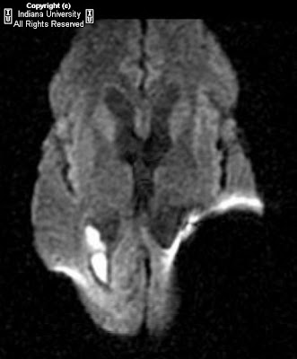
There is a left parieto-occipital ventriculostomy catheter is present entering the left occipital horn. There is diffuse dilation of the lateral ventricles (left greater than right), third ventricle, and fourth ventricle. There is focal, marked enhancement of the occipital horns of the ventricles bilaterally, with debris noted dependently in the lateral ventricles. This debris demonstrates restricted diffusion. There is T2 prolongation in the white matter surrounding the occipital horns bilaterally.
Differential diagnosis:
- Bilateral lateral ventriculitis
- Lymphoma
- Intraventricular hemorrhage
- Ependymal tumor spread (primary or metastatic)
- Prominent ependymal veins
Diagnosis: Ventriculitis
Key points
Ventriculitis is infection of the ependyma lining the ventricles of varying etiologies.
May be caused by rupture of brain abscess into the ventricle, as a complication of meningitis (30% of cases), or as a complication of neurosurgical devices (most common ventriculostomy).
Can be caused by viral, fungal, bacterial, or parasitic organisms. Most common – bacterial (Staphylococcus, Streptococcus, Enterobacter). Viral or fungal – immuno-compromised patients.
High mortality – 40-80%
Treatment – surgical irrigation and drainage, treatment of infectious etiology (antibiotics)
Imaging findings:
- CT: Ventriculomegaly with diffuse enhancement of the ventricular walls. Usually layering debris within the ventricles. Subtle low density surrounding the ventricles (edema).
- MR: Ventriculomegaly with layering debris in the ventricles (hyperintense on T1WI and FLAIR). Debris will demonstrate restricted diffusion on DWI (pus). Bright enhancement of ventricular walls. May have associated findings of choroid plexitis (uncommon).
Nessun commento:
Posta un commento