giovedì 22 aprile 2010

Pars flaccida cholesteatoma


Findings

There is an 8mm mass in Prussak’s space with erosion of the malleus and scutum.

Differential diagnosis:
- Pars flaccida cholesteatoma
- Cholesterol granuloma
- Paraganglioma
- Pars tensa cholesteatoma


Diagnosis: Pars flaccida cholesteatoma


Key points

A pars flaccida cholesteatoma often occurs when a patient has chronic middle ear inflammation and/or TM perforation. The cholesteatoma forms when there is an accumulation of stratified epithelial cells in Prussak's space. The cholesteatoma can be seen in all age groups, but tend to be more aggressive in children. Patients can present with aural discharge, conductive hearing loss, and otalgia. Early treatment with surgery can preserve hearing.


Radiologic overview of the diagnosis

A pars flaccida cholesteatoma appears as a mass in Prussak's space with erosion of the scutum and/or adjacent ossicle. Ossicle erosion is seen ~70% of the cases. There is no enhancement of the cholesteatoma itself, though surrounding granulation tissue may enhance.

High resolution temporal bone CT is the best modality to evaluate a suspected cholesteatoma. A pars tensa cholesteatoma is far less common and involves the sinus tympanum. A cholesterol granuloma appears blue on otoscopy and may have similar bony erosions as that of a cholesteatoma. A paraganglioma appears as a cherry red mass on otoscopy and usually does not erode bone.

In this case, there is an 8mm mass in Prussak's space with erosion of the malleus and scutum.

lunedì 19 aprile 2010

Leptomeningeal carcinomatosis from esophageal adenocarcinoma


Findings

Figure 1, Figure 2, Figure 3, and Figure 4: Postgadolinium T1 weighted images demonstrate linear contrast enhancement in the subarachnoid space, most notably interdigitating between the folia of the superior cerebellum and in the sulci of the parietal and occipital lobes. The T1 weighted precontrast images are unremarkable.
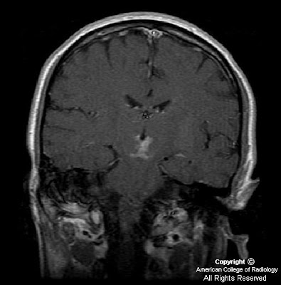
On FLAIR imaging in Figure 5, there is corresponding hyperintensity in the subarachnoid space.
Chest TC (not shown) demonstrates a high-attenuation central mass within the esophagus, which is expanding the lumen.


Diagnosis: Leptomeningeal carcinomatosis from esophageal adenocarcinoma


The leptomeninges consist of two layers; the pia mater and the arachnoid mater, which enclose the subarachnoid space and the CSF. The leptomeninges are a frequently missed site of metastastic involvement, especially for non-hematologic primary malignancies. There are many proposed routes of entry for tumor cells into the CSF including hematogenous spread via the arachnoid vessels or choroid plexus, direct extension from the skull, vertebrae, dura or retrograde perineural spread via the peripheral or cranial nerves. Once tumor cells reach the CSF, rapid dissemination can occur.

Up to 50% of patients with leptomeningeal metastases present with signs of increased intracranial pressure and/or hydrocephalus including headaches, back pain, nausea, vomiting and dizziness. These symptoms are most likely secondary to obstruction of CSF flow by tumor cells. Other clinical manifestations include seizures, focal cranial nerve deficits and meningeal signs such as nuchal rigidity and photophobia.

CSF cytology is the definitive test for diagnosis of leptomeningeal involvement. However, while highly specific, cytology is often falsely negative. The accuracy of a single lumbar puncture is approximately 50%, which increases to 90% with three LPs. Cytology remains negative in 10-20% of patients, presumably in situations in which the malignant cells are more adherent to the leptomeninges.

Contrast enhanced MR imaging is the diagnostic test of choice as an adjunct to CSF cytology when leptomeningeal metastases are suspected. Gadolinium enhanced MRI is more sensitive than a single lumbar puncture, but is less specific. The most common imaging findings include diffuse leptomeningeal contrast enhancement, multiple masses or nodules within the subarachnoid space and/or hydrocephalus. The diffuse leptomeningeal enhancement pattern has been referred to as sugar-coating or zuckerguss (German for icing or sugar-coating). Studies have shown that contrast-enhanced T1-weighted MR imaging is the most sensitive single sequence for detection of leptomeningeal metastases.

Prognosis for leptomeningeal metastases is poor. Without treatment, the average survival is 1-2 months. Treatment options include corticosteroids, intrathecal chemotherapy and radiation therapy. Despite aggressive therapy, the median survival in most randomized-controlled trials is 3-4 months.

martedì 6 aprile 2010

Acute infarction secondary to occlusion of artery of Percheron


Findings

Figure 1: Coronal DWI image demonstrates restricted diffusion within bilateral medial inferior thalami and superior midbrain.
Figure 2: Coronal DWI image, obtained 12 days later demonstrates relatively decreased diffusion hyperintensity.
Figure 3: Coronal post-contrast coronal T1 weighted image demonstrates new enhancement involving bilateral inferior medial thalami and superior midbrain, secondary to breakdown of blood-brain barrier.
Figure 4: Diagram illustrating normal paramedian thalamic mesencephalic arterial supply, with many perforating vessels arising from bilateral P1 segments of the PCA. (Reprinted with permission from AJNR)
Figure 5: Diagram illustrating the variant, “artery of Percheron” a single perforating blood vessel arising from one P1 segment.


Diagnosis: Acute infarction secondary to occlusion of artery of Percheron


The thalami and the midbrain receive their blood supply from both the anterior and posterior circulations, and several variations in this supply are known to exist. The anterior circulation usually supplies the anteroinferior aspects of the thalami and midbrain, with thalamoperforator arteries arising from the posterior communicating arteries. The posterior circulation usually supplies the medial aspects of the thalami and midbrain via branches arising from P1 segments and the lateral and superior aspects with branches arising from P2 segments of the posterior cerebral arteries. Most of the perforating branches from the P1 segments have an ipisilateral distribution (78%); bilateral or even contralateral distributions may be observed in 22% of individuals.

Percheron studied the variations of this arterial supply and its distributions and described three different variations involving the paramedian thalamic-mesencephalic arterial supply: (1) small branches arising from both P1 segments, (2) an asymmetrical common trunk arising from a P1 segment (this variation is called the artery of Percheron), (3) or an arterial arcade emanating from an artery bridging the two P1 segments. In the second type, a common trunk arising from one of the P1 segments provides bilateral distribution. Occlusion of this trunk results in bilateral infarctions in the middle aspects of thalami and brain stem.

The thalami contain strategic nuclei and integrate several important cortical functions. Thus, infarcts at the mesencephalic-diencephalic junctions may result in complex clinical syndromes, with patients exhibiting a wide range of symptoms varying from motor deficits to behavioral and sensory alterations. The third nerve palsy demonstrated in this particular patient may have been due to involvement of the midbrain at the level of the Edinger-Westphal nucleus.

Performing conventional angiography may not be indicated, because lack of visualization of the artery does not exclude its presence (because it is occluded).