martedì 25 maggio 2010

Skull base Langerhans cell histiocytosis (LCH)


Findings:

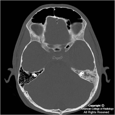
Figure 1and Figure 2: There is diffuse sclerosis of the left mastoid with near complete opacification of the mastoid air cells.
Figure 3 and Figure 4: There is diffuse sclerosis of the mastoid with a soft tissue thickening identified in the cavernous sinus. There is additional dural extension suggested along the left leaf of the tentorium.


Diagnosis: Langerhans cell histiocytosis (LCH), skull base


Langerhans cell histiocytosis (LCH) is the term used to describe the spectrum of diseases characterized by proliferation of specialized bone marrow-derived dendritic cells, called Langerhans cells (LC) with bone and/or soft tissue involvement. Although the etiology and pathogenesis of LCH is poorly understood, studies suggest that it is a reactive disease caused by immune dysregulation. The disorders once collectively called Histiocytosis X (eosinophilic granuloma, Hand-Schüller-Christian disease, and Letterer-Siwe disease) are now part of this category. LCH is defined in terms of whether it involves a single site, multiple sites, a single organ, or multiple organs. Prognosis and treatment are closely linked to the extent of disease at presentation and whether or not “risk” organs (ie: liver, spleen, lung, bone marrow) are involved. In general, patients with LCH localized to the bone have a favorable prognosis whereas patients with multisystem involvement incur a poor prognosis.

Langerhans cell histiocytosis of the bone is a relatively rare disorder with an incidence of one case per 2 million persons per year. The skull is the most commonly involved bony site in the pediatric age group. Otologic involvement usually occurs during the course of multisystemic disease, and may be the only symptom in up to 25% of patients. Because the otologic symptoms are similar to otomastoiditis, diagnosis of LCH of the skull is often delayed. The most frequent otologic symptom is otorrhea resistant to medical treatment. Other findings include mastoid swelling, aural polyps, periauricular eczema, sagging of the canal wall skin from erosion of the posterior bony external auditory canal, conductive hearing loss, and otalgia. Neurosensorial deafness, vertigo from involvement of bony labyrinth, and paralysis of cranial nerves (particularly VII and VIII) have been described. The classic clinical profile is a young man with otalgia, otorrhea, and post-auricular mass. If the diagnosis is suspected, a full examination and work-up is warranted to exclude the possibility of multisystemic or multiostotic disease.

The best imaging modality to diagnose LCH of the skull base is dedicated temporal bone CT. The skull base may show focal or diffuse bone destructive change and may present as: 1) sharply-defined “punched out” appearance, 2) irregular sclerotic lesions or 3) fragments of bone within soft tissue component. Other locations that are affected include the petrous apex, mandible, maxilla, and vertebral body. Ossicular and otic capsule destruction are common. Bilateral disease occurs in up to 30% of patients. The radiologic differential diagnosis for LCH of the skull base includes mastoiditis, cholesteatoma, cholesterol granuloma, and rhabdomyosarcoma. Rhabdomyosarcoma may exactly mimic unilateral LCH- biopsy may be required to differentiate the two entities.

Contrast-enhanced examination may help differentiate inflammatory mastoid lesions from LCH. Findings include a heterogeneously enhancing mass in the temporal bone, including mastoid complex, middle ear, extracalvarial, intracranial, and extra-dural compartments. The skull base tends to show a variable amount of enhancing soft tissue. On MR, T1 weighted images reveal iso- to hypointense mastoid, middle ear or skull base mass. T2 weighted images reveal hyper- to isointense soft tissue mass. Contrast-enhanced T1 weighted images show a heterogeneously-enhancing mastoid, middle ear, or skull base mass. MR may be obtained to delineate boundaries of soft tissue component which generally displays poorly defined borders.

The prognosis of LCH localized to the temporal bone is excellent, with a cure rate of 90%. The soft tissue component resolves initially, followed by reossification of the bone lesion. The treatment of choice for LCH localized to the mastoid-middle ear consists of surgical curettage or mastoidectomy. Low dose radiotherapy is used for larger, destructive lesions. Systemic disease is treated with chemotherapy in combination with radiation therapy.

lunedì 24 maggio 2010

Multiple bilateral infarcts secondary to IV drug abuse-related cerebral vasculitis


Additional clinical history: Woman in acute heroin withdrawal who also has bilateral lower extremity weakness.


Findings:

Brain MRI shows restricted diffusion within the bilateral parietal and occipital lobes as well as over the motor cortex of the frontal lobes bilaterally, left greater than right. T2 prolongation is noted in all the locations where restricted diffusion is seen. Coronal and sagittal reformatted CTA images show areas of subtle beaded appearance of arteries, with focal narrowing and dilatation.


Diagnosis: Multiple bilateral infarcts secondary to IV drug abuse-related cerebral vasculitis


Key points

Increased T2 signal lesions with restricted diffusion have a broad differential
Clinical history, distribution (vascular territories involved, unilateral vs. bilateral), number (single vs. multiple), the presence or absence of mass effect, and the presence or absence of a peripheral ring or central necrosis are key diagnostic clues.
Abscesses may be single or multiple, may be unilateral or bilateral, often have a peripheral ring, and often display mass effect.
Tumors often have mass effect, often have a peripheral ring, may be single or multiple, may be unilateral or bilateral, and may show central necrosis.
Hemorrhage is often in the subdural or subarachnoid space, but may be intraparenchymal, especially if there is history of head trauma or associated fracture.
In acute infarct, there is no mass effect, no peripheral ring, and no central necrosis and the lesions are usually in a single vascular distribution and are usually- but not always- unilateral.
Examples of bilateral acute infarcts, as seen in this patient (note multiple bilateral lesions without mass effect, peripheral ring or central necrosis), are watershed infarcts occurring in typical watershed zones and infarcts occurring secondary to cardiac emboli or vasculitis, both of which are not distributed with respect to single vascular territories (as in this patient).
Besides IV drug abuse (as in this patient), cerebral vasculitis with secondary infarct can be seen in bacterial meningitis, tuberculous meningitis, viral, mycotic, syphilitic or post radiation arteritis, cell mediated arteritis, collagen vascular disease, sarcoid, Wegener's granulomatosis, and Moyamoya disease.

lunedì 17 maggio 2010

Focal cortical dysplasia


Findings:

Figure 1 and Figure 2: Axial FLAIR and T2W MR images demonstrate blurring of the grey-white junction with high signal intensity in the subcortical white matter of the right frontal lobe. Adjacent cortical ribbon thickening is also present. These findings are characteristic of focal cortical dysplasia.


Diagnosis: Focal cortical dysplasia


Focal cortical dysplasia (FCD) is a congenital disease in which the neurons arrange abnormally in focal areas of the cerebral cortex. It is a common cause of intractable and drug resistant epilepsy, and while seen mostly in the pediatric population, it is not uncommon to see the disease manifest itself in adulthood. Radiologic diagnosis is important for pre-surgical planning and can have prognostic implications.

FCD can be further sub-typed based on histopathology. In Type I FCD there is architectural distortion of the cerebral cortex alone without any abnormal cells. In Type II FCD, however, pathology will demonstrate both architectural distortion and dysmorphic neurons. This includes the characteristic elliptically shaped neuron with a displaced nucleus and lack of axons, known as the “balloon cell”. While the pathologic diagnosis is made retrospectively, clinically those with Type I FCD generally are responsive to medical therapy, while those with Type II FCD are resistant.

Regardless of the pathologic type of FCD, patients that demonstrate preoperative imaging findings tend to have a better outcome after surgery than those who do not demonstrate any findings. The characteristic MR abnormalities in FCD include blurring of the grey-white junction, abnormal high T2 signal in the subcortical white matter, and adjacent focal cortical thickening. Often these findings are very subtle and may not be detected on MR imaging alone. In such cases MR/FDG-PET fusion has been utilized for more sensitive detection. Surgery is the mainstay of treatment for these patients.


Extra: Balloon cells are large elliptical shaped cells with displaced nuclei and are characteristic of focal cortical dysplasia.

giovedì 13 maggio 2010

Poorly differentiated orbital malignant lymphoma


Findigs:

Figure 1: Axial CT through the orbital floors at the level of the inferior orbital foramen demonstrates an erosive mass, containing calcifications, with expansion through the floor of the orbit and into the maxillary sinus.
Figure 2: Contrast enhanced coronal CT image demonstrates a heterogeneously enhancing, soft tissue mass involving the floor of the orbit, with bony invasion.


Diagnosis: Poorly differentiated orbital malignant lymphoma (Non-Hodgkin's type) (biopsy proven)


In the adult population the differential diagnosis of malignant orbital tumors differs from that of children. Whereas, carcinoma is the frequently enountered in adults (often from contiguous spread from the paranasal sinus, etc), metastatic neuroblastoma is the more frequently encountered malignant neoplasm in children.

Neoplasms of the paranasal sinuses are uncommon, but frequently extend to involve the orbit when they do occur. Benign tumors tend to push the periorbital tissues aside, while malignant lesions tend to invade the periosteum.

Evaluation of the paranasal sinus mass is best done utilizing multidetector CT, because of its ability to detect bony destruction and possible intracranial extension of disease. CT usually demonstrates a homogeneous mass with well-defined borders that demonstrates heterogeneous enhancement.

Lymphoid tumors are one of the most common orbital tumors despite the orbit not containing lymph nodes or a well defined lymphatic vasculature. Ocular malignant lymphomas are uncommon. The incidence ranges from 1-4%. Involvement of the orbit as a presenting sign is rare and few such cases are seen in the literature. Although most orbital lymphomas are localized to the orbit at diagnosis, many patients will develop systemic lymphoma over time. Orbital lymphoma is an adult disease, usually presenting in patients between the age of 50-70 years. The course is usually of an anterior mass that causes painless proptosis over several weeks to months.

Radiation therapy is the mainstay of treatment since surgical resection plays a limited role. Response rate and prognosis depends upon cell type, the extent of disease and whether the orbital disease represents the primary site or the site of spread of disease from an extra-ocular primary. Five year survival rates range from 54-78%.

lunedì 10 maggio 2010

Parapharyngeal abscess


Findings

The right parapharyngeal space contains a hypo attenuating collection near simple fluid in attenuation. Collection extends from just below the level of the hyoid to just below the right mandibular condyle, is anterior to the carotid space inferiorly, anteromedial to the carotid space superiorly. No significant rim enhancement. No inflammatory fat stranding is present. Right submandibular gland is along the anterolateral margin of this collection. Multiple prominent lymph nodes were visible.

Differential diagnosis:
- Abscess
- Lymphangioma
- Second branchial cleft cyst
- Ranula


Diagnosis: Parapharyngeal abscess

venerdì 7 maggio 2010

Aplasia of right submandibular gland with compensatory parotid and sublingual gland hypertrophy


Findings

There is aplasia of the right submandibular gland, and severe atrophy of the left submandibular gland. There is hypertrophy of the sublingual glands bilaterally, with herniation of the right sublingual gland through the mylohyoid boutonniere. There is also hypertrophy of the parotid glands bilaterally, without a discrete mass and without ductal dilatation or sialolithiasis.

Differential diagnosis:
- Parotiditis
- Parotid gland tumor
- Parotid hyperplasia
- Lymphadenopathy
- Amyloidoma


Diagnosis: Aplasia of right submandibular gland with compensatory parotid and sublingual gland hypertrophy


Key points

Congenital absence of a major salivary gland is extremely rare. It is reported to be associated with other congenital craniofacial anomalies and absence of all four major salivary glands is the most common pattern.
Most patients are asymptomatic. Hypertrophy of other salivary glands may result in an asymptomatic neck mass.
Symptoms such as dysphagia, xerostomia, dental caries, and local pain have been reported. Symptomatic patients generally have aplasia of multiple glands.
Agenesis of the major salivary glands may be associated with other first and second branchial arch defects.
The cause is not known , but is thought to be a result of a fetal development disturbance in the 4th-8th weeks of gestation
Enlargement of the other salivary glands is felt to be compensatory in etiology.

lunedì 3 maggio 2010

IVC thrombosis with enlarged epidural veins causing a lumbar radiculopathy


Findings

Figure 1 and Figure 2: Sagittal T1 and T2 weighted images reveal the presence of signal in the IVC and iliac veins indicative of thrombus. Prominent epidural veins are appreciated here as well.
Figure 3 and Figure 4: Axial T2 weighted sequences reveal enlarged epidural veins as demonstrated by prominent flow voids in the epidural space. Note absent flow void in the IVC and iliac veins. Normal flow voids are seen in the associated arterial structures on these images.


Diagnosis: IVC thrombosis with enlarged epidural veins causing a lumbar radiculopathy


Lumbar radiculopathy and low back pain is a frequently encountered complaint in the general population. The most common etiology is structural spine disease, including disc herniation and spondylosis. However, nonstructural diseases, such as cytomegalovirus polyradiculopathy in immunocompromised individuals as well as other infections, neoplasms, and inflammatory conditions have also been shown to cause lumbar radiculopathy. Vascular causes are unusual as patients with vascular pathology tend to present with myelopathy.

Enlarged epidural veins are commonly a result of arteriovenous malformations, fistulas, and varicose veins. In the cervical spine, the epidural veins function as collateral pathways and become enlarged as they receive more blood flow when the jugular veins are compromised. Similarly, it can be inferred that in the case of an IVC obstruction or occlusion, the lumbar epidural veins become enlarged by the same mechanism; when IVC blood flow is compromised, blood is able to return to the heart through the azygos and hemiazygos veins via collateral pathways including the epidural venous system. While many epidural veins become enlarged as a result of this process, the vein implicated in causing the radiculopathy is the vein below the pedicle. This vein lies in close proximity to the existing nerve root and as the vein becomes larger, it will impinge directly upon the nerve root leading to the radiculopathy. Therefore, the best way to eliminate the radicular symptoms is by treating the underlying cause of the IVC thrombosis.

Risk factors for the development of IVC thrombosis and venous thromboembolism (VTE) include hypercoaguable states such as factor V Leiden mutation, prothrombin gene mutation, protein S deficiency, protein C deficiency, antithrombin deficiency, and dysfibrinogenemia. Acquired risk factors include a prior thrombotic event, recent major surgery, presence of a central venous catheter, trauma, immobilization, malignancy, pregnancy, oral contraceptives, heparin, myeloproliferative disorders, and antiphospholipid syndrome.