Visualizzazione post con etichetta Nuclear. Mostra tutti i post

mercoledì 25 marzo 2009

Reflex sympathetic dystrophy (RSD)


Findings


Three phase technetium 99m labeled MDP bone scan is shown. There is abnormal radiopharmaceutical uptake in the periarticular regions of the left hand and wrist on both immediate and delayed images.

Differential diagnosis:
- Reflex sympathetic dystrophy
- Inflammatory arthritis


Diagnosis: Reflex sympathetic dystrophy (RSD)


Key points

RSD is a poorly understood chronic abnormality of the sympathetic nervous system in a regional distribution (usually the distal extremities) that follows trauma, surgery, infection, casting, or splinting.
Clinically, it presents as severe, protracted pain in the affected limb that is out of proportion to the original injury. Additionally, there may be vasomotor disturbances. Progresses to skin and muscular atrophy.
Synonyms: Sudeck's atrophy, Complex regional pain syndrome type 1, Causalgia.

Three phases have been described:
1) Non focal pain with decreased range of motion, soft tissue edema, and increased skin temperature (lasts weeks to months)
2) Decreasing pain and skin temperature with skin thickening. Atrophy and osteoporosis may become apparent (lasts 3-6 months)
3) Continued pain with increasing atrophy and joint stiffness. Contractures may occur.

May occur in up to 5% of all injuries; most often in the upper extremity.
Typically, affects females more, often less than 50 years of age.
Plain radiographs only 60% sensitive and non-specific, showing osteoporosis of the affected limb with associated soft tissue swelling or atrophy (depending on the stage).
CT also insensitive and non-specific.
MR may show patchy bone marrow edema with stage dependent soft tissue changes.
Nuclear medicine three phase bone scan is the best study for diagnosis of RSD, showing diffuse increased uptake in the affected region on flow images; continued diffuse uptake on blood pool images, beginning to localize to periarticular regions; and continued uptake in the periarticular regions on delayed images.
Delayed images are the best for diagnosis, with a reported sensitivity of near 100%.
A "reversed" pattern of decreased relative uptake in the affected extremity may be seen on bone scan, however. This is most common, but not exclusively, in pediatric patients.
Treatment: medications (steroids, opiates, anti-epileptics, etc.); physical therapy; sympathetic nerve block; surgical sympathectomy.

lunedì 26 gennaio 2009

Brain death from meningitis secondary to ear infection


Findings

Initital head / temporal CT: Pneumocephalus along the left falx of unknown etiology. No evidence of intracranial lesion, midline shift, or intracranial hemorrhage. No fracture of the temporal bones. Widening of the left lambdoid suture with well corticated borders, inconsistent with fracture. Fluid is present in the mastoid air cells bilaterally.

Follow-up CT: Unchanged pneumocephalus. Loss of the suprasellar and quadrigeminal plate cisterns consistent with herniation. Decreasing differentiation between the gray and white matter consistent with edema. Fluid again was seen in the mastoid air cells bilaterally.

Nuclear medicine study: No evidence of flow/perfusion to brain either on the early dynamic images or on the delayed images.

Differential diagnosis:
- Skull fracture
- Meningitis

Epilogue: Patient's girlfriend reported he was diagnosed with an ear infection, but did not fill the prescription. After toxicology consult, possiblity of meningitis was raised. While still in ER the second head CT was done which showed changes of diffuse brain hypoxia with cerebral edema and herniation. Examination at this point revealed fixed and dilated pupils with no brainstem reflexes. ICP monitor was placed showing pressure was significantly elevated, unable to be controlled with hyperosmolar therapy. A nuclear medicine scan was obtained and showed no evidence of flow or perfusion to the brain, findings consistent with brain death when taken in correlation with clinical findings.


Diagnosis: Brain death from meningitis secondary to ear infection


Key points

Differential diagnosis of Pneumocephalus
- Traumatic 74%
May be found within any compartment from skull, skull base, paranasal sinus, or mastoid fracture
3% of all skull fractures
8% of all paranasal sinus fractures
- Neoplasm involving sinus 13%
Osteoma, pituitary adenoma, mucocele, epidermoid, paranasal sinus malignancy
- Iatrogenic 4%
Lumbar puncture, craniotomy, craniectomy, ventriculostomy, ICP monitor placement
- Infectious 9%
Rare sequela of gas-producing infection
Typically sinusitis or mastoiditis
- Regardless, pneumocephalus itself is not a problem—what's causing it?

Epidural
- Remains localized
- Air will not necessarily move with changes in head position

Subdural
- Air-fluid levels
- Moves with changes in head position
- Confluent
- Tension pneumocephalus may result in "Mount Fuji sign"—subdural air separates/compresses frontal lobes, creating widened interhemispheric space between frontal lobe tips—mimics silhouette of Mt Fuji.

Subarachnoid
- Multifocal
- Non-confluent

CT: Imaging tool of choice
MRI: Foci of absent signal on all sequences


giovedì 9 ottobre 2008

CSF leak


Findings

CT shows dehiscence of the cribriform plate bilaterally. Right gyrus rectus encephalocele. Nuclear medicine cisternogram shows abnormal radiotracer localization in the nasal cavities bilaterally.


Diagnosis: CSF leak


Key points

Causes include:
- Blunt head trauma
- Sequelae of skull-base surgery
Commonly functional endoscopic sinus surgery (FESS)
Transsphenoidal pituitary surgery
Translabyrinthine acoustic schwannoma
Mastoid surgery with intact tympanic membrane
- Destructive skull-base lesions, including neoplasms (both benign and malignant), and empty sella
- Developmental defects of the ethmoid, sphenoid, frontal, or petrous temporal bones with the formation of a meningocele or meningoencephalocele (with an intact tympanic membrane)
- Fracture of the petrous temporal bone or other destructive processes in which CSF in the middle ear drains to the nose in the presence of an intact tympanic membrane
- Less than 5% of all cases of CSF rhinorrhea are spontaneous

Most cases of CSF rhinorrhea begin soon after a head injury and cease spontaneously within 7-180 days.
Radionuclide cisternography is used to confirm a suspected CSF leak.
Leaks are most common in the region of the cribriform plate and ethmoid sinuses.
CSF leaks may be intermittent leading to a false negative study.
The study is performed in conjunction with ENT and possibly neurosurgery, in addition to neurointerventional radiology, who performs the lumbar puncture.
Typically Tc-99m sulfur colloid is administered into the thecal sac by lumbar puncture. Additionally cotton pledgets are placed in the superior and middle turbinates by ENT. Imaging of the spinal canal and head are obtained between 1 and 3 hours. The cotton pledgets are removed from 4 to 24 hours later and are individually placed in a well counter. Serum samples are obtained at the same time and counted for activity. Pledget to serum ratios of more than 1.5 may be interpreted as evidence of CSF leak.
CT cisternography after the administration of iodinated contrast within the thecal sac may be performed for further anatomic delineation.
Treatment is primarily surgical.

martedì 30 settembre 2008

Sialadenitis with sialolithiasis


Findings

Uptake image series: Normal thyroid uptake. Symmetric radiotracer visualized in the sublingual glands. Expected radiopharmaceutical by the bilateral parotid glands and right submandibular gland. Increased uptake in the left submandibular gland. Increased radiopharmaceutical visualized in the oral cavity.
L lat washout and R lat washout image: Unchanged symmetric activity in the thyroid gland. Expected washout and decreased radiopharmaceutical in the parotid glands and right submandibular gland. Decreased washout in the left submandibular gland.
15 min post lemon image: Decreased washout of the radiopharmaceutical in the left submandibular gland with normal washout in the remaining salivary glands.

Differential diagnosis:
- Sialadenitis
- Warthin's tumor
- Oncocytoma
- Oxyphilic adenomas


Diagnosis: Sialadenitis with sialolithiasis

Follow-up: Patient subsequently had her left submandibular gland surgically removed. In the operating room they found a calcified stone in the parenchyma of the gland.


Key Points

Nuclear sialography is used to assess the function of the salivary glands. CT and US are used for structural information. CT and US can be used to differentiate masses from inflammation and benign from malignant. US-guided fine needle biopsy has a high degree of accuracy.
The salivary glands consist of three paired exocrine glands. The parotid glands empty into the oral cavity through Stenson's duct. The submandibular glands empty through Wharton's ducts. The sublingual glands are the third type and connect to the oral cavity through multiple small ducts.
Rinsing the mouth with water before Tc99m-pertechnetate increases retention in the salivary gland and delays its secretion into the oral cavity. Patient is placed in the seated position with the head tilted back to prevent superimposition of the thyroid gland. A dynamic blood flow study should be performed, followed by sequential 1 minute images for 60 minutes. This should demonstrate simultaneous, symmetric uptake of the three paired exocrine glands.
The salivary gland is then subjected to gustatory stimulation with a lemon or with a 1:1 lemon juice to water mix (rinse for 5 seconds and spit). Normally see rapid symmetrical and complete resolution of radiopharmaceutical from the salivary glands. It is useful to obtain lateral views of the head and neck to confirm radiotracer in the saliva of the oral cavity after gustatory stimulation.
Warthin's tumor (papillary cystadenoma) appears as a focal region of increased uptake. Oncocytomas and oxyphilic adenomas are other tumors which may have increased radiopharmaceutical uptake. Increased uptake could also be seen with acute inflammation.
Metastatic lesions appear as focal regions of decreased uptake. Also seen with cysts, enlarged lymph nodes, and chronic inflammatory disease. Could also result from congenital aplasia, obstructive sialolithiasis, trauma, or radiotherapy.
Mixed benign tumors are the most common type of tumor of the salivary glands and may present as focal areas of increased or decreased signal.
With Sjögren's syndrome, there may be asymmetric arrival or delayed accumulation of radiotracer. There may be absent or decreased response to gustatory stimulation, especially the submandibular glands. The presence of high Ga-67 concentration in the lacrimal and salivary glands in pathognomonic for Sjögren's syndrome. A poor response to gustatory stimulation can also be seen with systemic connective tissue disease and viral parotitis or mumps, or following radiotherapy. Failure to excrete radiotracer is seen in stenosis or blockage of the salivary duct.

martedì 9 settembre 2008

Brain death due to subarachnoid hemorrhage


Findings

Figure 1: Noncontrast cranial CT with high density material in the subarachnoid spaces and fourth ventricle compatible with hemorrhage. Loss of the gray matter/white matter differentiation consistent with diffuse cerebral edema is also identified.
Figure 2: Noncontrast cranial CT with high density material in the subarachnoid space and layering dependently in the occipital horns of the lateral ventricles compatible with hemorrhage. Loss of the gray matter/white matter differentiation consistent with severe cerebral edema is again identified.
Figure 3: This coronal image from a CT angiogram demonstrates absent intracranial arterial blood flow compatible with brain death. The superficial temporal arteries (branches of the external carotid arteries) are patent. High density material can be seen in the subarachnoid spaces compatible with hemorrhage.
Figure 4: This static image obtained following the administration of 99mtechnetium-ECD demonstrates absent intracranial radiotracer accumulation compatible with brain death. Note dense accumulation of tracer at the level of the midface, consistent with the "hot nose" sign.


Diagnosis: Brain death


The diagnosis of brain death is predominantly a clinical diagnosis made at the bed side. The criteria require that certain clinical conditions be met, such as a core temperature above 32°C, as well as a neurologic examination demonstrating the absence of brain function. In cases where these criteria cannot be met other means of diagnosis may be employed. These methods include an electroencephalogram (EEG), cerebral angiography, magnetic resonance angiography (MRA), computed tomographic angiography (CTA), and brain scintigraphy.

Documentation of the absence of cerebral perfusion is one method of diagnosing brain demise when clinical criteria are not met. This can be accomplished in several ways including, conventional angiography, CTA, and brain scintigraphy. The pathophysiology behind the findings demonstrated on these studies has to do with the cerebral edema associated with brain demise. This edema causes an elevation in intracranial pressure (ICP) such that it exceeds the mean arterial pressure (MAP), and thus cerebral blood flow is impeded. This finding is well demonstrated in the following equation: cerebral perfusion pressure = MAP - ICP. False negatives may occur with either examination in the presence of a ventricular shunt, trauma, a recent surgical procedure to lower ICP, and open cranial sutures. Each of these lower ICP and thus allow cerebral perfusion.

Images obtained from a CTA will demonstrate absent blood flow beyond the extracranial internal carotid and vertebral arteries; the external carotid arteries and its branches should remain patent. Likewise, brain scintigraphy may also be performed to confirm the diagnosis. Technetium 99m-labeled hexamethylpropyleneaminoxime (99mTc-HMPAO) or 99mTc-labeled ethylene L-cysteinate dimer (99mTc-ECD) are two perfusion agents that made be used. As their names imply, these perfusion agents demonstrate uptake in perfused tissues. In the presence of cerebral demise, the dynamic and static images will demonstrate the absence of radiotracer above the skull base. The bolus of radiotracer will fail to perfuse the intracranial internal carotid and cerebral arteries. Relatively increased flow through the maxillary branch of the external carotid artery will cause relatively increased radiotracer accumulation in the nasal region, resulting in the “hot nose” sign, which is best seen on the anterior static images. While this finding has been observed in as many as 52% of patients diagnosed with brain death, it cannot in itself indicate brain death.

In conclusion the diagnosis of brain demise is important in the care of patients and their families. While this diagnosis is primarily one made clinically, imaging studies can allow for accurate diagnosis when clinical criteria cannot be met.

lunedì 25 agosto 2008

Graves disease


Findings

0.1 mCi of I-123 in the form of sodium iodide capsule was administered orally. 4 and 24 hour radioiodine uptakes were determined. The following day, 5.4 mCi of Tc 99m sodium pertechnetate was injected intravenously for the perfusion part of the scan followed by routine planar and pinhole imaging of the neck.

There is increased flow to the thyroid gland bilaterally. Planar and pinhole imaging of the neck demonstrate a symmetrically enlarged gland with homogenous radiotracer uptake. Thyroid to salivary gland ratio is increased and there is suppression of the background. Four-hour radioactive iodine uptake (RAIU) is 22.2% and 24-hour radioactive iodine uptake is 55.3%.


Diagnosis: Graves disease


Graves disease is the most common etiology of thyrotoxicosis and predominantly occurs in middle-age females. Binding of immunoglobulins to TSH receptors results in autonomous, inappropriate hyperfunctioning of the thyroid gland. This result in suppression of TSH levels, which are typically less than 0.01 mU/L.

Thyroid scintigraphy and RAIU determination are useful for distinguishing Graves disease from other causes of thyrotoxicosis in ambiguous cases. Thyroid scan radiotracers include Tc99m pertechnetate, I123 and I131. I123 is preferred over I131 for most applications due to the shorter half-life and lower peak energy of the I123 gamma photon, which results in more optimal image quality and lower radiation dose to the patient. Imaging is performed 2-6 hours after I123 is administered. Radioiodine distributes to the salivary glands, stomach and choroid plexus and is only stored within the thyroid. The classic thyroid scan findings associated with Graves disease include homogeneously increased activity in an enlarged thyroid gland, increased thyroid to salivary gland activity and suppression of the background. RAIU measurements can be performed at 4-6 hours and 24 hours. Graves disease usually yields RAIU values of 40 to 80% (normal 10 to 30%) at 24 hours. However, high-turn over varieties may manifest only as elevated 4-6 hour RAIU values (normal 4 to 15%), as 24 hour RAIU results may be normal or only mildly elevated.

Patients with Graves disease are initially treated with beta-blockers and temporarily with thyroid specific medications, such as PTU and methimazole. Radioiodine therapy, rather than surgery, represents the definitive treatment of choice. Indeed, high cure rates result from I131 treatment doses of at least 15 mCi. Side-effects for Graves treatment doses include hypothyroidism, radiation induced thyroiditis, xerostomia, sialadenitis, change in taste, and worsening of ophthalmopathy. Leukemia and other secondary malignancies are rare in any setting and not relevant to the Graves/hyperthyroid treatment doses

mercoledì 23 agosto 2006

Pick's disease


Findings

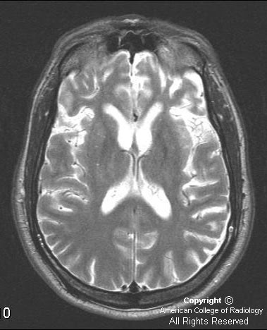
Figure 1, Figure 2, Figure 3, and Figure 4: MR images demonstrate marked atrophy involving the frontal and temporal lobes on T1, T2, and FLAIR.
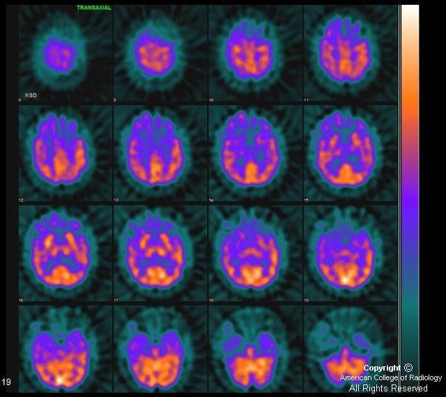
Figure 5: Selected axial SPECT images after the intravenous administration of Tc-99m Neurolite demonstrate decreased uptake in the frontal and temporal lobes, right slightly greater that left.


Diagnosis: Pick's disease


Pick's disease is described as a rare, progressive, degenerative disease, mostly involving the frontal and temporal lobes. This was first described by Arnold Pick in 1892. It is not thought to be inherited. Women are more affected than men. There is no cure.

This disease is more common in a younger age group (40 to 60) with an average of 54. Alzheimer's disease would be more likely in older patients, over 70.

Behavioral and physical changes include repetitive or compulsive behaviors, mood changes, apathy, echolalia, aphasia, apraxia, rigidity, and a multitude of other behaviors that range from not speaking or moving to rapid pacing. It will always progress to a vegetative state.

MRI will demonstrate atrophy of the frontal and temporal lobes with preservation of the pre- and postcentral gyrus on all sequences. There is no abnormal enhancement associated with this disease.

SPECT imaging will demonstrate decreased radiotracer uptake in the frontal and temporal lobes.

Histologic evaluation, which is usually postmortem, demonstrates a "ballooned" appearance of the neurons related to swelling. These are called Pick bodies. This is different than Alzheimer's type dementia, which demonstrates amyloid plaques and neurofibrillary tangles. In multi-infarct dementia, there is myelin and axonal loss with associated astrocytosis and generalized necrosis.