lunedì 31 dicembre 2007

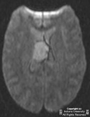
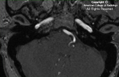
Occlusion of the left internal carotid artery with subsequent large left MCA acute infarction


Findings

CT head demonstrates increased density of the left MCA, compatible with a dense MCA sign. Additionally, there is subtle hypodensity of the left lentiform nucleus and left sub insular cortex. MR head demonstrates restricted diffusion in a left MCA distribution. MR angiography demonstrates absence of flow in the left internal carotid artery and left middle cerebral artery.


Diagnosis: Occlusion of the left internal carotid artery with subsequent large left MCA acute infarction


Key points

Cerebrovascular infarction most commonly involves the MCA distribution.
Ischemic stroke is far more common than hemorrhagic stroke, accounting for approximately 85% of cases.
Third leading cause of death in the United States.
Non-contrast head CT is the initial study of choice to evaluate for signs of hemorrhage.
Therapeutic window for t-PA (tissue plasminogen activator) thrombolysis is 3 hours from symptom onset.
Non-contrast CT usually is negative within the first 6 hours of onset. From 6-12 hours, sufficient tissue edema occurs to cause regional hypodensity.
Diffusion-weighted MRI can show changes of ischemic stroke in as little as 30 minutes after symptom onset.
Signs of MCA infarction include the dense MCA sign, and insular ribbon sign.

venerdì 21 dicembre 2007

Fusiform basilar artery aneurysm


Findings

CT shows a basilar artery aneurysm at the level of the pons. Subtle calcification is present within the aneurysm wall (Figure 1 and Figure 2).

Axial GRE (Figure 5), T1 sagittal (Figure 3), T1 post gadolinium axial (Figure 6), and T2 axial (Figure 4) demonstrate a fusiform aneurysm of the mid to distal basilar artery that does not involve the basilar tip. An area of slightly increased signal intensity within the aneurysm suggests thrombus formation (blue arrow in Figure 4 and Figure 6). No subarachnoid space or fourth ventricle hemorrhage is present.


Diagnosis: Fusiform basilar artery aneurysm


Basilar artery aneurysms comprise approximately 10% of all intracranial aneurysms. Fusiform aneurysms comprise only 1% of vertebrobasilar aneurysms. The fusiform aneurysm is a long segment of irregular fusiform or ovoid arterial dilatation. It is more common in the vertebrobasilar circulation than in the carotid circulation. In older adults, atherosclerosis is the most common cause of fusiform aneurysms in the basilar artery. Nonatherosclerotic fusiform aneurysms usually occur in younger patients with underlying vasculopathy or immune disorders. Fusiform aneurysms usually progressively enlarge over time. The pathologic etiology of fusiform aneuryms is partial or total absence of the internal elastic lamina and/or media, either congenital or acquired, which reduces elastic wall tension and allows subsequent expansion of the vessel diameter over time.

The saccular aneurysm is a focal dilatation of the arterial wall. The pathogenesis of saccular aneurysms reflects a combination of congenital, acquired, and hereditary factors. Large (>5 mm) aneurysms are found in 5% of the population, characteristically distributed at the arterial bifurcations. Association with polycystic kidney disease, Ehlers-Danlos syndrome, and other connective tissue disorders implicates hereditary factors.

Most intracranial aneurysms (~90%) are saccular and arise in the carotid circulation. 30-35% of aneurysms arise from the anterior communicating artery, 30-35% from the posterior communicating artery origin, 20% from the middle cerebral artery bifurcation, 5% from the basilar artery bifurcation or tip and the remaining 1-5% arise from other posterior circulation vessels. Intracranial aneurysms are multiple in 15-20% of cases. 80-90% of nontraumatic subarachnoid hemorrhage (SAH) is due to aneurysms. The risk of bleeding is approximately 2.5% per year for lesions >6mm in diameter. Complications of SAH include hydrocephalus, rebleeding, and vasospasm.

On non-enhanced CT, the aneurysm is hyperdense, and calcification in the wall may sometimes be detected. Aneurysmal SAH may be detected in 90-95% of cases on non-contrast head CT. CTA may detect aneurysms greater than 3 mm, providing detailed evaluation of morphology such as the relationship to the parent vessel and the neck width. CTA can detect more than 95% of aneurysms identified on conventional angiography. MRI demonstrates variable signal on T1 weighted images, hypointense lumen and clot on T2 weighted images, and strong enhancement of the residual lumen on post-contrast T1 weighted images. CTA or dynamic contrast-enhanced 3D-TOF MRA are the studies of choice for evaluation as non contrast 3D-TOF images are suboptimal due to flow saturation and phase dispersion. Conventional angiography is the definitive procedure for the detection and characterization of cerebral aneurysms. Aneurysm location, size, and morphology may be evaluated in the acute or chronic setting with this modality.

Management of fusiform aneurysms is difficult due to their morphology. Occlusion of the parent arteries, “Hunterian ligation,” is advocated as the preferred method of treatment. The “Alskock Test” is usually performed to evaluate patency of the posterior communicating (PCOM) arteries by compressing the cervical carotid artery during contrast injection of the vertebral artery. A PCOM greater than 1 mm can be visualized and suggests the possibility of collateral flow after occlusion of the vertebrobasilar system. Endovascular occlusion of basilar and vertebral arteries with balloons and coils is used in nonatherosclerotic fusiform aneurysm cases with good results. There are reports of successful treatment of ruptured basilar aneurysms with stent-graft placement from the vertebrobasilar junction to the midbasilar artery.

giovedì 20 dicembre 2007

Central neurocytoma


Findings

CT: Ovoid 3.0 cm intraventricular mass arising from the frontal horn of the right lateral ventricle. Arises just above the foramen of Monroe and mildly displaces the septum pellucidum. There is a faint contrast enhancement, and a few small, irregular vascular structures are demonstrated.
MR: Mass is isointense on T1W sequences and mildly T2W hyperintense to gray matter. Internal microcystic change and diffusion restriction is noted. No hydrocephalus.

Differential Diagnosis:
- Intraventricular (central) neurocytoma
- Intraventricular oligodendroglioma (can appear identical)
- Ependymoma
- Others
Meningioma
Astrocytoma
Subependymoma
Germ cell tumors
Colloid cyst
Choroid plexus papilloma


Diagnosis: Central neurocytoma


Discussion

Intraventricular neurocytomas are extra-axial brain tumors seen in young adults and older children with an age range of 17 to 53 years. Patients frequently develop hydrocephalus and symptoms related to elevated intracranial pressure. In addition, patients have also been reported to present with seizures, visual symptoms, or coma. On gross exam, these tumors are near or attach to the septum pellucidum and are well circumscribed. Microscopic evaluation reveals cellular tissue with fine to coarse calcifications being present more than half the time. Central neurocytoma can appear similar to an oligodendroglioma radiographically and histologically, but can be differentiated by a positive immunostain for synaptophysin. Clinically, intraventricular neurocytomas have a good prognosis and generally behave in a benign fashion. Complete resection is the therapy of choice and patients with incomplete resections often still have a good prognosis. Radiation therapy is utilized only in rare malignant cases.


Radiologic overview

CT: Demonstrates a large mass involving the lateral and third ventricles. There is usually associated hydrocephalus. The mass is isodense to brain with fine to course calcification. Moderate enhancement with IV contrast is seen in the noncalcified portions. The tumor frequently abuts or is attached to the septum pellucidum. Extension into the fourth ventricle and extraventricular tissue is associated with anaplastic histologic appearance.

MR: The tumor has a very heterogeneous appearance on MR secondary to its calcifications, which appear hypointense. Noncalcified portions are hyperintense to isointense to gray matter on T2 images. Similarly, T1 images demonstrate the tumor as isointense to hyperintense to gray matter. There is moderate enhancement with gadolinium.

martedì 18 dicembre 2007

Synovial cyst


Findings

Figure 1: An axial T2 image demonstrates a high signal round structure, adjacent to the right facet and postero-lateral to the thecal sac with a low signal rim.
Figure 2 and Figure 3: Axial and sagittal T2-weighted images demonstrate a cystic structure with a low signal rim adjacent to the right facet with mass effect on the right lateral recess at the L4-L5 level.


Diagnosis: Synovial cyst


Synovial cysts are formed by degeneration of the facet joint. Ninety percent of synovial cysts occur in the lumbar spine with 70-80% occurring at L4-L5. The cysts themselves are usually 1-2 cm in size and appear round or lobulated with sharp margins. Synovial cysts are postero-lateral extradural cystic masses and are adjacent to the facet joint. Direct communication with the facet joint confirms the diagnosis but is not always visualized.

Differential diagnosis of synovial cysts include:
- Extruded disk fragments
- Ganglion cysts
- Nerve sheath tumors
- Septic facet arthritis
- Asymmetric ligamentum flavum hypertrophy.

Characteristic imaging findings of synovial cysts include a low T1 signal round lesion adjacent to the postero-lateral aspect of the thecal sac with a rim that is iso-signal on T1 and low signal on T2. Synovial cysts are bright on T2-weighted images and may demonstrate communication with the facet joint. If proteinaceous fluid or hemorrhage is present within the cyst, the signal may be more heterogeneous. The wall of the cyst may enhance with contrast.

Synovial cysts may be incidental findings or may cause varying degrees of central canal, subarticular or lateral recess narrowing. Synovial cysts are usually seen in patients greater than 60 years of age and are more common in females. Clincal presentation may result secondary to chronic low back pain, acute pain from hemorrhage into the cyst or radicular symptoms. Synovial cysts may spontaneously regress with conservative management. Conservative management includes bed rest and analgesia. Additional minimally invasive treatment includes facet injection with steroids and or percutaneous aspiration of the cyst material under CT guidance. Surgical treatment includes laminectomy with cyst excision or hemilaminectomy and flavectomy of the affected side.

venerdì 14 dicembre 2007

Vascular Loop Compression of CN VII


Findings

Figure 1: Source image from an MR angiogram of the brain demonstrates a tortuous high-riding left PICA knuckling the root exit zone of CN VII.
Figure 2: 3D FIESTA demonstrates the vascular loop and CN VII.
Figure 3: MIP of the MR angiogram demonstrating the vascular loop (PICA) arising from the left vertebral artery.


Diagnosis: Vascular Loop Compression of CN VII


Vascular loops resulting from aberrant tortuous vessels in the cerebellopontine angle can result in symptoms referable to irritation of cranial nerves V or VII. Vascular loop compression of CN V at its root entry zone or proximal preganglionic segment results in trigeminal neuralgia (TN) with the offending vessels in order of most to least likely being the superior cerebellar artery, posterior inferior cerebellar artery, and vertebrobasilar system. Vascular loop compression of the CN VII at its root exit zone or in the mid-CPA cistern results in hemifacial spasms (HFS) with the offending vessels in order of most to least likely being the anterior inferior cerebellar artery, posterior inferior cerebellar artery, and vertebral artery.

The “kindling theory” of the etiology of vascular loop syndrome hypothesizes that vessel contact results in ectopic excitation with antidromic impulses traveling back to the brainstem nuclei. Reorganization within the nucleus results in increased discharge with hyperactivity traveling orthodromically down the CN nerves causing the aforementioned symptoms. Microscopic evaluation demonstrates breach of the myelin cover of the affected CN. Other etiologies of vascular loop syndromes that are less likely include atherosclerotic vertebrobasilar dolichoectasia or fusiform aneurysms impinging on the CNs.

High resolution T2 MR shows serpiginous asymmetric signal void representing the vessel in the CPA. The offending vessels may be extremely small and may not be resolved by imaging. MR angiogram may be of benefit in delineating the vertebrobasilar tree. FLAIR MR is useful to assess for MS plaques in the brainstem nuclei. DWI can be performed to assess for epidermoids in the CPA causing compression of the CNs. T1 contrast enhanced sequence can be used to assess for an asymmetric venous cause as well as neuritis, perineural tumor or cisternal tumor.

Patients with CN V involvement present with episodic lancinating pain along the V2 or V3 distributions. The pain can occur spontaneously or in response to gentle tactile stimulation of a trigger point. CN VII involvement causes unilaterial involuntary facial spasms, which usually begins with orbicularis oculi spasms. Demographically, patients are generally greater than 65 years of age with no gender predominance. Some patients can be managed conservatively on medications with microvascular decompression (MVD) performed when the symptoms are disabling in spite of drug therapy. Prognosis for TN after MVD is fairly good with 70% pain-free on no medications 10 years after surgery. Recurrences usually happen within the first two years postoperative. One percent have permanent post-MVD complications with unilateral deafness being most common.

Prognosis for HFS after MVD is good with 90% achieving greater than five year symptom relief. Ten percent, however, have permanent post-MVD complications with unilateral deafness being most common.

lunedì 10 dicembre 2007

Acute disseminated encephalomyelitis (ADEM)


Additionale clinical history: A 23-month-old male with 2 weeks of viral symptoms presenting with progressive weakness and inability to walk.


Findings

Figure 1, Figure 2, and Figure 3: Axial FLAIR images demonstrate increased signal intensity in the centrum semiovale (Figure 3), putamen (Figure 2) and thalamus (Figure 2) as well as the dentate nuclei (Figure 1).
Figure 4: T1 weighted post gadolinium axial image demonstrates no evidence of enhancement.


Diagnosis: Acute Disseminated Encephalomyelitis (ADEM)


ADEM is thought to be a postviral leukoencephalopathy of children and young adults mediated by an allergic or autoimmune cross-reaction with an antigen. The patient typically presents with confusion, headaches, seizures and/or focal neurological symptoms occurring within 2 or 3 weeks of a recent viral infection, vaccination, respiratory infection or exanthematous disease of childhood. Epstein-Barr virus, cytomegalovirus and Mycoplasma pneumoniae are the most common pathogens associated with ADEM. The diagnosis can be made by clinical history and CSF analysis. CSF analysis may demonstrate an increase in white cells with lymphocyte predominance and an increase in myelin basic protein.

On MR, ADEM demonstrates multiple foci of high signal intensity on FLAIR and T2 weighted images in the white matter, especially subcortically. The cortical gray matter is usually spared though deep gray matter lesions are occasionally observed. Gadolinium T1 weighted images may demonstrate enhancement in a nodular or ring like pattern. ADEM can also affect the spinal cord or brain stem. The main differential to consider is multiple sclerosis as the imaging characteristics and clinical presentation may overlap. Because ADEM is generally considered a monophasic disease, a follow-up MR 6 months after the start of the disease should not reveal any new lesions. Although patients with ADEM are at a slight increased risk for recurrence, if new lesions are found multiple sclerosis should be strongly considered.

The treatment of ADEM is the administration of corticosteroids. The resolution of neurological deficits is often seen within one month of onset of the disease (80-90%), however, permanent neurological damage may occur (10-20%).

venerdì 7 dicembre 2007

Choanal atresia


Findings

There is bony obstruction of the right choana (Figure 1 and Figure 2) with an air fluid level in the meatus. Note the thickening of the vomer (Figure 1) and the hypoplastic right inferior turbinate (Figure 1).


Diagnosis: Choanal atresia


Choanal atresia is a predominately bony occlusion of the posterior choanae (the posterior nasal cavity transition to the nasophayrnx).
Unilateral choanal atresia presents with unilateral purulent rhinorrhea and a mild breathing obstruction. Patients with bilateral choanal atresia present with severe respiratory distress in the immediate newborn period.
Findings of choanal atresia include unilateral or bilateral narrowing of the posterior nasal cavity at the choanae with a membranous-osseous obstruction.

Choanal atresia results secondary to failure of perforation of the oronasal membrane during the seventh week of gestation. Bony choanal atresia (85-90%) is caused by incomplete canalization of the choanae while membranous choanal atresia (10-15%) results secondary to incomplete resorption of epithelial plugs. Unilateral choanal atresia is twice as common as bilateral choanal atresia. Choanal atresia is twice as common in females.

Patients with bilateral choanal atresia present with severe respiratory distress in the immediate newborn period. This respiratory distress is exacerbated because infants are obligate nasal breathers up until 6 months of age. Feeding aggravates the respiratory distress, while crying, which results in increased oral breathing, relieves it. Unilateral choanal atresia (or choanal stenosis) results in purulent unilateral rhinorrhea with mild respiratory obstruction and may present later in childhood. Failure to pass a nasogastric tube is diagnostic. A number of other associated abnormalities may be associated with choanal atresia, including CHARGE syndrome.

Thin-axial section CT of the maxillofacial bones is recommended to make the diagnosis. Narrowing of the posterior choana less then .34 mm in children less than 2 years old is diagnostic. Associated findings include thickening of the vomer and medial bowing of the posterior maxilla. The nasal cavity may be filled with soft tissue or fluid. If the diagnosis is suspected, suctioning of the child before scanning is recommended. A thick or thin obstructing membrane may also be present.

Membranous choanal atresia may be treated with passage of a nasogastric tube which perforates the obstruction. An oral airway should be established immediately upon suspicion of bilateral choanal atresia. Transnasal endoscopic surgical correction of bilateral choanal atresia should be initiated promptly after the diagnosis. Transpalatal resection of the vomer with choanal reconstruction may be required for bilateral bony atresia.

lunedì 3 dicembre 2007

Multiple fungal abscesses secondary to blastomycosis dermatitidis


Findings

Figure 1 and Figure 2: Noncontrast axial CT images demonstrate two focal areas of subtle hyperattenuation in the right parietal and left superior temporal lobe with adjacent edema.
T2 (Figure 3 and Figure 4), FLAIR (Figure 5 and Figure 6), and post-gadolinium T1 weighted (Figure 7 and Figure 8) MR images demonstrate two lesions located near the grey white junction with a small surrounding zone of edema and ring enhancement. No additional lesions were seen. There was no evidence of restricted diffusion.
Figure 9: The spectrum from the lesion shows no significant elevation of the choline peak, no depression of the NAA, and no lactate peak.


Diagnosis: Multiple fungal abscesses secondary to blastomycosis dermatitidis


The differential diagnosis for multiple ring enhancing lesions includes
- Metastatic disease
- Primary neoplastic processes
- Infection
- Demyelination

MRI spectroscopy is a powerful tool to help differentiate these entities in cases where the clinical and conventional imaging findings are equivocal.

Blastomycosis is an endemic organism in the southeast region of the United States usually contained within moist soil. Infection results from inhalation of the organisms which then transform to the yeast. Acutely cases may mimic bacterial pneumonia. Chronically, the disease can present in the lungs, urinary tract, skin, and occasionally with CNS involvement. CNS and severe pulmonary infections are treated with Amphotericin B.

Ring enhancing lesions on MRI have a long differential. Some use the mneumonic “Magical Dr” to help remember the etiologies. These include: Metastatic disease, Abscess, Glioblastoma multiforme, Infarction, Contusion, Aids (toxo), Lymphoma, Demyelination, and Radiation. Each of these entities can have similar imaging features on conventional MRI, however, the constellation of findings can help differentiate between the cases. In cases where the findings are equivocal, advanced techniques such as MR spectroscopy and perfusion imaging may help arrange the differential diagnosis appropriately.

MR spectroscopy of a tumor characteristically shows elevation of the choline to creatine ratio, elevation of the choline to NAA ratio, and frequently will have a lactate peak suggesting necrosis. While some inflammatory lesions can have a similar spectra such as multiple sclerosis, the lack of any significant spectral abnormalities in this case suggests a benign process. Very few processes that have post contrast ring enhancement have a relatively normal MR spectrum.

giovedì 29 novembre 2007

Dacryocystitis


Findings

Ring enhancing lesion in the region of the right lacrimal sac, contiguous with the nasolacrimal duct. Surrounding preseptal edema. Scattered relatively mild ethmoid air cell mucosal thickening and bilateral maxillary sinus mucosal thickening. Bones are unremarkable. A mucous retention cyst is seen in the nasopharynx.

Clinical differential diagnosis:
- Cellulitis
- Abscess
- Dacryocystitis


Diagnosis: Dacryocystitis


Stagnation of tears in a pathologically closed lacrimal drainage system can result in dacryocystitis.
Acquired dacryocystitis can be acute or chronic.
There is also a form of congenital dacryocystitis, thought to be related to lacrimal excretory system embryogenesis, specifically incomplete canalization of the nasolacrimal duct.
Acute dacryocystitis is manifested by the sudden onset of pain, erythema, and edema overlying the lacrimal sac region.
Complications include abscess formation and spread of infection into the adjacent orbit.

mercoledì 28 novembre 2007

Medulloblastoma


Findings

Large solid and cystic midline heterogeneously enhancing cerebellar mass effacing the fourth ventricle and causing lateral and third marked ventriculomegaly with transependymal edema. Two foci of leptomeningeal contrast enhancement in the posterior fossa, possibly metastases.


Differential Diagnosis:
- Pilocytic astrocytoma
- Medulloblastoma
- Ependymoma
- Brainstem glioma


Diagnosis: Medulloblastoma, undifferentiated type (WHO Grade IV)


Discussion

Medulloblastoma is somewhat more common in males than females with a ratio of approximately 1.5 to 1. This tumor accounts for approximately 7-8% of all intracranial tumors but over 30% of pediatric brain tumors. 75% of the cases occur in children, but the tumor can be seen at any age. The median age of presentation is 9 years. Presentation usually is secondary to hydrocephalus which develops because of mass effect from the tumor on the normal cerebrospinal fluid pathway. Truncal ataxia and trouble walking can be seen secondary to tumor invasion in to the cerebellum. Medulloblastomas can be seen in syndromes such as Gorlin's and Turcot's syndromes.

Medulloblastoma is a primitive neuroectodermal tumor which means it is a small round blue cell tumor. These types of tumors typically are tightly packed which results in a higher density than surrounding soft tissues. This fact helps with the diagnosis as these tumors will appear dense on noncontrasted head CT. This tumor will typically intensely and homogenously enhance. Calcification can be seen but it is uncommon.

Medulloblastoma can become metastatic via CSF spread. This a poor prognostic factor. Preoperative MRI can help identify metastatic lesion so they can be removed at initial surgery if possible. Surgery is the first line therapy for medulloblastoma. Postoperative chemotherapy and radiation are employed as well. The radiation therapy is extensive with the entire spine and brain being irradiated. With the combination of these three therapies the patients can reach an 80% 5-year survival rate excluding the metastatic at time of diagnosis patients.

mercoledì 21 novembre 2007

Hydranencephaly


Findings

Figure 1: Noncontrast CT of the head reveals complete absence of supratentorial brain tissue which is replaced by cerebrospinal fluid. The falx is present.
Figure 2: Noncontrast CT of the head reveals complete absence of supratentorial brain tissue with residual pons and cerebellum.
Figure 3: Axial T2 reveals complete absence of supratentorial brain tissue which is replaced by cerebrospinal fluid. The falx is present.
Figure 4: Axial T2 reveals complete absence of supratentorial brain tissue with residual pons and cerebellum.
Figure 5: Sagittal T1 reveals the presence of the tentorium cerebelli and structures of the midbrain along with previous findings.


Diagnosis: Hydranencephaly


Hydranencphaly is a congenital malformation of the brain in which there is near total to complete absence of the cerebral cortex and basal ganglia. There is usually preservation of the thalami, cerebral peduncles, pons, midbrain and cerebellum. Although the main etiology is unknown, it is thought to be secondary to an in-utero event. Possibilities include infection (CMV, HSV, Toxoplasmosis), vascular accident, specifically occlusion of the anterior circulation, or diffuse hypoxic-ischemic necrosis due to maternal exposure to carbon monoxide or butane gas.

Hydranencephaly can be difficult to distinguish from extreme hydrocephalus, alobar holoprosencephaly and porencephaly. The presence of residual cerebral cortex helps to distinguish hydranencephaly from the other diagnoses. The absence of the third ventricle and presence of the falx also aids in differentiation since the third ventricle is present in the other diagnoses and the falx is absent in holoprosencephaly.

In utero ultrasound is helpful in diagnosing hydranencephaly. This usually presents as a large cystic mass filling the entire cranial cavity with absence or discontinuity of the cerebral cortex. The appearance of the thalami and brainstem protruding inside a cystic cavity is also characteristic. CT and MRI findings are similar with the absence of most supratentorial structures and preservation of the brainstem and cerebellum. MRI is superior for the detection of cortical remnants.

Prognosis in these children is very poor with most deaths occurring within the first year. Although children with hydranencephaly may appear healthy, developmental delay occurs within a few weeks. Irritability, abnormal muscle tone and seizures are common manifestations.