giovedì 28 dicembre 2006

Septo-optic dysplasia/de Morsier's syndrome


Findings

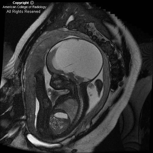
Figure 1: Axial T2 image shows micro-ophthalmia with an irregular-appearing left globe, suggesting a colobomatous deformity.
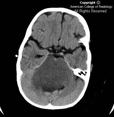
Figure 2: Axial FLAIR shows absence of the septum pellucidum.
Figure 3: Coronal T1 postcontrast shows absence of the septum pellucidum, a “point-down” appearance to the inferior aspect of the frontal horns of the lateral ventricles.
Figure 4: Axial SPGR: Bilateral atretic optic nerves are seen.
Figure 5: Sagittal T1 FLAIR sequence shows atretic optic chiasm.


Diagnosis: Septo-optic dysplasia/de Morsier's syndrome


Septo-optic dysplasia is characterized by malformations of the optic nerves and tracts, pituitary deficiency, and absence of the septum pellucidum. A small pituitary gland is often identified with an ectopic posterior lobe. Embryologically, the disease is a disorder of midline prosencephalic development. There is secondary degeneration of optic nerve fibers due to cerebral lesions.

The epidemiology of the disease is 1:50,000, with the affected male:female ratio equal to 1. The genetic pattern of inheritance may be either autosomal dominant or recessive.

The most common signs and symptoms in newborns include: hypoglycemia, seizures, apnea, cyanosis, hypotonia, and prolonged conjugated jaundice. Multiple pituitary hormone deficiencies can lead to various presentations. For example, short stature may result from deficiency of growth hormone. Other corresponding endocrinopathies are seen, depending on which hormone is absent. Symptoms may also include color blindness, visual loss, nystagmus, strabismus, mental retardation, spasticity, microcephaly, and anosmia.

Septo-optic dysplasia is frequently associated with other cerebral anomalies. The most commonly associated entity is schizencephaly. Others include perisylvian cortical dysplasia, midline malformation (callosal dysgenesis), ocular malformation (coloboma, anopthalmia, micropthalmia), olfactory tract/bulb hypoplasia and hypoplasia, of the pituitary and olfactory lobes.

CT findings of septo-optic dysplasia include absent septum pellucidum, large lateral ventricles, and small bony optic foramina. MRI is the best imaging tool for diagnosis. Imaging in three planes is crucial to identify all findings. Coronal MRI images demonstrate absent septum pellucidum, flat roof of frontal horns, and a “point down” appearance of the inferior aspect of frontal horn. A small optic chiasm/nerve, thin pituitary stalk, and posterior pituitary ectopia can also be seen when fat saturation sequences are used. T1 postcontrast imaging demonstrates enhancement of infundibulum and an ectopic posterior pituitary lobe.

Differential diagnosis includes syndromes overlapping with septo-optic dysplasia, such as optic infundibulum dysplasia (OID) with schizencephaly but normal septum pallucidum, Kallmann's syndrome, and lobar holoprosencephaly.

Untreated septo-optic dysplasia can lead to hypothalamic and pituitary crisis or sudden death from hypocortisolism. Treatment of septo-optic dysplasia consists of hormonal replacement therapy.

Post radiation white matter injury. Metastatic disease to the calvarium.


Findings

Initial MRI of the brain shows numerous enhancing lesions scattered throughout the calvarium, consistent with metastatic bony involvement. There is also diffuse enhancement of the pachymeninges. There are no enhancing parenchymal lesions. Newer MRI of the brain shows confluent periventricular deep white matter FLAIR and T2 hyper intensities. There are no abnormal parenchymal lesions or enhancement. There is mild volume loss. There was no restricted diffusion (images not shown) to suggest infarct. Again seen are heterogeneous and enhancing calvarial lesions.

Differential diagnosis:
- Post radiation white matter disease
- Microvascular disease
- Age related white matter changes
- Tumor edema from metastatic or primary neoplasm
- Diffuse infectious process
- Diffuse inflammatory process


Diagnosis: Post radiation white matter injury. Metastatic disease to the calvarium.

Diagnosis is presumed based on this history: The patient has a history of widely metastatic breast cancer, with dural and calvarial involvement. The patient received chemotherapy and underwent whole brain radiotherapy. The white matter changes are nonspecific. However, given the history of radiotherapy, the abnormalities are likely sequelae of radiation treatment:


Key points - Radiation injury to the brain

Clinically:
- Many patients are asymptomatic
- Common complaints include confusion, personality changes, memory loss, and dementia
- Injury occurs in 38% to 50 % of patients after whole brain radiation
- Poor correlation of clinical symptoms with severity of imaging findings
- Amount of injury depends on dose and fractionation methods
- Latent period for appearance on injury on imaging is 6 to 8 months


Radiology

Changes of symmetric white matter injury
Periventricular white matter is particularly susceptible
Long term results is cerebral atrophy

CT:
- Confluent white matter hypo densities
- Usually no enhancement

MRI:
- More sensitive than CT
- T2 and FLAIR white matter prolongation
- Ranges from punctate periventricular foci to confluent changes extending from the ventricles to the corticomedullary junction
- May show ring enhancement if there is radionecrosis
- Usually no significant mass effect or enhancement

martedì 26 dicembre 2006

Meningioma


Findings

CT Head demonstrates a calcified mass within the left frontal region with adjacent hyperostosis of the bone. MRI demonstrates an enhancing extra-axial dural-based mass over the left frontal lobe with minimal focal mass-effect on the adjacent cortex. No edema. There are associated dural tails and focal hyperostosis of the calvarium.


Diagnosis: Meningioma


Key poins

Nearly all meningiomas are benign, but that does not mean that they do not harm the patient. The tumors arise from arachnoid cap cells, which are cells of the meninges. Most meningiomas (90%) are categorized as benign tumors, with the remaining 10% being atypical or malignant. Meningiomas produce symptoms in many ways. They may irritate the underlying cortex, compress the brain or the cranial nerves, producing hyperostosis, invade the overlying soft tissues, or induce vascular injuries to the brain. The symptoms include seizures, headaches, and focal neurological defects, such as arm or leg weakness, or vision loss. Meningiomas may produce increased symptoms during pregnancy, but they often subside after the birth of the child.

Meningiomas are more common in females and more common in Africa. In the US they are responsible for about 20% of all primary intracranial neoplasms. The neoplasms are most often detected in middle age. Earlier detected meningiomas are often malignant. They are often associated with neurofibromatosis. Exposure to radiation has been found to be associated with a higher incidence of meningiomas. Meningiomas may occur intracranially or within the spinal canal.

Treatment options include surgery, radiation therapy, a combination of the two, or observation with periodic scans and examinations are then evaluated.


Radiology

Plain skull x-ray may reveal hyperostosis and increased vascular markings of the skull as well as intracranial calcifications.
On plain head CT scan, meningiomas are usually dural based, isodense to slightly hyperdense tumors. Homogenous and intense enhancement pattern. Perilesional edema may be present. Hyperostosis and calcification can be seen. They can have mass affect.
On MRI, the T1- and T2-weighted signals are variable. Giving contrast is key. Meningiomas enhance intensely and homogeneously after injection of gadolinium gadopentetate. Perilesional edema may be present. Enhancing dural tail is very helpful.

Endovascular angiography allows the surgeon to determine preoperatively the vascularization of the tumor and its encroachment on vital vascular structures. Angiography can be used to embolize feeding vessels prior to surgery. Blood supply is sunburst in appearance and is from external circulation.

giovedì 21 dicembre 2006

Vein of Galen aneurysm


Findings

The contrast-enhanced CT demonstrates a markedly dilated venous system on the axial view (Figure 1), including the Vein of Galen and the straight sinus, along with the torcular herophili. The ventricles are dilated and there are periventricular calcifications consistent with ischemic insults and/or thrombosed and calcified feeding vessels. The coronal view (Figure 2) demonstrates dilated jugular veins within the upper neck. The sagittal image demonstrates anterior displacement and mass effect (Figure 3) on the region of the superior cerebral aqueduct by the Vein of Galen.


Diagnosis: Vein of Galen Aneurysm


A Vein of Galen aneurysm is a rare congenital abnormality that is a misnomer. The “aneurysm” is actually one of a variety of arteriovenous malformations of the intracranial circulation with the Vein of Galen portion of the malformation more accurately described as a varix. These malformations are thought to result secondary to persistence of embryonic venous precursors involving the median vein of prosencephalon. There are multiple classification systems for Vein of Galen malformations (VOGM’s). Lasjaunias developed one of the most commonly used systems which divides the malformations into choroidal and mural subtypes. The choroidal subtype is characterized by multiple feeding vessels from the posterior choroidal and adjacent midbrain thalamoperforating arteries. The arterial feeders typically converge on a fistulous site anterior to the median vein and demonstrate a complex arterial maze which mimics a true AVM. The AVM nidus is usually near the thalamus or brainstem. Mural lesions are characterized by a fistulae in the wall of the median prosencephalic vein. Mural lesions typically have fewer feeding arteries and are less associated with heart failure and more commonly associated with delayed presentation. The Vein of Galen malformations (VOGMs) are rare anomalies which constitute 1% of all intracranial vascular malformations but represent 30% of vascular malformations in the pediatric age group.

The clinical presentation of Vein of Galen aneurysms depends on the age of presentation. Neonates classically present with high output cardiac failure. The steal phenomenon which occurs due to the fistulous connections can result in varying degrees of brain parenchymal loss. Neonates can also present with hydrocephalus and macrocephaly. Infants and older children usually present with developmental delay, ocular symptoms, and symptoms related to hydrocephalus. Focal neurologic deficits, seizures and hemorrhage can be seen at any age. Ultrasound is usually the first choice imaging modality and the diagnosis can be made prenatally by demonstrating a cystic structure posterior to the third ventricle with flow demonstrated on Doppler imaging. Contrast enhanced CT can aid in the diagnosis postnatally by demonstrating the anatomy. CT helps quantify cerebral parenchymal damage and ischemic changes that are manifested by encephalomalacia and parenchymal calcifications. Calcification of the aneurysm sac can be seen with delayed presentation, usually beyond 15 years of age. MRI is helpful in demonstrating the vascular anatomy and parenchymal damage present. Angiography is the gold standard with superior definition of small arterial feeders and also provides the vector for endovascular treatment.

Treatment of Vein of Galen aneurysms has evolved over the years, although there still is no simple cure. Surgery has traditionally been the mainstay treatment with overall dismal outcomes. Even with the recent advancements of microneurosurgery, the results have been dissatisfying with a reported mortality rate of almost 90% after surgery in neonates. Endovascular therapy has been proposed for some time, but only recently have technological advancements allowed subselective embolization of the inherently tortuous vessels. A number of temporary and permanent embolization agents have been used including various types of coils and acrylic agents. These have been used with varying success in venous, arterial, and combination approaches. There is some indication that staged arterial embolization of the AVM may be the most successful approach.

lunedì 18 dicembre 2006

Retinoblastoma


Findings

CT of the orbits with contrast demonstrates 7 x 4 mm mildly enhancing left retinal mass with associated focal, coarse calcification.

Differential diagnosis:
- Retinoblastoma
- Retinoma – benign variant
- Persistent hyperplastic primary vitreous
- Retrolental fibroplasia
- Toxocariasis (no calcification)
- Coat's disease


Diagnosis: Retinoblastoma


Key points

Rare, malignant, congenital tumor – arises from neuroectodermal cells of retina.
Clinical findings – leukocoria 60 % (reflection from white mass within the eye, giving the appearance of a white pupil)
Incidence – 1:15k – 1:34k live births. Most common intraocular neoplasm in childhood; 1% of all childhood malignancies.
70% < 3 years of age; mean age of presentation - 18 months.
30% bilateral, 30% multifocal within one eye
66% non heritable form; Heritable sporadic form (20-25%); 10% familial history of retinoblastoma


Radiology

Intraocular mass
High density (calcification, hemorrhage); enhancement usual
Dense vitreous humor (commonly)
Calcifications (90%); in absence of calcification, suspect other mass lesions

Primary role of imaging is to determine tumor spread:
- Optic nerve extension (25%)
- Scleral break-through
- Metastases; meninges, liver, lymph nodes

giovedì 14 dicembre 2006

Right ICA aneurysm with oculomotor impingement


Findings

CT shows a clinically insignificant mild frontal scalp hematoma. Also, and more importantly, a prominent right suprasellar internal carotid artery in the region of the Circle of Willis. MRI shows a large saccular aneurysm of the distal right supraclinoid ICA with probable posterior impingement of the right CN III.


Diagnosis: Right ICA aneurysm with oculomotor impingement


Key points

An aneurysm is a focal dilatation of an artery that involves the intima and adventia of the wall. Most aneurysms are saccular (round or lobulated) and occur at arterial bifurcations, postulated to be the result of congenital defects or degenerative changes the vessel wall. More than 90% of saccular aneurysms occur in the anterior circulation, with 30-35% occurring at the anterior communicating artery, 30-35% occurring at the posterior communicating artery, and 20% occurring at the middle cerebral artery. Approximately 10% of saccular aneurysms occur at the posterior or vertebrobasilar artery locations.

Giant aneurysms are defined as those greater than 2.5 cm. in diameter. They account for 5-13% of all intracranial aneurysms, most commonly seen in women by a ratio of 3:1. They are usually multilayered with thrombus of varying age. They usually have a residual patent lumen.

Intracranial aneurysms most commonly present with subarachnoid hemorrhage. Only approximately 1% are discovered incidentally. Prognosis after SAH is widely variable and usually correlates with the Hunt-Hess scale:
- Asymptomatic or mild headache
- Moderate or severe headache, nuchal rigidity, can have oculomotor palsy
- Confused, drowsiness, or mild focal signs
- Stupor or hemiparesis
- Coma, moribund, and/or extensor posturing

In general, the larger the aneurysm and the older the patient, the more likely to rupture with a cumulative risk of 1% per year. Mortality is approximately 50%. If untreated, 50% re-bleed within 6 months.


Radiology

Angiography remains the "gold standard.", however MR angiography is playing an increasingly important role. The entire intracranial circulation must be studied because of the 15-20% incidence of multiple aneurysms.
2-5 % of patients with SAH will have a normal initial angiogram. Of these, 10-20% will demonstrate an aneurysm on a second angiogram.
CT can detect aneurysms as small as 3-5 mm. as an intensely enhancing residual lumen often with a peripheral rim of calcification.
MRI and MRA is being used increasingly and can detect aneurysms as small as 3-4mm. The most sensitive MR sequence for detection of SAH is FLAIR.

The appearance of giant aneurysms on MR results from their multi-layered structure. On spin echo images, the residual lumen creates a flow void. Slowly flowing blood may demonstrate a high signal. The thrombosed portion resembles multilayered intracranial hematomas with layers of varying signal intensity depending on the state of hemoglobin. However, the high signal ring tends to be central rather than peripheral because methemoglobin formation is optimal adjacent to the flowing oxygenated blood.

mercoledì 13 dicembre 2006

Congenital Cytomegalovirus infection


Findings

Figures 1 and 2: Ultrasound images demonstrate multiple echogenic foci in a periventricular distribution. There is no hydrocephalus or evidence of callosal agenesis.
Figures 3, 4 and 5: Axial CT images show high attenuation within the subependymal region, consistent with calcifications.


Diagnosis: Congenital Cytomegalovirus infection


Differential diagnosis for congenital intracranial calcifications:
- Congenital TORCH infection
- Tuberous sclerosis
- Teratomas
- Sturge-Weber syndrome
- Venous sinus thrombosis


Cytomegalovirus is the most common congenital viral infection in the TORCH group, and occurs as a result of transplacental transmission of the virus from an infected mother. Most infections are asymptomatic; symptomatic infections can present with a variety of manifestations, such as seizures, mental retardation, optic nerve involvement, and sensorineural hearing loss. Timing of the maternal infection during gestation may play a role in determining the severity of symptoms, with early infection leading to a more severe presentation.

Central nervous system abnormalities associated with CMV infections include calcifications, ocular deficits, parenchymal atrophy, periventricular cyst formation (occipital lobes), deafness, cerebellar hypoplasia, and ventriculomegaly. Neuronal migrational anomalies are most common in CMV infection relative to other TORCH infections.

Periventricular and subependymal calcifications are a common manifestation of CMV infection. Periventricular calcifications can also be seen in toxoplasmosis and bacterial meningitis with ventriculitis. Calcifications in CMV tend to be limited to the subependymal region, whereas in toxoplasmosis, calcifications can also seen throughout the parenchyma. Calcifications in herpes simplex and rubella are much less common. Calcified subependymal nodules may also be seen in tuberous sclerosis, however calcifications tend to occur in adolescence, as opposed to the neonatal period.

Ultrasound may also play an adjunctive role in visualizing the manifestations of CMV. Sonographic findings suggestive of CMV infection include ventriculomegaly, multiple periventricular and intraparenchymal echogenic foci (representing intracranial calcifications), gyral abnormalities, occipital periventricular cysts, and hydrocephalus.

venerdì 8 dicembre 2006

Diffuse brainstem glioma


Findings

Figure 1: Axial postcontrast CT image shows non-enhancing diffuse mass causing expansion of the pons and engulfing the basilar artery.
Figure 2: Axial T2 image shows the hyperintense mass lesion causing marked expansion of pons more clearly and the flow void in basilar artery. Notice the mass effect on the fourth ventricle that is distorted and displaced posteriorly.
Figure 3: Sagittal post gad T1 image showing the cranio-caudal extent of mass lesion with involvement of entire brainstem.


Diagnosis: Diffuse brainstem glioma


Precise diagnosis of the different brainstem tumors by neuroimaging is very important in patient management. Focal brainstem tumors are treated surgically, whereas diffuse tumors are treated with chemotherapy and radiation.

Patients with NF1 have a higher incidence of brain tumors, with the brainstem commonly involved. Although the imaging appearance of these tumors is similar, they often have a relatively indolent clinical course.

Brainstem gliomas constitute about 15% of all pediatric CNS tumors with peak incidence between 3 and 10 years old. These are heterogeneous groups of tumors that may be diffuse, focal, dorsally exophytic, or cervicomedullary. These tumors commonly present with symptoms of double vision, weakness, unsteady gait, difficulty in swallowing, headache, dysarthria, nausea, and vomiting. Rarely, behavioral changes and seizures may also be seen in children with this tumor. Although no definite familial tendency is known, there is an increased incidence of brainstem gliomas in patients with neurofibromatosis Type 1.

Magnetic resonance imaging has emerged as the primary diagnostic modality for brainstem gliomas. MRI multiplanar images assist in the establishment of the tumor diagnosis, identification of tumor epicenter, and prediction of its biological behavior. Diffuse neoplasms tend to smoothly enlarge the affected area without focal areas of exophytic tumor. They are generally poorly marginated and involve more than 50% of the brainstem in the axial plane at the level of maximal involvement. Minimal or no contrast enhancement is seen after gadolinium, although enhancement is commonly seen after radiation therapy. It is difficult to biopsy these lesions because of the proximity of vital structures and thus imaging plays a crucial role in the diagnosis. Focal tumors are generally well marginated and involve less than 50% of brainstem in the axial plane. Focal neoplasms often enhance and have a better prognosis in general than diffuse neoplasms.

Diffuse gliomas are unfortunately the most common brainstem lesions, and they have the worst prognosis among the brainstem gliomas. Radiation and/or chemotherapy are the current mainstays of treatment for diffuse brainstem gliomas. The majority of focal neoplasms are amenable to surgery and longer survival.

martedì 5 dicembre 2006

Fetal hydrocephalus


Findings

The cerebellar hemispheres (Figure 1), vermis and cisterna magna (Figure 1) are normal.
There is hydrocephalus, manifested by the marked dilatation of lateral ventricles, a midline falx (Figure 2 and Figure 3), and thinning of the cerebral cortical mantle (Figure 2 and Figure 3).
The fourth ventricle (Figure 4) is normal, therefore, the obstruction is suspected to be at the level of the Acqueduct of Sylvius (Figure 4). No obstructing lesions are identified.
Aqueductal Stenosis is more common in males as seen in this case (Figure 5)


Diagnosis: Fetal hydrocephalus


Fetal MRI is used as a problem-solving tool in obstetrics with a wide variety of indications when ultrasound findings are equivocal or need further evaluation.
MRI in the first trimester is a relative contraindication. MRI poses no known risk to the fetus in the second and third trimester. Intravenous gadolinium is not approved for use in pregnancy.
Ventriculomegaly is the most common CNS abnormality identified on prenatal US and also the most common indication for fetal MRI. 70-84% of fetuses with ventriculomegaly show associated structural or chromosomal anomalies.

Ultrasound is the primary technique for fetal imaging for its proven utility, widespread availability and relative low cost. However, there are pitfalls in the evaluation of the brain and spine with US. Discrimination of normal and abnormal appearance of the CNS on US is based on the ability to produce high resolution images of the cerebrum, cerebellum, and spine. Maternal obesity, oligohydramnios, suboptimal fetal lie may cause inability to obtain adequate US images. MRI is less affected by these factors and over the past decade has emerged as a clinically useful supplement to US where US findings have been inconclusive or insufficient to guide treatment choices.


Indications for fetal MRI

Neurologic indications:
- Ventriculomegaly
- Agenesis of the corpus callosum
- Posterior fossa abnormalities
- Malformations of cerebral cortical development - neuronal migrational anomalies such as schizencephaly, lissencephaly, polymicrogyria, and gray matter heterotopia.

Nonneurologic indications:
- Congenital diaphragmatic hernia
- Pulmonary sequestration
- Congenital cystic adenomatoid malformation
- Airway obstruction
- Volumetric mesurments of individual fetal organs

Safety concerns arise for both mother and fetus and the FDA guidelines require labeling of MRI devices to indicate the the safety in pregnancy has not yet been established. Maternal safety concerns are addressed by standard MRI screening while fetal safety concerns are related to teratogenesis and acoustic damage.

In general, MRI in the first trimester is a relative contraindication owing to the higher risk of mutation during the period of organogenesis. MRI poses no known risk to the fetus in the second and third trimester. Intravenous gadolinium is not approved for use in pregnancy as it has been shown to cross the placenta and appears within the fetal bladder. The half life of the drug in the fetal circulation and its effect on the developing human fetus are unknown.


Fetal ventriculomegaly and hydrocephalus

As in the case exemplified here, ventriculomegaly is the most common CNS abnormality identified on prenatal US. Ventriculomegaly is defined as an atrial width >10 mm measured in an axial plane at the level of the thalami and at the posterior margin of the glomus of the choroid plexus. The atrial diameter of the lateral ventricles remain relatively constant in size from 15-35 weeks gestation while the surrounding brain continues to grow, thus the lateral ventricles appear proportionately larger earlier in gestation. From 70-84% of fetuses with ventriculomegaly show associated structural or chromosomal anomalies. Structural abnormalities include agenesis of the corpus callosum, Dandy-Walker syndrome, holoprosencephaly, cortical malformations, porencephaly, and intracranial hemorrhage.

In patients with mild ventriculomegaly (atrial width of 10-15 mm) with no chromosomal or structural abnormalities, the frequency of neurodevelopmental abnormality ranges from 0-36% compared with 84% in patients with additional abnormalities.

Congenital hydrocephalus, as in the case exemplified above, is most commonly (43%) caused by occlusive malformations of the Aqueduct of Sylvius. Etiologies for Aqueductal stenosis are developmental (forking, narrowing, transverse septum{X-linked resessive}), infectious (toxoplasmosis, CMV, syphilis, mumps, influenza), and very rarely, neoplastic (glioma, pinealoma, meningioma). It occurs more commonly in males (M: F = 2:1). Thumb deformities occur in 16% and there is a mortality of 11-30%.

On imaging, Aqueductal stenosis causes enlargement of the lateral and third ventricles with a normal fourth ventricle. These may become markedly dilated with absent septum pellucidum with compression of the central white matter and cortex. At a later stage, the cerebral cortex becomes atrophic and paper thin.

In-utero procedures such as ventriculoamniotic shunts and cephalocentesis have high morbidity and mortality and outcomes are generally worse than when shunting procedures are done in the neonatal and infancy period.

Pituitary microadenoma


Findings

In the right anterior pituitary gland there is a focus of subtle hypointensity on the precontrast sagittal T1 image. On the early dynamic post contrast coronal T1 there is a 4 mm focus of hypoenhancement compared to the remainder of the anterior pituitary. On the delayed sagittal post contrast T1 image, the previous area of hypoenhancement shows increased enhancement compared to the remainder of the anterior pituitary gland.

Differential diagnosis: None in this case. Findings are diagnostic of a pituitary microadenoma (prolactinoma).

The gamut of pituitary/sellar lesions includes:
- Pituitary microadenoma
- Pituitary macroadenoma
- Pituitary apoplexy
- Craniopharyngioma
- Rathke's cleft cyst
- Aneurysm
- Meningioma
- Lymphoma
- Metastasis
- Granulomatous disease


Diagnosis: Pituitary microadenoma


Pituitary microadenomas are defined as less than 10 mm in diameter.
Pituitary macroadenomas are larger than 10 mm and are often associated with symptoms secondary to mass effect or hemorrhage:
- Mass effect can compresses the optic chiasm giving bitemporal hemianopsia.
- Invasion of the cavernous sinus can cause other cranial neuropathies.
- Inferior breakdown of the sella can cause CSF rhinorrhea.

75% of pituitary adenomas have elevated hormonal secretion, most commonly prolactin.
CT can show the mass if the lesion is large. Sella enlargement and erosion can also be seen.
MR is the imaging modality of choice. Microadenomas are hypointense on precontrast T1 images, hypoenhance compared to the remainder of the pituitary gland on early post contrast images, and show delayed enhancement greater than the remainder of the gland. T2 characteristics are variable. With larger lesions, MR can define mass extent including cavernous sinus invasion and hemorrhage.
Prolactin secreting microadenomas are most often treated medically with bromocriptine.

venerdì 1 dicembre 2006

Eagle's syndrome


Findings

There is ossification and pseudoarticulation of the stylohyoid ligaments bilaterally, left greater than right (on these image focusing on the left side).

Differential diagnosis:
- Eagle's Syndrome
- Calcified thyroglossal duct cyst
- Foreign body
- Vascular calcifications
- Chronic Mastoiditis
- Post-traumatic calcifications


Diagnosis: Eagle's syndrome


Key points

Eagle’s syndrome is characterized by craniofacial or cervical pain due to an elongated styloid process or calcified stylohyoid ligament.
Eagle's Syndrome is due to elongation of the styloid process > 30 mm.
4% of the general population has elongation of the styloid process, but only 4% of this group have symptoms of Eagle's Syndrome.
Most common symptom is vague neck pain (41%), however other symptoms include foreign body sensation, throat pain, odynophagia, and pain upon changing head position.
Pain can be due to nerve compression (glossopharyngeal, vagus, or trigeminal), impingement on the carotid vessels, degenerative changes at the stylohyoid ligament insertion site, or rheumatic styloiditis from infection.
There is a 3:1 female preponderence, and most present between age 30 and 40.
Etiology is debated, but some have suggested prior trauma (e.g., tonsillectomy), congenital elongation, ossification of the stylohyoid ligament, and elongation at the cartilaginous junction of the tympanohyale and stylohyoid.
Surgical shortening is the best treatment, and the procedure can easily be performed intra- or extra- orally. Conservative management with local anesthesia or oral analgesia generally fails.