venerdì 24 luglio 2009

Brain abscess


Findings

There is an intra-axial mass, located in the posterior left frontal lobe, in the superior frontal gyrus just anterior to the precentral gyrus. There is surrounding vasogenic edema, which expands the left precentral gyrus. Mass shows a well-defined rim on MR, somewhat irregular, consistent with a capsule. Central portion shows pronounced diffusion restriction.

Differential diagnosis:
- Abscess
- Metastasis
- Glioblastoma
- Infarct
- Demyelinating disease


Diagnosis: Brain abscess


Key points

A brain abscess is a focal, suppurative infection within the brain parenchyma, typically surrounded by a vascularized capsule.
The most common symptom in patients with a brain abscess is headache, occurring in >75% of patients.
MRI is better than CT for demonstrating abscesses in the early (cerebritis) stages and is superior to CT for identifying abscesses in the posterior fossa.
On contrast-enhanced T1-weighted MRI, a mature brain abscess has a capsule that enhances surrounding a hypo dense center and surrounded by a hypo dense area of edema.
On T2-weighted MRI, there is a hyper intense central area of pus surrounded by a well-defined hypo intense capsule and a hyper intense surrounding area of edema.
The distinction between a brain abscess and other focal CNS lesions such as primary or metastatic tumors may be facilitated by the use of diffusion-weighted imaging sequences on which brain abscesses typically show increased signal and low apparent diffusion coefficient.

Ring-enhancing lesion differential (MAGIC DR) :
- Metastasis
- Abscess
- GBM
- Infarct
- Contusion
- Demyelination
- Radiation necrosis

Serial MRI or CT scans should be obtained on a monthly or twice-monthly basis to document resolution of the abscess.

mercoledì 22 luglio 2009

Chiari II Malformation


Findings

T1-weighted sagittal images (Figure 1 and Figure 2) and T2-weighted axial images (Figure 3, Figure 4, and Figure 5) of the brain demonstrate a small posterior fossa with herniation of the cerebellar tonsils through the foramen magnum and a beaked appearance of the tectum. There is associated hydrocephalus.

T2-weighted sagittal (Figure 6 and Figure 7) and axial (Figure 8 and Figure 9) images of the spine demonstrate a myelomeningocele at the level of the lower lumbar spine.


Diagnosis: Chiari II Malformation


The Chiari II malformation is a complex anomaly with skull, dural, brain, spinal and spinal cord manifestations. It is regarded as a disease of primary neurulation and is almost invariably associated with a myelomeningocele. There are multiple associated findings, to include; protrusion of the cerebellar tonsils through the foramen magnum, extension of the cerebellar hemispheres and vermis above the incisura of the tentorium, beaked appearance of the tectum, fenestration of the falx, hydrocephalus, a prominent massa intermedia, colpocephaly, and dysgensis of the corpus callosum.

The Chiari II malformation is the most common serious malformation of the posterior fossa, with a frequency of approximately 1 case per 1000 population. Hindbrain dysfunction is the major cause of mortality. The mortality rate is 15% in the first few years of life and is reported to be as high as 50% for the long term rate, regardless of the treatment strategy.

MRI is currently the best diagnostic method for detecting the full constellation of findings associated with Chiari II malformations. CT scans are commonly performed to assess for hydrocephalus as well as to follow up infants who have undergone a ventriculoperitoneal shunt procedure. Many of the typical findings described by using cranial CT scans and MRIs also can be demonstrated on cranial sonograms. Ultrasound has the benefit of not using ionizing radiation, however it is heavily operator dependent and some of the associated abnormalities may be missed.

martedì 21 luglio 2009

Subacute combined degeneration


Findings

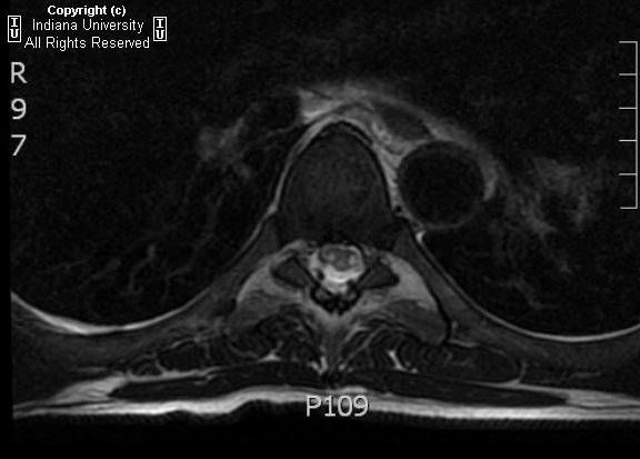
MRI of the thoracic and cervical spine show increased T2 signal within the posterior columns and the lateral corticospinal tracts on axial imaging. On sagital imaging, a longitudinal T2 signal abnormality is noted within the dorsal cord. There was no significant contrast enhancement status post gadolinium administration.

Differential diagnosis:
- Subacute combined demyelination
- Multiple sclerosis
- Astrocytoma
- Spinal cord infarction
- Infectious myelitis


Diagnosis: Subacute combined degeneration.


Vitamin B12 deficiency is the result of a malabsorption syndrome and can affect the brain, optic nerves, peripheral nerves and spinal cord. When patients present with myelopathy, such as sensory disturbances, weakness, and spasticity, it is known as subacute combined degeneration (SCD).

Clinical presentation of SCD is caused by dorsal column, lateral corticospinal tract, and sometimes lateral spinothalamic tract dysfunction. Patients initially present with paresthesia in the hands and feet, which can progress to sensory loss, gait ataxia, and distal weakness particularly within the legs. If the disease goes untreated, ataxic paraplegia may evolve. On physical exam, there is a loss of vibratory and joint position sense, weakness, spasticity, hyperreflexia, and extensor plantar responses. The diagnosis of B12 deficiency is made by serologic studies showing low serum B12 level; if the B12 level is borderline, elevated levels of homocysteine and methylmalonic acid help cinch the diagnosis.


Neuroradiology

Radiologic manifestations of SCD may be seen on MRI imaging, primarily within the cervical and thoracic spine, and include the following spinal manifestations:
- Mild spinal cord expansion and hypointensity on T1 weighted imaging.
- Increased T2 signal intensity primarily within the dorsal columns +/- lateral columns.
- Longitudinal dorsal cord T2 signal abnormality.
- Inverted "V" or "rabbit ears" T2 signal intensity within the dorsal spinal cord on axial imaging.
- Possible mild dorsal column contrast enhancement, with enhancement signifying breakdown of the blood nerve barrier.


Differential considerations

Although these MRI findings are consistent with SCD, they are nonspecific and include a broad differential diagnosis: demyelinating disorders, infectious etiologies, inflammatory conditions, ischemia, contusion, and neoplasms. However, SCD can be distinguished from other differentials given its bilateral nature of T2 signal abnormality over multiple levels that is confined to specific white matter spinal tracts. There can be partial to full reversal of MRI abnormalities following B12 therapy, with fifty percent of patients fully recovering and with the greatest recovery occurring when treatment is began in the early stages of the disease.

Multiple sclerosis is a demyelinating disorder with multiple lesions separated in time and space. MRI spine findings included increased T2 signal intensity and hypo or isointense T1 signal within lesions. These lesions, however, are more focal and well-circumscribed in nature compared to the contiguous lesions of SCD and show homogenous, nodular, ring enhancement of acute and subacute lesions status post contrast administration. Multiple sclerosis lesions rarely spans more than one or two vertebral segments and are usually not symmetric in nature.

Astrocytoma of the spine is an intramedullary glioma, more often located in the cervical than thoracic spine. This lesion presents as a hyperintensity on proton density and T2 weighted imaging, and almost always enhances. On T1 imaging, there is cord expansion, usually less than four segments. Again this is distinguished from SCD by location (intramedullary T2 signal intensity versus dorsal and lateral columns) and contiguity (SCD is contiguous over multiple vertebral segments).

Spinal cord infarction causes permanent tissue loss in the spinal cord secondary to vessel occlusion and usually presents hyper acutely. On MRI imaging, focal T2 hyperintensities can be seen in the gray matter, gray matter with adjacent white matter, or an entire cross section of the cord. These lesions are usually within the thoracic cord as it is an arterial border zone. T2 hyperintensities may also bee visualized within the anterior vertebral body bone marrow or deep medullary portion near the endplate secondary to vertebral body infarction. Diffusion weighted imaging shows restricted diffusion in the affected areas of the cord. Slight cord expansion and decreased signal is noted on T1 imaging. The MRI lesion distribution, bony involvement and diffusion changes differentiate spinal cord infarction from SCD.

Infectious myelitis can be secondary to HIV vacuolar myelopathy, varicella-zoster/herpes, or Lyme disease. On T1 imaging, there is cord expansion that nearly fills the spinal canal and variable, nonfocal enhancement status post contract administration. On T2 imaging, there is diffuse increased signal intensity throughout the involved segment, secondary to a swollen edematous cord. Unlike SCD, the signal abnormalities are not limited to the spinal tracts. However, MRI imaging findings may be identical to B12 deficiency and in such cases, can be distinguished by clinical presentation and laboratory findings.


venerdì 17 luglio 2009

Vagal Schwannoma


Findings

Figure 1, Figure 2, and Figure 3: Contrast enhanced axial CT images through the neck demonstrate a 4.5 X 6 cm circumscribed and moderately enhancing mass within the right carotid space with scattered areas of low density centrally within the mass. The internal and external carotid arteries as well as the parapharyngeal fat are displaced anteriorly. The parotid gland is separate from the lesion.
Axial and coronal T2 weighted images (Figure 4 and Figure 5) and axial and coronal T1 weighted post-gadolinium fat saturated images (Figure 6 and Figure 7) again show a well defined mass with heterogeneous T2 signal and areas of cystic degeneration. There is peripheral enhancement following gadolinium administration. Note that the mass is not contiguous with the parotid gland.


Diagnosis: Vagal Schwannoma


Schwannomas are benign neoplasms which arise from the nerve sheath and consist of schwann cells in a collagenous matrix. Histologically, the terms Antoni type A and Antoni type B are used to describe varying growth patterns. Type A tissue has elongated spindle cells and is compact in nature while type B has a looser organization with cystic spaces intermixed within the tissue. These cystic spaces result in high signal intensity on T2 weighted MR sequences.

Vestibular schwannomas are the most common cranial nerve (CN) schwannoma, followed by trigeminal and facial and then glossopharyngeal and vagus schwannomas. CN schwannomas are usually isolated lesions, except when they are associated with neurofibromatosis type 2 (NF2). NF2 is characterized by bilateral acoustic schwannomas but other CN schwannomas also occur at an increased frequency in these patients.

The majority of carotid space masses are benign, with the two most common lesions being the vagus schwannoma and the glomus tumor. Neoplasms of the vagus nerve include paragangliomas (50%), schwannomas (31%), neurofibromas (14%), and neurofibrosarcomas (6%). Most cases of schwannomas manifest between the third and sixth decades of life as a firm and painless mass in the neck.

Schwannomas of the vagus nerve are usually well-defined, rounded structures which are hypodense to muscle on CT and enhance moderately. On MR, there is variable T2 signal intensity depending on its content of Antoni A and Antoni B tissue. Hemorrhage and cystic degeneration are not uncommon. They result in anterior displacement of the carotids. In contrast, glomus tumors are highly vascular and demonstrate dramatic enhancement. Flow voids are usually visualized, resulting in the characteristic salt-and-pepper appearance. Carotid body tumors tend to splay the internal and external carotid arteries away from each other, while glomus vagale tumors displace the carotid artery anteriorly. The distinction between a vagus schwannoma and the glomus vagale tumor has to be made on the basis of flow voids and vascular flow curves. Also, glomus vagale tumors are relatively uncommon.

giovedì 9 luglio 2009

Pituitary microadenoma


Findings

Figure 1: Contrast enhanced T1 weighted image of the sella demonstrate normal enhancement of the pituitary gland with a small round hypoenhancing focus consistent with microadenoma which fills in on later sequences.
Figure 2: Coronal thin section T2 weighted image demonstrates a bright focus on the left side of the pituitary gland, in the region of previously noted hypoenhancement, consistent with a microadenoma.


Diagnosis: Pituitary microadenoma


Pituitary microadenomas account for 10-15% of all intracranial tumors, though most are found incidentally during autopsy. Pituitary microadenomas are usually benign and slow-growing. Most are asymptomatic. There is 10-20% prevalence in the general population. Prolactin-secreting adenomas or prolactinomas are the most common type, accounting for 30-40% of symptomatic microadenomas. One percent of microadenomas are multiple.

The typical presentation of prolactinoma is a young female with primary or secondary amenorrhea and infertility, with associated galactorrhea. Though usually seen in females, prolactinomas can occur in males with delayed puberty or primary hypogonadism. Growth hormone secreting adenomas, another type of pituitary microadenoma, present with acromegaly in adults and gigantism in adolescents. Prolactinomas typically occur in the 25-30 year age group, while growth hormone-secreting adenomas occur between 30-50 years of age.

Pituitary microadenoma is an intrapituitary lesion surrounded by cresecentic rim of compressed anterior pituitary. Though predominately located within the sella turcica they rarely can have an ectopic origin within the sphenoid or cavernous sinus, pituitary stalk, or third ventricle. By definition, microadenomas are less than 10 mm in diameter. The differential for pituitary microadenoma includes non-neoplastic cysts, such as Rathke cleft cyst and pars intermedia cyst; craniopharyngioma; and pituitary hyperplasia.

On imaging studies, the microadenoma is classically well-circumscribed and enhances less than surrounding normal gland. Microadenomas are isodense to normal pituitary and cannot be seen on non-contrast enhanced CT if uncomplicated by hemorrhage or cyst. On dynamic contrast-enhanced CT, two-thirds of pituitary microadenomas typically appear as hypodense masses surrounded by normally enhancing pituitary. On MR, non-contrast T1 and T2 weighted images may not show microadenomas as they appear isointense to the adjacent pituitary. Dynamic contrast-enhanced MR is the diagnostic imaging modality of choice because microadenomas enhance more slowly than normal pituitary. With contrast-enhanced MR, 70-90% of lesions appear as a relatively hypoenhancing lesion surrounded by enhancing pituitary. Scans should be obtained at 10-15 second intervals following rapid bolus injection of contrast. It is important to to note that 10-30% of microadenomas are only seen on dynamic contrast enhanced MR imaging.

Conservative treatment is recommended for “incidentalomas,” including clinical and imaging follow-up, unless there is a change in size in which case ophthalmological and endocrinological evaluation is recommended. For functioning microadenomas, medical therapy, such as bromocriptine and dopamine agonists, i.e. cabergoline, may reduce prolactin secretion to normal in 80% of cases. Surgical treatment is curative in 60-90% of cases.

lunedì 6 luglio 2009

Intramedullary spinal cord metastasis from ovarian cancer


Findings

The T1-WI shows an intramedullary lesion (Figure 1), with heterogeneous enhancing in the post-contrast sequence (Figure 2). In the T2-WI the lesion appears as heterogeneously hyperintense. The lesion is surrounded by edema.


Diagnosis: Intramedullary spinal cord metastasis from ovarian cancer


Intramedullary spinal cord metastases are rare, estimated at only 0.9-2.1% of all cancer patients at autopsy. However, only 0.1 – 0.4% of all cancer patients have clinically significant intramedullary metastatic disease. Lung metastases are most common, followed by breast cancer, renal cell carcinoma, melanoma and lymphoma. Ovarian carcinoma metastatic to the spinal cord is extremely rare with only a handful of cases reported in the literature.

Lesions are usually single (75%), two lesions found in 20% of cases and multiple in 5%. However, the majority of patients will have systemic metastases at the time of diagnosis. The cervical cord is most commonly involved; however, there can be involvement of all portions of the cord.

Common presenting symptoms include weakness, sensory loss, sphincter dysfunction, backache and radicular pain. One-quarter of patients may present with Brown-Sequard syndrome.

Pathogenesis is poorly understood. The most accepted route is hematogenous spread, both arterial and through Batson’s venous plexus. Other proposed mechanisms include meningeal carcinomatosis with infiltration of cells into the cord and direct invasion from contiguous structures.

Treatment is generally palliative and may consist of radiation, steroids and chemotherapy. Microsurgical resection has been attempted in select cases. Prognosis is uniformly poor with only 10% of patients living longer than 6 months after presentation.

giovedì 2 luglio 2009

Subperiosteal cephalohematoma


Findings

Lenticular left parietal calvarial mass-like lesion with peripheral sclerosis/calcification and central lucency. The lesion does not cross suture lines.

Differential diagnosis:
- Calcified cephalohematoma
- Caput succedaneum
- Subgaleal hematoma
- Subperiosteal osteoid osteoma
- Leptomeningeal cyst


Diagnosis: Subperiosteal cephalohematoma.


Cephalhematomas are subperiosteal hematomas that frequently occur after using instruments (forceps) during delivery. They are not associated with skull fractures and are limited by suture lines. They typically occur in the parietal region and may not appear until 2-3 days after birth. Treatment is usually not necessary. Complications include infection. Caput succedaneum is a subcutaneous hemorrhage that occurs after vaginal delivery, does cross suture lines and requires no intervention. A subgaleal hemorrhage covers a much larger area than a cephalohematoma and is potentially life threatening. It usually occurs from rupture of emissary veins in the subaponeurotic space, often secondary to vacuum suctioning.


Radiologic overview

Often seen as soft tissue subperiosteal elevation on conventional radiography. Frequency of location: parietal > occipital > frontal. The outer border may calcify. The skull at the hematoma site my remain thickened for years. It does not cross suture lines as it is bounded by the periosteum.

mercoledì 1 luglio 2009

Tuberous sclerosis with subependymal giant cell astrocytoma


Findings


Head CT shows a heterogeneous soft tissue mass in right lateral ventricle at right foramen of Monro. Heterogeneous contrast enhancement. Nodular ependymal calcifications. Hydrocephalus.
MR confirms heterogeneously enhancing mass at R foramen of Monro. T2 hyper intensity surrounding R frontal horn representing transependymal CSF resorption. Additional sub-ependymal nodule in L frontal horn.


Diagnosis: Tuberous sclerosis with subependymal giant cell astrocytoma.


Key points

TS is an inherited disorder of multiple hamartomatous lesions.
Classic clinical triad: Mental retardation, facial angiofibroma, seizure.
Subependymal nodules (SEN) (98%) 50% calcify. 30-80% enhance.
Enlarging SEN at foramen of Monro = Subependymal giant cell astrocytoma (SGCA)
Ventriculomegaly common, even in absence of obstructing SEN / SGCA.
Cortical / subcortical tubers (70-95%) cause thickening of cortex, gyral enlargement.
Number of tubers corresponds with degree of mental retardation.
White matter lesions – linear, wedge-shaped T2/FLAIR hyper intensities.
Extra cerebral TS: Renal angiomyolipoma / cysts (40-80%), cardiac rhabdomyoma (50-65%), lung LAM, subungual fibromas (15-20%), skin "ash-leaf spots," shagreen patches.

Bilateral carotid body tumors with a third mediastinal paraganglioma


Findings

Figure 1: Neck CT angiography shows bilateral enhancing, hypervascular masses, causing splaying of the carotid and jugular vessels, consistent with bilateral carotid body tumors.
Figure 2: Neck CT angiography characterizes the radioactivity focus in the superior mediastinum as a hypervascular soft tissue mass adjacent to the left common carotid artery, consistent with a third paraganglioma.
Figure 3: 3D computed tomographic reconstruction image shows the relationship of these masses to adjacent vascular structures.
Figure 4: There is bilateral focal radiotracer (In111-DTPA-octreotide)uptake in the neck corresponding to the clinical masses and also a third focus of activity in the left upper mediastinum.


Diagnosis: Bilateral carotid body tumors with a third mediastinal paraganglioma


Carotid body tumors, also known as chemodectomas or paragangliomas, are benign, slow growing neoplasms, so named because they arise from glomus cells at the carotid junctions. They have overall good prognosis, but a propensity for local invasion. They follow the rule of tens: 10% are bilateral, 10% malignant, 10% familial, and 10% multicentric (although in cases with genetic predisposition 32%-78% are multi-centric). Hereditary phenotypes typically occur earlier than sporadic ones, during the second and third decade of life, and have been associated with tumor syndromes including: MEN II, von Hippel-Lindau disease, and neurofibromatosis type I.

Somatostatin receptor scintigraphy (SRS) assists diagnosis of carotid body tumors by confirming overexpression of somatostatin receptors and screens the entire body for metastatic disease with excellent image quality. Although other head and neck tumors may also overexpress somatostatin receptors (such as lymphoma, medullary and differentiated thyroid cancers) these are usually less avid. The hypervascular nature of these masses and their proximity to the carotid junctions can be ascertained with neck CT angiography or ultrasound. Schwannomas should be included in the differential diagnosis however they are usually not octreotide-avid.

Several authors have reported that SRS has a superior sensitivity compared to MIBG imaging for detection of head and neck paragangliomas. For example, Muros and colleagues, in a group of 8 patients with head and neck paragangliomas, found MIBG imaging detected only 50% of tumors compared to 100% sensitivity with octreotide scintigraphy. MIBG remains a sensitive modality for evaluation of paragangliomas elsewhere in the body. Tissue confirmation of carotid body tumors is generally avoided due to high risk of bleeding and cranial nerve damage, which are also the complications of surgical resection. Therefore imaging modalities play an important role in the diagnosis and surgical planning for carotid body tumors.